



MS9427
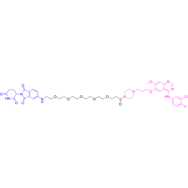
MS9427 is a potent PROTAC EGFR degrader with Kds of 7.1 nM and 4.3 nM for EGFR WT and EGFR L858R, respectively. MS9427 selectively degrades the mutant but not the WT EGFR through both the ubiquitin/proteasome system (UPS) and autophagy/lysosome pathways. MS9427 potently inhibits the proliferation of NSCLC cells. MS9427 can be used for researching anticancer.
(Pink: EGFR ligand (HY-W109039); Blue: Cereblon ligand (HY-W023573); Black: linker).
For research use only. We do not sell to patients.
- Purity : 95.29%
- CAS No.: 2772613-37-1
- Formula: C48H58ClFN8O12
- Molecular Weight:993.47
-
Storage:
4°C, protect from light, stored under nitrogen
* In solvent : -80°C, 6 months; -20°C, 1 month (protect from light, stored under nitrogen)

To place orders, for customer services and technical support, please contact: MedChemExpress USA
Tel: 609-228-6898 E-mail: [email protected] [email protected]


-
 Biological Activity
-
Chemical Information
-
Solvent & Solubility
- Protocols
- Purity & Documentation
- References
-
 Help & FAQs




Biological Activity
Description
IC50 & Target
[1]|
EGFR (WT) 7.1 nM (Kd) |
EGFR L858R 4.3 nM (Kd) |
Cellular Effect
|
Cell Line

|
Type | Value | Description | References |
|---|---|---|---|---|
| HCC827 | GI50 |
0.87 μM
Compound: 72; MS9427
|
Antiproliferative activity against human HCC827 cells assessed as cell growth inhibition incubated for 3 days by CCK-8 assay
Antiproliferative activity against human HCC827 cells assessed as cell growth inhibition incubated for 3 days by CCK-8 assay
|
[PMID: 35675209] |
In Vitro
MS9427 has antiproliferative activity against HCC-827 cells, with a GI50 of 0.87 ± 0.27 μM[1].
MS9427 (0-10 μM, 16 h) potently induces EGFRDel19 degradation (DC50=82 ± 73 nM) and inhibits EGFR phosphorylation (p-EGFR) in a concentration-dependent manner in HCC-827 cells[1].
MedChemExpress (MCE) has not independently confirmed the accuracy of these methods. They are for reference only.
-
Cell Line:HCC-827 cells
-
Concentration:1, 10, 50, 100, 200, 500, 100, and 10000 nM
-
Incubation Time:16 h
-
Result:Inhibited EGFR phosphorylation (p-EGFR) in a concentration-dependent manner in HCC-827 cells.
-
Cell Line:HCC-827 cells
-
Concentration:100 nM
-
Incubation Time:1, 2, 4, 6, 12, 24, 48 h
-
Result:Induced EGFR degradation in a time-dependent manner and through the UPS and autophagy/lysosome system.


Chemical Information
-
CAS No. 2772613-37-1
-
Appearance Solid
-
Molecular Weight 993.47
-
Formula C48H58ClFN8O12
-
Color Off-white to light yellow
-
SMILES
O=C1N(C(CC2)C(NC2=O)=O)C(C3=C1C=CC(NCCOCCOCCOCCOCCOCCC(N4CCN(CCCOC5=CC6=C(NC7=CC=C(F)C(Cl)=C7)N=CN=C6C=C5OC)CC4)=O)=C3)=O
-
Shipping
Room temperature in continental US; may vary elsewhere.
-
Storage
4°C, protect from light, stored under nitrogen
* In solvent : -80°C, 6 months; -20°C, 1 month (protect from light, stored under nitrogen)


Solvent & Solubility
In Vitro:
DMSO : 70 mg/mL (70.46 mM; Need ultrasonic; Hygroscopic DMSO has a significant impact on the solubility of product, please use newly opened DMSO)
Please refer to the solubility information to select the appropriate solvent. Once prepared, please aliquot and store the solution to prevent product inactivation from repeated freeze-thaw cycles.
Storage method and period of stock solution: -80°C, 6 months; -20°C, 1 month (protect from light, stored under nitrogen). When stored at -80°C, please use it within 6 months. When stored at -20°C, please use it within 1 month.
Please refer to the solubility information to select the appropriate solvent. Once prepared, please aliquot and store the solution to prevent product inactivation from repeated freeze-thaw cycles.
Storage method and period of stock solution: -80°C, 6 months; -20°C, 1 month (protect from light, stored under nitrogen). When stored at -80°C, please use it within 6 months. When stored at -20°C, please use it within 1 month.
Concentration (start) × Volume (start) = Concentration (final) × Volume (final)


Purity & Documentation
-
Data Sheet (268 KB)
-
SDS (252 KB)
- English - EN (252 KB)
- Français - FR (252 KB)
- Deutsch - DE (252 KB)
- Norwegian - NO (252 KB)
- Español - ES (252 KB)
- Swedish - SV (252 KB)
- Italian - IT (252 KB)
- Korean - KR (252 KB)
- Portuguese - PT (252 KB)
-
Handling Instructions (2659 KB)
References


Complete Stock Solution Preparation Table
Please refer to the solubility information to select the appropriate solvent. Once prepared, please aliquot and store the solution to prevent product inactivation from repeated freeze-thaw cycles.
Storage method and period of stock solution: -80°C, 6 months; -20°C, 1 month (protect from light, stored under nitrogen). When stored at -80°C, please use it within 6 months. When stored at -20°C, please use it within 1 month.
| Optional Solvent | Concentration Solvent Mass | 1 mg | 5 mg | 10 mg | 25 mg |
|---|---|---|---|---|---|
| DMSO | 1 mM | 1.0066 mL | 5.0329 mL | 10.0657 mL | 25.1643 mL |
| 5 mM | 0.2013 mL | 1.0066 mL | 2.0131 mL | 5.0329 mL | |
| 10 mM | 0.1007 mL | 0.5033 mL | 1.0066 mL | 2.5164 mL | |
| 15 mM | 0.0671 mL | 0.3355 mL | 0.6710 mL | 1.6776 mL | |
| 20 mM | 0.0503 mL | 0.2516 mL | 0.5033 mL | 1.2582 mL | |
| 25 mM | 0.0403 mL | 0.2013 mL | 0.4026 mL | 1.0066 mL | |
| 30 mM | 0.0336 mL | 0.1678 mL | 0.3355 mL | 0.8388 mL | |
| 40 mM | 0.0252 mL | 0.1258 mL | 0.2516 mL | 0.6291 mL | |
| 50 mM | 0.0201 mL | 0.1007 mL | 0.2013 mL | 0.5033 mL | |
| 60 mM | 0.0168 mL | 0.0839 mL | 0.1678 mL | 0.4194 mL |


MS9427 Related Classifications
HY-147941 Related Classifications
 Powered by Bioz