



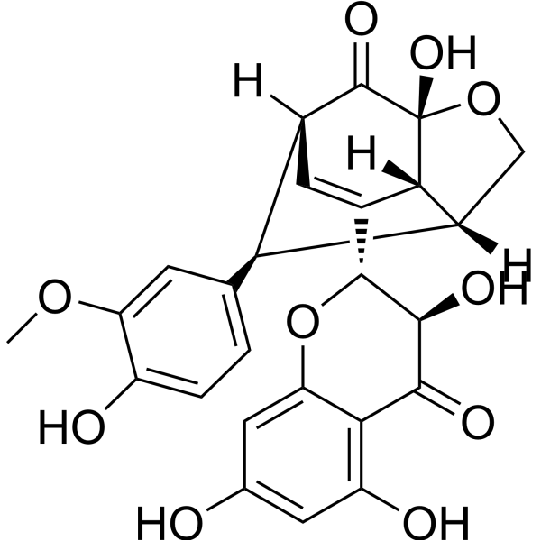
Silydianin
Based on 1 publication(s) in Google Scholar
Silydianin is a flavonolignan. Silydianin can be obtained from Silybum marianum. Silydianin inhibits PTP1B with an IC50 of 17.38 μM. Silydianin inhibits both monophenolase and diphenolase of tyrosinase significantly, with IC50s of 2.6 μM and 16.5 μM, respectively. Silydianin induces Apoptosis and reduces cytokines (IL-4 and IL-5). Silymarin has antioxidant, cytoprotective and immunomodulatory effects. Silydianin has antitumor activity against prostate cancer. Silymarin can be used in allergic asthma research.
Nos produits utilisent uniquement pour la recherche. Nous ne vendons pas aux patients.
- Pureté : 99.71%
- CAS No.: 29782-68-1
- Formule: C25H22O10
- Masse moléculaire:482.44
-
Stockage:Powder -20°C, 3 years , 4°C, 2 years ; In solvent -80°C, 6 months , -20°C, 1 month

To place orders, for customer services and technical support, please contact: MedChemExpress USA
Tel: 609-228-6898 E-mail: [email protected] [email protected]


-
 Activité biologique
-
Chemical Information
-
Solvant et solubilité
- Protocole
- Pureté et documentation
- Références
-
 Help & FAQs


-
HY-N0647
SilychristinMCT8 Inhibitor -
HY-N1181
TamarixetinAnti-inflammatory And Antioxidant -
HY-121936
Yohimbic acidDrug Derivative -

HY-N0646R
Silydianin (Standard)Standard of Silydianin -
HY-100463
TPI-1SHP-1 Inhibitor -
HY-115502
BCIDUSP6 Inhibitor -
HY-B0579
Cyclosporin ACalcineurin Inhibitor -
HY-15832
GSK 2830371Wip1 Phosphatase Inhibitor -
HY-100625
BVT948PTP Inhibitor -
HY-100462
PTP1B-IN-2PTP1B Inhibitor -
HY-B0879A
Suramin sodium saltPTPases Inhibitor -
HY-D0886
β-Glycerophosphate disodium salt pentahydratePhosphatase Inhibitor
Publications Citing Use of MedChemExpress (MCE) Silydianin
MoreVoir tous les produits spécifiques à Isoform Endogenous Metabolite
More

Activité biologique
Description
IC50 & Target
IC50s of 2.6 μM and 16.5 μM for monophenolase and diphenolase, respectively
Cellular Effect
|
Cell Line

|
Type | Value | Description | References |
|---|---|---|---|---|
| Huh-7 | IC50 |
>130 μM
Compound: 13
|
Cytotoxicity against human Huh7.5.1 cells after 72 hrs
Cytotoxicity against human Huh7.5.1 cells after 72 hrs
|
[PMID: 23673225] |
In Vitro
Silydianin inhibits both monophenolase and diphenolase of tyrosinase significantly, with IC50s of 2.6 μM and 16.5 μM for monophenolase and diphenolase, respectively[1].
Silydianin (0.1-100 μM; 2-24 h) slightly induces apoptosis of stimulated and unstimulated neutrophils[2].
Silydianin (10-100 μM; 30 min) displays concentration-dependent cytoprotection against the toxic effects of Allyl alcohol and Carbon tetrachloride (HY-Y0298) in primary human hepatocytes[3].
Silydianin (30-90 μM; 72 h) is slightly growth inhibitory in DU145 and PC3 cells[4].
MedChemExpress (MCE) has not independently confirmed the accuracy of these methods. They are for reference only.
In Vivo
MedChemExpress (MCE) has not independently confirmed the accuracy of these methods. They are for reference only.
-
Animal Model:Female Balb/c mice (6-8 weeks old) with allergic asthma model[5]
-
Dosage:1% solution
-
Administration:Aerosolized by ultrasonic nebulizer, 3 times, on days 25, 27, 29
-
Result:Reduced eosinophilic infiltration in the lungs, decreased IL-4 and IL-5 levels in BAL fluid, downregulated IL-4 and IL-5 gene expressions.
Alleviated AHR.
Did not significantly affect IL-13 level and mucus hyper-secretion.


Chemical Information
-
CAS No. 29782-68-1
-
Appearance Solid
-
Masse moléculaire 482.44
-
Formule C25H22O10
-
Color White to off-white
-
SMILES
O=C1[C@]2([H])C=C([C@@H]3[C@@H](O)C(C4=C(O)C=C(O)C=C4O3)=O)[C@@]5([H])[C@]([C@H]2C6=CC=C(O)C(OC)=C6)([H])CO[C@]51O
-
Structure Classification
-
Initial Source
-
Livraison
Room temperature in continental US; may vary elsewhere.
-
Stockage
Powder -20°C 3 years 4°C 2 years In solvent -80°C 6 months -20°C 1 month


Publications (1)
-
Journal Impact Factor
-
Most Recent


Solvant et solubilité
In Vitro:
DMSO : 125 mg/mL (259.10 mM; Need ultrasonic; Hygroscopic DMSO has a significant impact on the solubility of product, please use newly opened DMSO)
Please refer to the solubility information to select the appropriate solvent. Once prepared, please aliquot and store the solution to prevent product inactivation from repeated freeze-thaw cycles.
Storage method and period of stock solution: -80°C, 6 months; -20°C, 1 month. When stored at -80°C, please use it within 6 months. When stored at -20°C, please use it within 1 month.
Please refer to the solubility information to select the appropriate solvent. Once prepared, please aliquot and store the solution to prevent product inactivation from repeated freeze-thaw cycles.
Storage method and period of stock solution: -80°C, 6 months; -20°C, 1 month. When stored at -80°C, please use it within 6 months. When stored at -20°C, please use it within 1 month.
Concentration (start) × Volume (start) = Concentration (final) × Volume (final)
In Vivo:
Select the appropriate dissolution method based on your experimental animal and administration route.
- For the following dissolution methods, please ensure to first prepare a clear stock solution using an In Vitro approach and then sequentially add co-solvents:
- To ensure reliable experimental results, the clarified stock solution can be appropriately stored based on storage conditions. As for the working solution for In Vivo experiments, it is recommended to prepare freshly and use it on the same day.
- The percentages shown for the solvents indicate their volumetric ratio in the final prepared solution. If precipitation or phase separation occurs during preparation, heat and/or sonication can be used to aid dissolution.
Add each solvent one by one: 10% DMSO 40% PEG300 5% Tween-80 45% Saline
Solubility: ≥ 2.08 mg/mL (4.31 mM); Clear solution
This protocol yields a clear solution of ≥ 2.08 mg/mL (saturation unknown).
Taking 1 mL working solution as an example, add 100 μL DMSO stock solution (20.8 mg/mL) to 400 μL PEG300, and mix evenly; then add 50 μL Tween-80 and mix evenly; then add 450 μL Saline to adjust the volume to 1 mL.
Preparation of Saline: Dissolve 0.9 g sodium chloride in ddH₂O and dilute to 100 mL to obtain a clear Saline solution.
Add each solvent one by one: 10% DMSO 90% (20% SBE-β-CD in Saline)
Solubility: ≥ 2.08 mg/mL (4.31 mM); Clear solution
This protocol yields a clear solution of ≥ 2.08 mg/mL (saturation unknown).
Taking 1 mL working solution as an example, add 100 μL DMSO stock solution (20.8 mg/mL) to 900 μL 20% SBE-β-CD in Saline, and mix evenly.
Preparation of 20% SBE-β-CD in Saline (4°C, storage for one week): 2 g SBE-β-CD powder is dissolved in 10 mL Saline, completely dissolve until clear.
In Vivo Dissolution Calculator
Please enter the basic information of animal experiments:
-
-
-
-
Recommended: Prepare an additional quantity of animals to account for potential losses during experiments.
Please enter your animal formula composition:
-
%DMSO +
Recommended: Keep the proportion of DMSO in working solution below 2% if your animal is weak.
-
%+
-
+%Tween-80 + +
-
%Saline +
The co-solvents required include: DMSO, . All of co-solvents are available by MedChemExpress (MCE). , Tween 80. All of co-solvents are available by MedChemExpress (MCE).
Working solution concentration: 0.22 mg/mL
Method for preparing stock solution: mg drug dissolved in μL DMSO. Stock solution concentration: mg/mL.
1. Take μL DMSO stock solution;
2. Add μL .
μL , mix evenly;
3. Then add μL Tween 80, mix evenly;
4. Then add μL
Please ensure that the stock solution in the first step is dissolved to a clear state, and add co-solvents in sequence. You can use ultrasonic heating (ultrasonic cleaner, recommended frequency 20-40 kHz), vortexing, etc. to assist dissolution.


Pureté et documentation
-
Fiche technique (293 KB)
-
SDS (396 KB)
- English - EN (396 KB)
- Français - FR (396 KB)
- Deutsch - DE (396 KB)
- Norwegian - NO (396 KB)
- Español - ES (396 KB)
- Swedish - SV (396 KB)
- Italian - IT (396 KB)
- Korean - KR (396 KB)
- Portuguese - PT (396 KB)
-
Instruction de manipulation (2659 KB)
Références
[1]. Kim JY, et al. Tyrosinase inhibitory study of flavonolignans from the seeds of Silybum marianum (Milk thistle). Bioorg Med Chem. 2019 Jun 15;27(12):2499-2507. [Content Brief]
[2]. Zielińska-Przyjemska M, et al. An in vitro study of the protective effect of the flavonoid silydianin against reactive oxygen species. Phytother Res. 2006 Feb;20(2):115-9. [Content Brief]
[3]. Dvorák Z, et al. Primary cultures of human hepatocytes as a tool in cytotoxicity studies: cell protection against model toxins by flavonolignans obtained from Silybum marianum. Toxicol Lett. 2003 Feb 3;137(3):201-12. [Content Brief]
[4]. Davis-Searles PR, et al. Milk thistle and prostate cancer: differential effects of pure flavonolignans from Silybum marianum on antiproliferative end points in human prostate carcinoma cells. Cancer Res. 2005 May 15;65(10):4448-57. [Content Brief]
[5]. Nasab EM, et al. Immunomodulatory effects of two silymarin isomers in a Balb/c mouse model of allergic asthma. Allergol Immunopathol (Madr). 2020 Nov-Dec;48(6):646-653. [Content Brief]
[6]. Qin N, et al. Identification of flavonolignans from Silybum marianum seeds as allosteric protein tyrosine phosphatase 1B inhibitors. J Enzyme Inhib Med Chem. 2018 Dec;33(1):1283-1291. [Content Brief]
[7]. Vostálová J, et al. Skin Protective Activity of Silymarin and its Flavonolignans. Molecules. 2019 Mar 14;24(6). [Content Brief]


Complete Stock Solution Preparation Table
Please refer to the solubility information to select the appropriate solvent. Once prepared, please aliquot and store the solution to prevent product inactivation from repeated freeze-thaw cycles.
Storage method and period of stock solution: -80°C, 6 months; -20°C, 1 month. When stored at -80°C, please use it within 6 months. When stored at -20°C, please use it within 1 month.
| Optional Solvent | Concentration Solvent Mass | 1 mg | 5 mg | 10 mg | 25 mg |
|---|---|---|---|---|---|
| DMSO | 1 mM | 2.0728 mL | 10.3640 mL | 20.7280 mL | 51.8199 mL |
| 5 mM | 0.4146 mL | 2.0728 mL | 4.1456 mL | 10.3640 mL | |
| 10 mM | 0.2073 mL | 1.0364 mL | 2.0728 mL | 5.1820 mL | |
| 15 mM | 0.1382 mL | 0.6909 mL | 1.3819 mL | 3.4547 mL | |
| 20 mM | 0.1036 mL | 0.5182 mL | 1.0364 mL | 2.5910 mL | |
| 25 mM | 0.0829 mL | 0.4146 mL | 0.8291 mL | 2.0728 mL | |
| 30 mM | 0.0691 mL | 0.3455 mL | 0.6909 mL | 1.7273 mL | |
| 40 mM | 0.0518 mL | 0.2591 mL | 0.5182 mL | 1.2955 mL | |
| 50 mM | 0.0415 mL | 0.2073 mL | 0.4146 mL | 1.0364 mL | |
| 60 mM | 0.0345 mL | 0.1727 mL | 0.3455 mL | 0.8637 mL | |
| 80 mM | 0.0259 mL | 0.1295 mL | 0.2591 mL | 0.6477 mL | |
| 100 mM | 0.0207 mL | 0.1036 mL | 0.2073 mL | 0.5182 mL |


Silydianin Related Classifications
HY-N0646 Related Classifications
 Powered by Bioz