



BMS-986365
Based on 3 publication(s) in Google Scholar
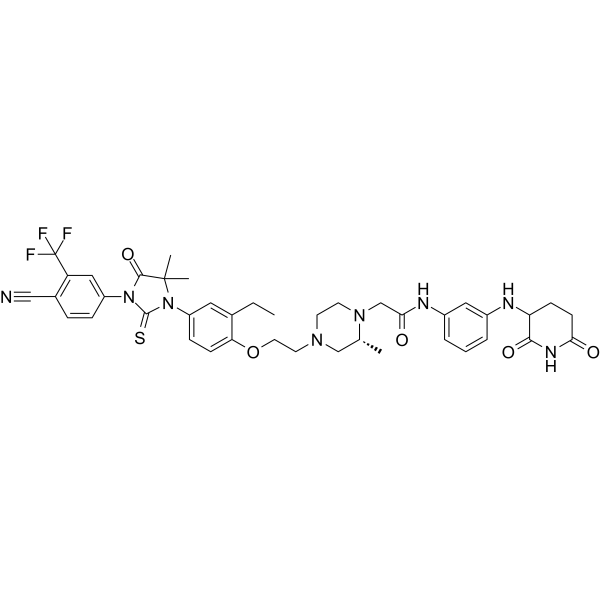
BMS-986365 (CC-94676) is an orally active and selective targeted androgen receptor (AR) PROTAC degrader (DC50 of 10-40 nM). BMS-986365 is capable of inducing cereblon (CRBN) E3 ligase-dependent ubiquitination and degradation of the androgen receptor (AR), as well as various AR mutants. BMS-986365 shows no degradation of the close AR family members estrogen receptor (ER), progesterone receptor (PR), and glucocorticoid receptor (GR). BMS-986365 shows significant in vivo potency, degrading AR, inhibiting AR signaling, and restricting tumor growth in animal models of advanced prostate cancer. BMS-986365 can be used for the study of metastatic castration-resistant prostate cancer (mCRPC).
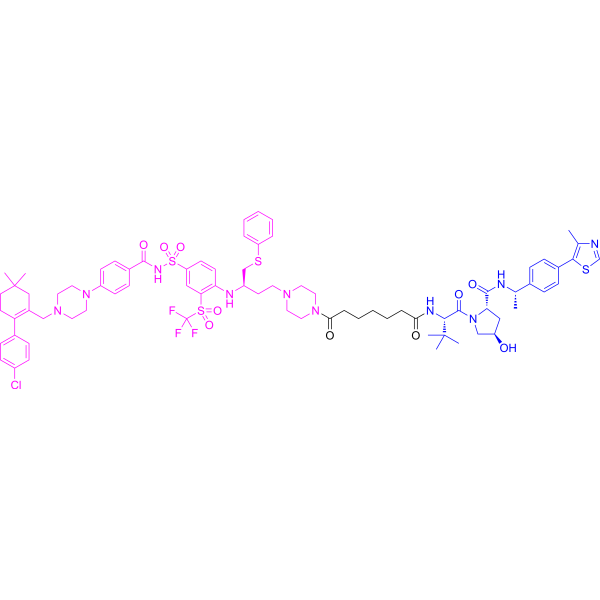
(Pink: Androgen Receptor ligand (HY-168697); Blue: Cereblon ligand (HY-W247437); Black: linker).
For research use only. We do not sell to patients.
- Purity : 99.92%
- CAS No.: 2446928-30-7
- Formula: C41H45F3N8O5S
- Molecular Weight:818.91
-
Storage:Powder -20°C, 3 years , 4°C, 2 years ; In solvent -80°C, 6 months , -20°C, 1 month

To place orders, for customer services and technical support, please contact: MedChemExpress USA
Tel: 609-228-6898 E-mail: [email protected] [email protected]


-
 Biological Activity
-
Chemical Information
-
Solvent & Solubility
- Protocols
- Purity & Documentation
- References
-
 Help & FAQs


-
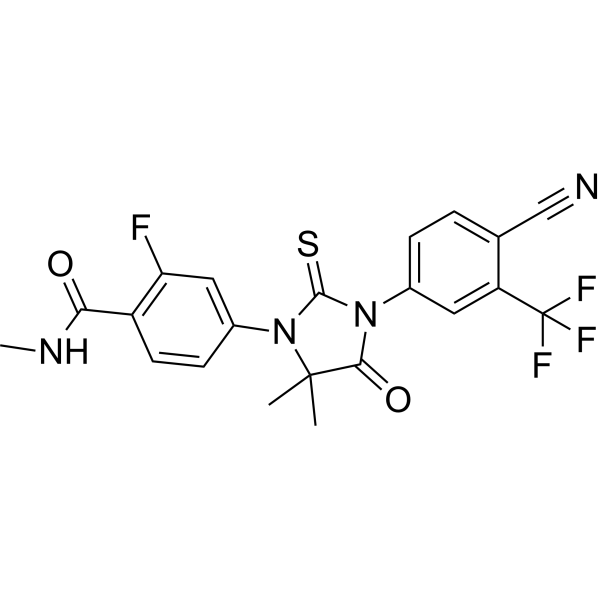
HY-70002
EnzalutamideAndrogen Receptor Antagonist -
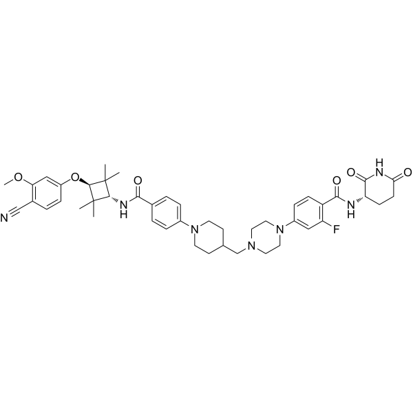
HY-153342
LuxdegalutamidePROTAC -
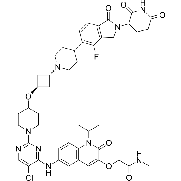
HY-158105
ARV-393BCL6 PROTAC Degrader -
HY-16985
DarolutamideAndrogen Receptor Antagonist -
HY-153220
ZelebrudomideBTK PROTAC Degrader -
HY-138641
BavdegalutamideAR PROTAC Degrader -
HY-138642
VepdegestrantPROTAC ER Degrader -
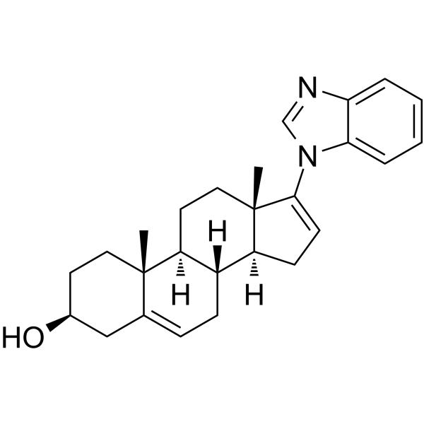
HY-70006
GaleteroneMolecular Glue Degrader -
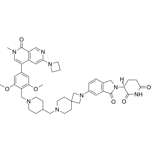
HY-153367
FHD-609BRD9 PROTAC Degrader -
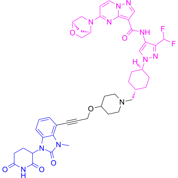
HY-145483
KT-474PROTAC IRAK4 Degrader -
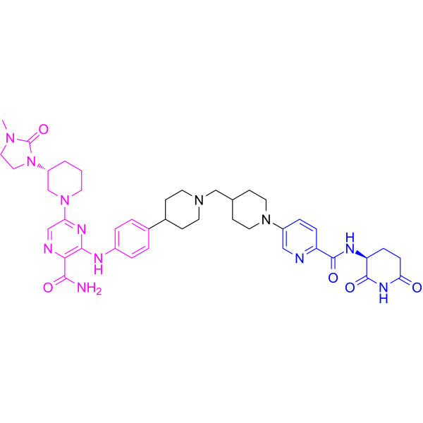
HY-153321
BexobrutidegBTK Degrader -
HY-130604
DT2216PROTAC BCL-XL Degrader
Publications Citing Use of MedChemExpress (MCE) BMS-986365
MoreAll PROTACs Isoforms
More

Biological Activity
Description
In Vitro
BMS-986365 (0.05-5 μM; 6 h) maintains low levels of AR protein despite increased AR transcript levels in VCaP and LNCaP cells[3].
BMS986365 (0.1-10 μM; 6-72 hours) inhibits AR target gene transcription and AR-dependent proliferation in VCaP and LNCaP cells[3].
BMS-986365 exhibits AR degradation potency in VCaP (ARWT amplified) cells with a GI50: 753 nM)[3].
BMS-986365 shows a difference in degradation potency in VCaP cells and LNCaP cells, with DC50 values of 7 nM and 29 nM, respectively[3].
BMS986365 potently inhibits androgen-stimulated proliferation of VCaP and LNCaP cells but showed little antiproliferative effect in 22Rv1 or PC3 cells[3].
BMS986365 inhibits R1881-stimulated proliferation of VCaP and LNCaP cells with GI50 values of 11 and 4 nM, respectively[3].
MedChemExpress (MCE) has not independently confirmed the accuracy of these methods. They are for reference only.
-
Cell Line:VCaP and LNCaP cells
-
Concentration:0.05 μM, 0.5 μM, and 5 μM
-
Incubation Time:6 h
-
Result:Indicated strong degradation of AR.
The T1/2 for AR degradation at 500 nM was ~0.6 and ~1.3 hours for VCaP and LNCaP cells, respectively.
-
Cell Line:VCaP and LNCaP cellsVCaP and LNCaP cells treated with R1881
-
Concentration:0.1 μM, 1 μM, or 10 μM
-
Incubation Time:6, 24, or 72 hours
-
Result:Exerted a potent inhibitory effect on androgen upregulated FKBP5 expression, with IC50 values of 1 and 4 nM for VCaP and LNCaP, respectively.
Parmacokinetics
In Vivo
BMS-986365 (p.o.; for 7 days once daily) shows dose-dependent degradation of AR in male beagle dogs and sexually immature cynomolgus monkeys[3].
BMS986365 (30 mg/kg; p.o.; once daily; for 45 consecutive days) exhibits significant antitumor activity in a VCaP mouse xenograft model[3].
MedChemExpress (MCE) has not independently confirmed the accuracy of these methods. They are for reference only.
-
Animal Model:Male 6- to 8-week-old intact or castrated NOD.Cg-Prkdcscid Il2rgtm1Wjl/SzJ (NSG) were inoculated with 5 x 10⁶ VCaP or HR-VCaP cells[3]
-
Dosage:30 mg/kg
-
Administration:p.o.; once daily; for 3 consecutive days
-
Result:Evaluation of plasma and tumor samples indicated good compound exposure in both the circulation and within tumors at 2, 6, and 24 hours after the last dose.
Reduced AR to 91% and 83% of control levels at 6 and 24 hours, respectively, after the last dose.
-
Animal Model:Male NSG mice were inoculated with 5 x 10⁶ HR-VCaP tumor cells into the right flank[3].
-
Dosage:30 mg/kg
-
Administration:p.o.; once daily for 45 consecutive days.
-
Result:Exhibited significant antitumor activity in a VCaP xenograft model, exhibiting 81% tumor volume reduction in intact mice.
Clinical Trial
| NCT Number | Sponsor | Condition | Start Date |
Phase

|
|---|---|---|---|---|
| NCT01329991 | Plexxikon| | 2011-05 | PHASE1 |


Chemical Information
-
CAS No. 2446928-30-7
-
Appearance Solid
-
Molecular Weight 818.91
-
Formula C41H45F3N8O5S
-
Color White to off-white
-
SMILES
N#CC1=C(C(F)(F)F)C=C(N2C(N(C3=CC=C(OCCN4C[C@@H](C)N(CC(NC5=CC=CC(NC6C(NC(CC6)=O)=O)=C5)=O)CC4)C(CC)=C3)C(C)(C)C2=O)=S)C=C1
-
Synonyms
CC-94676
-
Shipping
Room temperature in continental US; may vary elsewhere.
-
Storage
Powder -20°C 3 years 4°C 2 years In solvent -80°C 6 months -20°C 1 month


Publications (3)
-
Journal Impact Factor
-
Most Recent
-
Anal Chem
Hydrogen/Deuterium Exchange for Chiral Stability Assessment in Acidic Methine-Containing Compounds. [Abstract]2025 Dec 2;97(47):26097-26107. PMID: 41243541 -
-


Solvent & Solubility
In Vitro:
DMSO : 100 mg/mL (122.11 mM; Need ultrasonic; Hygroscopic DMSO has a significant impact on the solubility of product, please use newly opened DMSO)
Please refer to the solubility information to select the appropriate solvent. Once prepared, please aliquot and store the solution to prevent product inactivation from repeated freeze-thaw cycles.
Storage method and period of stock solution: -80°C, 6 months; -20°C, 1 month. When stored at -80°C, please use it within 6 months. When stored at -20°C, please use it within 1 month.
Please refer to the solubility information to select the appropriate solvent. Once prepared, please aliquot and store the solution to prevent product inactivation from repeated freeze-thaw cycles.
Storage method and period of stock solution: -80°C, 6 months; -20°C, 1 month. When stored at -80°C, please use it within 6 months. When stored at -20°C, please use it within 1 month.
Concentration (start) × Volume (start) = Concentration (final) × Volume (final)
In Vivo:
Select the appropriate dissolution method based on your experimental animal and administration route.
- For the following dissolution methods, please ensure to first prepare a clear stock solution using an In Vitro approach and then sequentially add co-solvents:
- To ensure reliable experimental results, the clarified stock solution can be appropriately stored based on storage conditions. As for the working solution for In Vivo experiments, it is recommended to prepare freshly and use it on the same day.
- The percentages shown for the solvents indicate their volumetric ratio in the final prepared solution. If precipitation or phase separation occurs during preparation, heat and/or sonication can be used to aid dissolution.
In Vivo Dissolution Calculator
Please enter the basic information of animal experiments:
-
-
-
-
Recommended: Prepare an additional quantity of animals to account for potential losses during experiments.
Please enter your animal formula composition:
-
%DMSO +
Recommended: Keep the proportion of DMSO in working solution below 2% if your animal is weak.
-
%+
-
+%Tween-80 + +
-
%Saline +
The co-solvents required include: DMSO, . All of co-solvents are available by MedChemExpress (MCE). , Tween 80. All of co-solvents are available by MedChemExpress (MCE).
Working solution concentration: 0.22 mg/mL
Method for preparing stock solution: mg drug dissolved in μL DMSO. Stock solution concentration: mg/mL.
1. Take μL DMSO stock solution;
2. Add μL .
μL , mix evenly;
3. Then add μL Tween 80, mix evenly;
4. Then add μL
Please ensure that the stock solution in the first step is dissolved to a clear state, and add co-solvents in sequence. You can use ultrasonic heating (ultrasonic cleaner, recommended frequency 20-40 kHz), vortexing, etc. to assist dissolution.


Purity & Documentation
-
Data Sheet (277 KB)
-
SDS (252 KB)
- English - EN (252 KB)
- Français - FR (252 KB)
- Deutsch - DE (252 KB)
- Norwegian - NO (252 KB)
- Español - ES (252 KB)
- Swedish - SV (252 KB)
- Italian - IT (252 KB)
- Korean - KR (252 KB)
- Portuguese - PT (252 KB)
-
Handling Instructions (2659 KB)
References
[2]. Rathkopf DE, et al. Safety and clinical activity of BMS-986365 (CC-94676), a dual androgen receptor ligand-directed degrader and antagonist, in heavily pretreated patients with metastatic castration-resistant prostate cancer. Ann Oncol. 2024 Sep 16:S0923-7534(24)04001-8. [Content Brief]
[3]. Nayak S, et al. Discovery of BMS-986365, a first-in-class dual androgen receptor ligand-directed degrader (AR LDD) and antagonist, for the treatment of advanced prostate cancer. Clin Cancer Res. 2025 Aug 11. [Content Brief]


Complete Stock Solution Preparation Table
Please refer to the solubility information to select the appropriate solvent. Once prepared, please aliquot and store the solution to prevent product inactivation from repeated freeze-thaw cycles.
Storage method and period of stock solution: -80°C, 6 months; -20°C, 1 month. When stored at -80°C, please use it within 6 months. When stored at -20°C, please use it within 1 month.
| Optional Solvent | Concentration Solvent Mass | 1 mg | 5 mg | 10 mg | 25 mg |
|---|---|---|---|---|---|
| DMSO | 1 mM | 1.2211 mL | 6.1057 mL | 12.2114 mL | 30.5284 mL |
| 5 mM | 0.2442 mL | 1.2211 mL | 2.4423 mL | 6.1057 mL | |
| 10 mM | 0.1221 mL | 0.6106 mL | 1.2211 mL | 3.0528 mL | |
| 15 mM | 0.0814 mL | 0.4070 mL | 0.8141 mL | 2.0352 mL | |
| 20 mM | 0.0611 mL | 0.3053 mL | 0.6106 mL | 1.5264 mL | |
| 25 mM | 0.0488 mL | 0.2442 mL | 0.4885 mL | 1.2211 mL | |
| 30 mM | 0.0407 mL | 0.2035 mL | 0.4070 mL | 1.0176 mL | |
| 40 mM | 0.0305 mL | 0.1526 mL | 0.3053 mL | 0.7632 mL | |
| 50 mM | 0.0244 mL | 0.1221 mL | 0.2442 mL | 0.6106 mL | |
| 60 mM | 0.0204 mL | 0.1018 mL | 0.2035 mL | 0.5088 mL | |
| 80 mM | 0.0153 mL | 0.0763 mL | 0.1526 mL | 0.3816 mL | |
| 100 mM | 0.0122 mL | 0.0611 mL | 0.1221 mL | 0.3053 mL |


BMS-986365 Related Classifications
HY-158101 Related Classifications
 Powered by Bioz