



Valencene
Based on 1 publication(s) in Google Scholar
Valencene is a sesquiterpene that can be isolated from Cyperus rotundus. Valencene possesses antiallergic, antimelanogenesis, anti-infammatory, and antioxidant activitivies. Valencene inhibits the exaggerated expression of Th2 chemokines and proinflammatory chemokines through blockade of the NF-κB pathway. Valencene inhibits the production and expression of proinflammatory cytokines IL-1β and IL-6 in LPS-stimulated RAW 264.7 cells. Valencene can reduce atopic dermatitis symptoms and recover decreased expression of filaggrin in DNCB-sensitized mouse model.
For research use only. We do not sell to patients.
- Purity : 80.24%
- CAS No.: 4630-07-3
- Formula: C15H24
- Molecular Weight:204.35
-
Storage:
-20°C, protect from light
* In solvent : -80°C, 6 months; -20°C, 1 month (protect from light)

To place orders, for customer services and technical support, please contact: MedChemExpress USA
Tel: 609-228-6898 E-mail: [email protected] [email protected]


-
 Biological Activity
-
Chemical Information
-
Solvent & Solubility
- Protocols
- Purity & Documentation
- References
-
 Help & FAQs



Publications Citing Use of MedChemExpress (MCE) Valencene
More Customer Validation & Images

-
Cell Proliferation/Viability Assay

-
IF

-
WB

-
IHC

-
In Vivo Imaging


Biological Activity
Description
In Vitro
Valencene (10-100 μM, 1 h pre-treatment) significantly inhibits TNF-α/IFN-γ-stimulated chemokines secretion in HaCaT cells[1].
Valencene (10-100 μM, 24 h) significantly increases involucrin mRNA/protein expression[1].
MedChemExpress (MCE) has not independently confirmed the accuracy of these methods. They are for reference only. Further protocols information, click here.
-
Cell Line:TNF-α/IFN-γ-stimulated HaCaT cells
-
Concentration:10, 50, 100 μM
-
Incubation Time:1 h pre-treatment
-
Result:Significantly inhibited the mRNA expression of the AD-related chemokines CCL17/TARC. CCL22/MDC, CXCL8, MCP-1 and GM-CSF.
In Vivo
MedChemExpress (MCE) has not independently confirmed the accuracy of these methods. They are for reference only.
-
Animal Model:DNCB-sensitized male NC/Nga mice (6-week-old)[1]
-
Dosage:200 μL of 0, 50 or 100 μM
-
Administration:Topical, three times a week for 10 weeks
-
Result:Resulted in increased expression of filaggrin.
Reduced stratching behavior.
Decreased the tissue levels of IL-1β, IL-6, and IL-13.
Markedly suppressed the DNCB-induced increase of IL-1β, IL-6, and IL-13 levels in spleen tissue.


Chemical Information
-
CAS No. 4630-07-3
-
Appearance Liquid (Density: 0.92 g/cm3)
-
Molecular Weight 204.35
-
Formula C15H24
-
Color Colorless to light yellow
-
SMILES
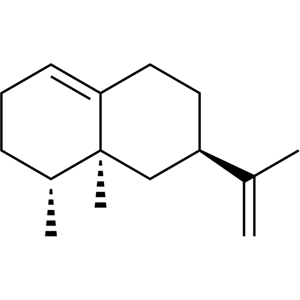
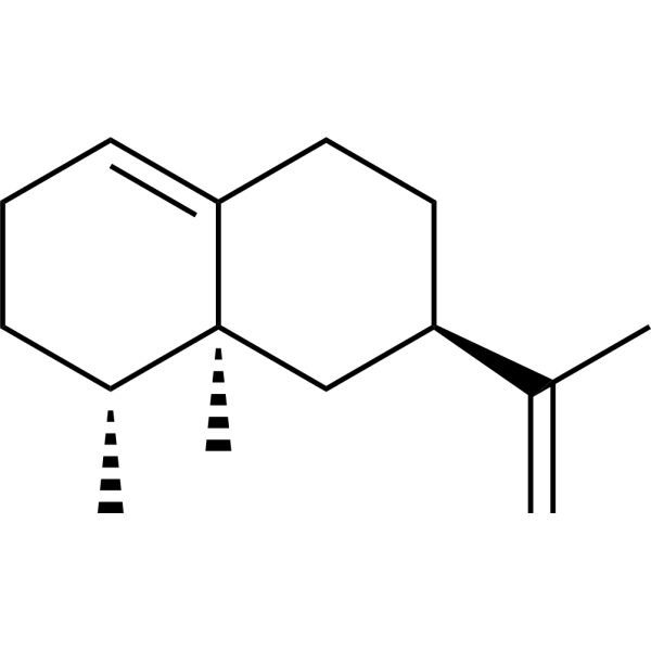
C=C([C@@H]1CCC2=CCC[C@@H](C)[C@]2(C)C1)C
-
Structure Classification
-
Initial Source
-
Shipping
Room temperature in continental US; may vary elsewhere.
-
Storage
-20°C, protect from light
* In solvent : -80°C, 6 months; -20°C, 1 month (protect from light)


Publications (1)
-
Journal Impact Factor
-
Most Recent
-
Int Immunopharmacol
An in vitro and in vivo study: Valencene protects cartilage and alleviates the progression of osteoarthritis by anti-oxidative stress and anti-inflammatory effects. [Abstract]2023 Oct:123:110726. PMID: 37536183
Valencene purchased from MedChemExpress. Usage Cited in: Int Immunopharmacol. 2023 Oct:123:110726. [Abstract]
Cell viability was determined by CCK-8 assay treated with Valencene (VA) (10, 25, 50 μg/mL).
Valencene purchased from MedChemExpress. Usage Cited in: Int Immunopharmacol. 2023 Oct:123:110726. [Abstract]
ROS levels under fluorescence microscopy treated with Valencene (50 μg/mL).
Valencene purchased from MedChemExpress. Usage Cited in: Int Immunopharmacol. 2023 Oct:123:110726. [Abstract]
Protein levels of Keap1,NRF2, HO-1, NQO1 were detected by Western blot treated with Valencene (10, 25, 50 μg/mL).
Valencene purchased from MedChemExpress. Usage Cited in: Int Immunopharmacol. 2023 Oct:123:110726. [Abstract]
Immunohistochemistry staining of Aggrecan, Col2 and MMP13 treated with Valencene (10, 15 mg/kg, i.p.).
Valencene purchased from MedChemExpress. Usage Cited in: Int Immunopharmacol. 2023 Oct:123:110726. [Abstract]
Micro-ct three-dimensional reconstruction of mouse right knee joint treated with Valencene (VA) (10, 15 mg/kg, i.p.).


Solvent & Solubility
In Vitro:
DMSO : 50 mg/mL (244.68 mM; Need ultrasonic; Hygroscopic DMSO has a significant impact on the solubility of product, please use newly opened DMSO)
Please refer to the solubility information to select the appropriate solvent. Once prepared, please aliquot and store the solution to prevent product inactivation from repeated freeze-thaw cycles.
Storage method and period of stock solution: -80°C, 6 months; -20°C, 1 month (protect from light). When stored at -80°C, please use it within 6 months. When stored at -20°C, please use it within 1 month.
Please refer to the solubility information to select the appropriate solvent. Once prepared, please aliquot and store the solution to prevent product inactivation from repeated freeze-thaw cycles.
Storage method and period of stock solution: -80°C, 6 months; -20°C, 1 month (protect from light). When stored at -80°C, please use it within 6 months. When stored at -20°C, please use it within 1 month.
Concentration (start) × Volume (start) = Concentration (final) × Volume (final)
In Vivo:
Select the appropriate dissolution method based on your experimental animal and administration route.
- For the following dissolution methods, please ensure to first prepare a clear stock solution using an In Vitro approach and then sequentially add co-solvents:
- To ensure reliable experimental results, the clarified stock solution can be appropriately stored based on storage conditions. As for the working solution for In Vivo experiments, it is recommended to prepare freshly and use it on the same day.
- The percentages shown for the solvents indicate their volumetric ratio in the final prepared solution. If precipitation or phase separation occurs during preparation, heat and/or sonication can be used to aid dissolution.
Add each solvent one by one: 10% DMSO 40% PEG300 5% Tween-80 45% Saline
Solubility: ≥ 2.5 mg/mL (12.23 mM); Clear solution
This protocol yields a clear solution of ≥ 2.5 mg/mL (saturation unknown).
Taking 1 mL working solution as an example, add 100 μL DMSO stock solution (25.0 mg/mL) to 400 μL PEG300, and mix evenly; then add 50 μL Tween-80 and mix evenly; then add 450 μL Saline to adjust the volume to 1 mL.
Preparation of Saline: Dissolve 0.9 g sodium chloride in ddH₂O and dilute to 100 mL to obtain a clear Saline solution.
Add each solvent one by one: 10% DMSO 90% (20% SBE-β-CD in Saline)
Solubility: ≥ 2.5 mg/mL (12.23 mM); Clear solution
This protocol yields a clear solution of ≥ 2.5 mg/mL (saturation unknown).
Taking 1 mL working solution as an example, add 100 μL DMSO stock solution (25.0 mg/mL) to 900 μL 20% SBE-β-CD in Saline, and mix evenly.
Preparation of 20% SBE-β-CD in Saline (4°C, storage for one week): 2 g SBE-β-CD powder is dissolved in 10 mL Saline, completely dissolve until clear.
In Vivo Dissolution Calculator
Please enter the basic information of animal experiments:
-
-
-
-
Recommended: Prepare an additional quantity of animals to account for potential losses during experiments.
Please enter your animal formula composition:
-
%DMSO +
Recommended: Keep the proportion of DMSO in working solution below 2% if your animal is weak.
-
%+
-
+%Tween-80 + +
-
%Saline +
The co-solvents required include: DMSO, . All of co-solvents are available by MedChemExpress (MCE). , Tween 80. All of co-solvents are available by MedChemExpress (MCE).
Working solution concentration: 0.22 mg/mL
Method for preparing stock solution: mg drug dissolved in μL DMSO. Stock solution concentration: mg/mL. * In solvent : -80°C, 6 months; -20°C, 1 month (protect from light)
1. Take μL DMSO stock solution;
2. Add μL .
μL , mix evenly;
3. Then add μL Tween 80, mix evenly;
4. Then add μL
Please ensure that the stock solution in the first step is dissolved to a clear state, and add co-solvents in sequence. You can use ultrasonic heating (ultrasonic cleaner, recommended frequency 20-40 kHz), vortexing, etc. to assist dissolution.

Protocols
-
RNA extraction experimental
By lysing cells, releasing RNA, and removing impurities such as proteins and DNA, high-purity RNA products are finally obtained. The commonly used traditional method is the guanidine isothiocyanate/phenol/chloroform method (Trizol), which is suitable for a variety of animal materials including animal tissues, microorganisms, cultured cells, etc., and most plant materials.
-
Naïve CD4+ T-cell subset differentiation/polarization
Naïve CD4+ T-cell subset differentiation/polarization is an in vitro assay in which purified naïve CD4+ T cells are activated through TCR and CD28 costimulation and cultured with defined cytokines and neutralizing antibodies to generate Th0, Th1, Th2, Th17, or induced Treg-like populations. Differentiation is detected by subset-associated cytokines and transcription factors: IFN-γ/T-bet for Th1, IL-4/GATA3 for Th2, IL-17A/RORγt for Th17, and Foxp3 for induced Treg cells. The assay readout is usually generated by intracellular cytokine staining after restimulation, transcription-factor staining by flow cytometry, ELISA of secreted cytokines, or gene-expression analysis. The result reflects cytokine-directed lineage commitment or polarization rather than antigen-specific immune protection by itself.
-
TPA/Croton Oil Ear Edema and Dermatitis
The TPA (12-O-tetradecanoylphorbol-13-acetate) and croton oil-induced mouse ear edema model is a well-established acute cutaneous inflammation system used to evaluate topical anti-inflammatory activity by measuring edema formation, neutrophil infiltration, vascular permeability, and cytokine-mediated skin responses in vivo. The inflammatory response is triggered by topical application of phorbol esters (TPA) or croton oil constituents, leading to rapid activation of protein kinase C signaling, leukocyte recruitment, and increased vascular permeability, which can be quantified by ear thickness, weight, dye extravasation, and biochemical markers such as myeloperoxidase (MPO) activity and pro-inflammatory mediators in ear tissue homogenates. This model is widely used for screening anti-inflammatory agents, where reductions in edema and inflammatory biomarkers reflect suppression of acute dermal inflammation and immune cell infiltration. Histological evaluation typically confirms epidermal
-
Research Protocol for Inflammation-related Diseases
The NLRP3 inflammasome is a cytosolic innate immune signaling platform that integrates priming signals and danger-signal activation to promote caspase-1 activation, maturation of IL-1β and IL-18, and gasdermin D-mediated pyroptotic cell death. The core experimental logic is to determine whether inflammatory disease phenotypes are driven by increased NLRP3 expression, ASC-containing inflammasome assembly, caspase-1 cleavage, GSDMD cleavage, and extracellular release of IL-1β/IL-18 rather than by nonspecific cell injury alone. The pathway is strongly linked to inflammation-related disease phenotypes because monosodium urate crystals activate NALP3/NLRP3 inflammasome signaling in gout-like crystal inflammation, cholesterol crystals activate NLRP3 inflammasomes in atherogenesis models, and DSS-induced intestinal inflammation has been reported to involve NLRP3 inflammasome activity. However, experimental colitis studies also show context-dependent protective effects of NLRP3 inflammasome co
-
Pyroptosis Solutions
Pyroptosis is a lytic inflammatory cell-death pathway executed by gasdermin pores, most classically through inflammasome-mediated activation of caspase-1, cleavage of gasdermin D, membrane pore formation, LDH release, and secretion of IL-1β and IL-18. The canonical pathway is commonly modeled by priming cells with an inflammatory signal such as LPS to induce pro-IL-1β and inflammasome components, followed by an activation signal such as ATP or nigericin to activate NLRP3, ASC speck formation, caspase-1 cleavage, GSDMD cleavage, cytokine release, and pyroptotic membrane rupture. The non-canonical pathway is triggered when cytosolic LPS activates mouse caspase-11 or human caspase-4/5, leading to GSDMD cleavage and pyroptosis, and this can secondarily activate NLRP3-dependent IL-1β release. Pyroptosis is linked to inflammatory injury, infection, cancer, liver disease, ocular disease, placental inflammation, and other disease phenotypes, but unresolved questions include which gasdermin fam
-
How to Choose the Right Model Animal
Choosing the right model animal is a validity-driven decision in which the species, strain, sex, age, genetic background, disease-induction method, outcome measures, and welfare burden must match the scientific question rather than laboratory tradition or convenience. A model should be selected by judging face validity, construct validity, and predictive validity: whether it resembles the human phenotype, whether it reproduces relevant mechanisms, and whether results are likely to predict human biology or treatment response. Animal studies often fail to translate because of species differences, weak disease resemblance, poor experimental design, inadequate reporting, publication bias, and underuse of randomization, blinding, and sample-size justification. Unresolved questions include how to rank competing models objectively, how much human-disease complexity must be reproduced for a given objective, and when non-animal systems such as organoids, ex vivo tissue, or computational models


Purity & Documentation
-
Data Sheet (276 KB)
-
SDS (480 KB)
- English - EN (480 KB)
- Français - FR (480 KB)
- Deutsch - DE (480 KB)
- Norwegian - NO (480 KB)
- Español - ES (480 KB)
- Swedish - SV (480 KB)
- Italian - IT (480 KB)
- Korean - KR (480 KB)
- Portuguese - PT (480 KB)
-
Handling Instructions (2659 KB)
References
[1]. Yang IJ, et al. Inhibitory Effect of Valencene on the Development of Atopic Dermatitis-Like Skin Lesions in NC/Nga Mice. Evid Based Complement Alternat Med. 2016;2016:9370893. [Content Brief]
[2]. Frohwitter J, et al. Production of the sesquiterpene (+)-valencene by metabolically engineered Corynebacterium glutamicum. J Biotechnol. 2014 Dec 10;191:205-13. [Content Brief]
[3]. Marques FM, et al. In vitro anti-inflammatory activity of terpenes via suppression of superoxide and nitric oxide generation and the NF-κB signalling pathway. Inflammopharmacology. 2019 Apr;27(2):281-289. [Content Brief]


Complete Stock Solution Preparation Table
Please refer to the solubility information to select the appropriate solvent. Once prepared, please aliquot and store the solution to prevent product inactivation from repeated freeze-thaw cycles.
Storage method and period of stock solution: -80°C, 6 months; -20°C, 1 month (protect from light). When stored at -80°C, please use it within 6 months. When stored at -20°C, please use it within 1 month.
| Optional Solvent | Concentration Solvent Mass | 1 mg | 5 mg | 10 mg | 25 mg |
|---|---|---|---|---|---|
| DMSO | 1 mM | 4.8936 mL | 24.4678 mL | 48.9356 mL | 122.3391 mL |
| 5 mM | 0.9787 mL | 4.8936 mL | 9.7871 mL | 24.4678 mL | |
| 10 mM | 0.4894 mL | 2.4468 mL | 4.8936 mL | 12.2339 mL | |
| 15 mM | 0.3262 mL | 1.6312 mL | 3.2624 mL | 8.1559 mL | |
| 20 mM | 0.2447 mL | 1.2234 mL | 2.4468 mL | 6.1170 mL | |
| 25 mM | 0.1957 mL | 0.9787 mL | 1.9574 mL | 4.8936 mL | |
| 30 mM | 0.1631 mL | 0.8156 mL | 1.6312 mL | 4.0780 mL | |
| 40 mM | 0.1223 mL | 0.6117 mL | 1.2234 mL | 3.0585 mL | |
| 50 mM | 0.0979 mL | 0.4894 mL | 0.9787 mL | 2.4468 mL | |
| 60 mM | 0.0816 mL | 0.4078 mL | 0.8156 mL | 2.0390 mL | |
| 80 mM | 0.0612 mL | 0.3058 mL | 0.6117 mL | 1.5292 mL | |
| 100 mM | 0.0489 mL | 0.2447 mL | 0.4894 mL | 1.2234 mL |


Valencene Related Classifications
HY-N6636 Related Classifications
 Powered by Bioz