



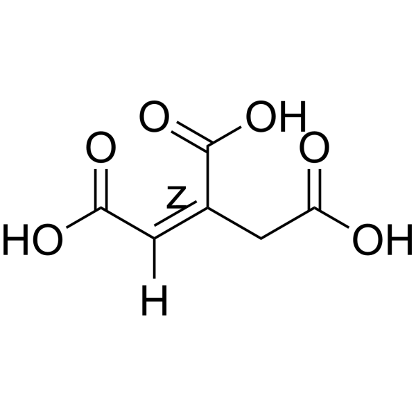
(Z)-Aconitic acid
Based on 2 publication(s) in Google Scholar
(Z)-Aconitic acid is an orally active glutamate decarboxylase inhibitor. (Z)-Aconitic acid reduces IκB-α phosphorylation. (Z)-Aconitic acid inhibits Antigen-induced arthritis and Monosodium urate (HY-B2130A)-induced gout.
For research use only. We do not sell to patients.
- Purity : 99.28%
- CAS No.: 585-84-2
- Formula: C6H6O6
- Molecular Weight:174.11
-
Storage:Powder -20°C, 3 years ; In solvent -80°C, 6 months , -20°C, 1 month

To place orders, for customer services and technical support, please contact: MedChemExpress USA
Tel: 609-228-6898 E-mail: [email protected] [email protected]


-
 Biological Activity
-
Chemical Information
-
Solvent & Solubility
- Protocols
- Purity & Documentation
- References
-
 Help & FAQs


-
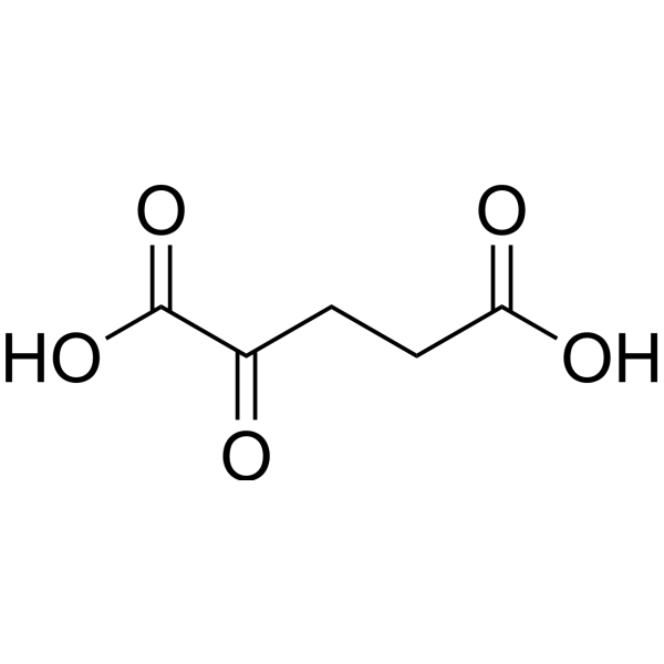
HY-W013636
2-Ketoglutaric acidKrebs Cycle Intermediate -
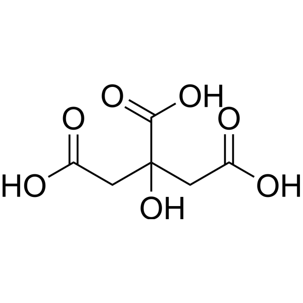
HY-N1428
Citric acidOrganic Acid -
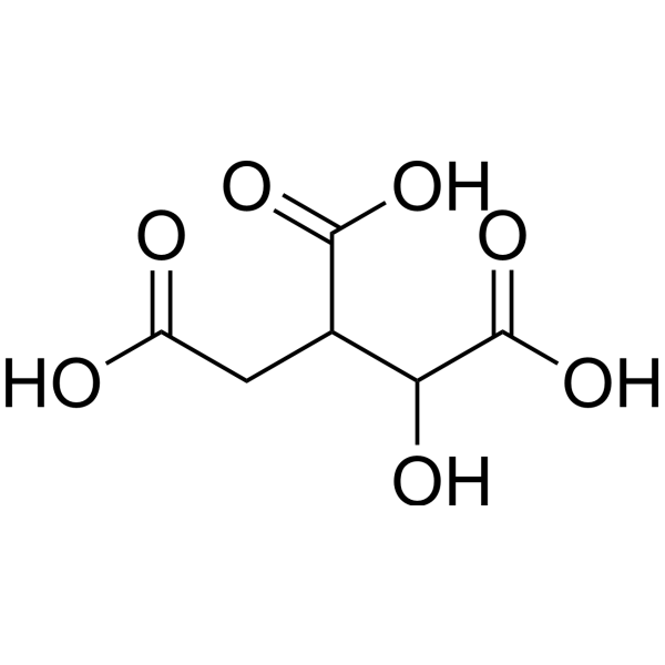
HY-113228
Isocitric acidEndogenous Metabolite -
HY-W010382
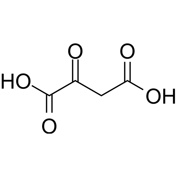
Oxaloacetic acidEndogenous Metabolite -
HY-W016813
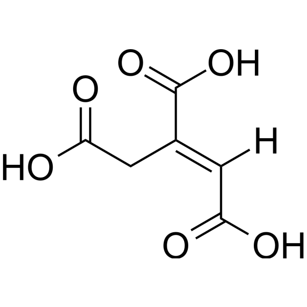
trans-Aconitic acidAconitase Inhibitor -
HY-N0215
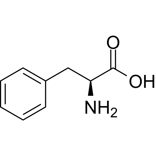
L-PhenylalanineCalcium Channels Antagonist -
HY-N0420
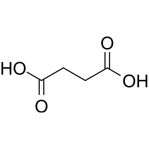
Succinic acidFood Additive, Platform chemical -
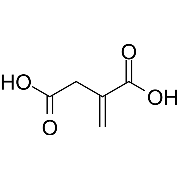
HY-Y0520
Itaconic acidFungi Metabolite -
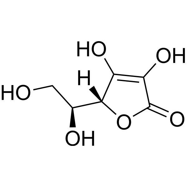
HY-B0166
L-Ascorbic acidAntioxidant -
HY-Y1311
Malic acidDicarboxylic Acid -
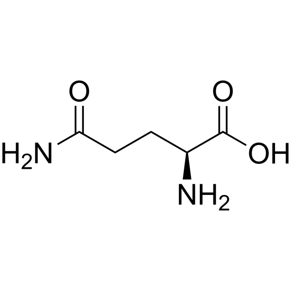
HY-N0390
L-GlutamineEndogenous Metabolite -
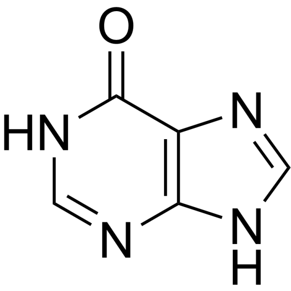
HY-N0091
HypoxanthineEndogenous Metabolite
Publications Citing Use of MedChemExpress (MCE) (Z)-Aconitic acid
MoreAll Endogenous Metabolite Isoforms
More

Biological Activity
Description
In Vitro
(Z)-Aconitic acid (10-90 μM; 2 h) reduces TNF-α release and IκB-α phosphorylation in LPS-stimulated THP-1 macrophages[1].
MedChemExpress (MCE) has not independently confirmed the accuracy of these methods. They are for reference only. Further protocols information, click here.
In Vivo
MedChemExpress (MCE) has not independently confirmed the accuracy of these methods. They are for reference only.
-
Animal Model:C57BL/6j male mice (8-10 weeks old) + antigen-induced arthritis (AIA) model or monosodium urate-induced gout model[1]
-
Dosage:10 mg/kg, 30 mg/kg, 90 mg/kg (0.5% w/v CMC suspension); 30 mg/kg (for gout model)
-
Administration:Oral gavage, once, 24 h after antigen challenge in AIA model; Oral gavage, once, 15 h after MSU injection in gout model
-
Result:Reduced total leukocytes and neutrophils in the synovial cavity and periarticular tissue, MPO activity, CXCL-1 levels in periarticular tissues, arthritis index, and hypernociception (AIA).
Significantly reduced total leukocyte and neutrophil accumulation into the synovial cavity at 30 mg/kg (gout model).


Chemical Information
-
CAS No. 585-84-2
-
Appearance Solid
-
Molecular Weight 174.11
-
Formula C6H6O6
-
Color White to light yellow
-
SMILES
O=C(O)/C([H])=C(C(O)=O)/CC(O)=O.
-
Synonyms
cis-Aconitic acid
-
Structure Classification
-
Initial Source
-
Shipping
Room temperature in continental US; may vary elsewhere.
-
Storage
Powder -20°C 3 years In solvent -80°C 6 months -20°C 1 month


Publications (2)
-
Journal Impact Factor
-
Most Recent
-
J Agric Food Chem
Pathway-Based Metabolomics Analysis Reveals Biosynthesis of Key Flavor Compounds in Mango. [Abstract]2022 Aug 31;70(34):10389-10399. PMID: 34792344 -
Life Sci
Homogentisic acid metabolism inhibits papillary thyroid carcinoma proliferation through ROS and p21-induced cell cycle arrest. [Abstract]2024 Jun 15:347:122682. PMID: 38702025


Solvent & Solubility
In Vitro:
H2O : 100 mg/mL (574.35 mM; Need ultrasonic)
DMSO : ≥ 100 mg/mL (574.35 mM; Hygroscopic DMSO has a significant impact on the solubility of product, please use newly opened DMSO)
* "≥" means soluble, but saturation unknown.
Please refer to the solubility information to select the appropriate solvent. Once prepared, please aliquot and store the solution to prevent product inactivation from repeated freeze-thaw cycles.
Storage method and period of stock solution: -80°C, 6 months; -20°C, 1 month. When stored at -80°C, please use it within 6 months. When stored at -20°C, please use it within 1 month.
* Note: If you choose water as the stock solution, please dilute it to the working solution, then filter and sterilize it with a 0.22 μm filter before use.
Please refer to the solubility information to select the appropriate solvent. Once prepared, please aliquot and store the solution to prevent product inactivation from repeated freeze-thaw cycles.
Storage method and period of stock solution: -80°C, 6 months; -20°C, 1 month. When stored at -80°C, please use it within 6 months. When stored at -20°C, please use it within 1 month.
* Note: If you choose water as the stock solution, please dilute it to the working solution, then filter and sterilize it with a 0.22 μm filter before use.
Concentration (start) × Volume (start) = Concentration (final) × Volume (final)
In Vivo:
Select the appropriate dissolution method based on your experimental animal and administration route.
- For the following dissolution methods, please ensure to first prepare a clear stock solution using an In Vitro approach and then sequentially add co-solvents:
- To ensure reliable experimental results, the clarified stock solution can be appropriately stored based on storage conditions. As for the working solution for In Vivo experiments, it is recommended to prepare freshly and use it on the same day.
- The percentages shown for the solvents indicate their volumetric ratio in the final prepared solution. If precipitation or phase separation occurs during preparation, heat and/or sonication can be used to aid dissolution.
Add each solvent one by one: 10% DMSO 40% PEG300 5% Tween-80 45% Saline
Solubility: ≥ 2.5 mg/mL (14.36 mM); Clear solution
This protocol yields a clear solution of ≥ 2.5 mg/mL (saturation unknown).
Taking 1 mL working solution as an example, add 100 μL DMSO stock solution (25.0 mg/mL) to 400 μL PEG300, and mix evenly; then add 50 μL Tween-80 and mix evenly; then add 450 μL Saline to adjust the volume to 1 mL.
Preparation of Saline: Dissolve 0.9 g sodium chloride in ddH₂O and dilute to 100 mL to obtain a clear Saline solution.
Add each solvent one by one: 10% DMSO 90% (20% SBE-β-CD in Saline)
Solubility: ≥ 2.5 mg/mL (14.36 mM); Clear solution
This protocol yields a clear solution of ≥ 2.5 mg/mL (saturation unknown).
Taking 1 mL working solution as an example, add 100 μL DMSO stock solution (25.0 mg/mL) to 900 μL 20% SBE-β-CD in Saline, and mix evenly.
Preparation of 20% SBE-β-CD in Saline (4°C, storage for one week): 2 g SBE-β-CD powder is dissolved in 10 mL Saline, completely dissolve until clear.
For the following dissolution methods, please prepare the working solution directly:
It is recommended to prepare fresh solutions and use them promptly within a short period of time.
The percentages shown for the solvents indicate their volumetric ratio in the final prepared solution. If precipitation or phase separation occurs during preparation, heat and/or sonication can be used to aid dissolution.
Add each solvent one by one: PBS
Solubility: 100 mg/mL (574.35 mM); Clear solution; Need ultrasonic
In Vivo Dissolution Calculator
Please enter the basic information of animal experiments:
-
-
-
-
Recommended: Prepare an additional quantity of animals to account for potential losses during experiments.
Working solution concentration: 0.22 mg/mL
This product has good water solubility, please refer to the measured solubility data in water/PBS/Saline for details.

Protocols
-
Kinase activity and phosphorylation assays
Kinase activity assays measure the ability of kinases to transfer phosphate groups from ATP to specific substrates, while phosphorylation assays detect the presence and levels of phosphorylated proteins. Common methods include radiolabeled ATP incorporation (e. g. ,), ADP release detection via bioluminescence (e. g. ,[3]), enzyme-linked immunosorbent assays (ELISA) for phospho-specific epitopes (e. g. ,[6]), and microtiter-based formats for high-throughput screening (e. g. ,[8]). The ADP-Glo assay quantifies kinase activity by measuring ADP produced during phosphorylation using a luciferase-based system. Radiometric assays involve autoradiography or scintillation counting after incorporation of 32P-labeled ATP into substrate proteins. ELISA-based approaches rely on phospho-specific antibodies to detect activated kinases in cell lysates or purified samples.
-
Western Blot
Western blotting (WB) is a commonly used experimental method in molecular biology, biochemistry, and immunogenetics for identifying and quantifying target proteins. It combines gel electrophoresis with immunoassay, enabling researchers to analyze protein expression, post-translational modifications, and molecular weight.
-
Collagen-Induced Arthritis
Collagen-induced arthritis (CIA) is an autoimmune murine model of rheumatoid arthritis in which immunization with type II collagen (CII) emulsified in an adjuvant induces a T cell- and autoantibody-driven inflammatory arthritis characterized by synovial hyperplasia, immune cell infiltration, and joint destruction. The model typically relies on genetically susceptible mouse strains (e. g. , DBA/1) and reproduces key features of human rheumatoid arthritis, including anti-collagen immune responses and progressive joint inflammation. Disease onset generally occurs within ~3-4 weeks after immunization, depending on antigen/adjuvant combinations and protocol variation. The immunopathology is driven by adaptive immune activation against CII, leading to systemic and local joint inflammation mediated by pro-inflammatory cytokines and effector immune cells, making CIA a standard preclinical platform for evaluating immunomodulatory and anti-arthritic interventions.
-
Protocol for Kinase activity and phosphorylation assays
Kinase activity assays measure transfer of phosphate from ATP to a protein or peptide substrate, generating phosphorylated substrate, ADP, or incorporated radiolabeled phosphate as the readout; phosphorylation assays measure site-specific phosphorylation in cells or tissues as a proxy for kinase-pathway activation, inhibition, or substrate regulation. Phosphorylation can be detected by phospho-specific Western blot, immunoprecipitation kinase assay, phospho-immunofluorescence, phospho-flow cytometry, luminescent ADP detection, radiolabeled ATP incorporation, or reporter-based pathway assays, and these readouts can be applied to cancer cells, primary neurons, mouse tumors, organoids, inflammatory macrophages, ferroptosis studies, and mitophagy studies when the kinase target is biologically relevant.


Purity & Documentation
-
Data Sheet (276 KB)
-
SDS (557 KB)
- English - EN (557 KB)
- Français - FR (557 KB)
- Deutsch - DE (557 KB)
- Norwegian - NO (557 KB)
- Español - ES (557 KB)
- Swedish - SV (557 KB)
- Italian - IT (557 KB)
- Korean - KR (557 KB)
- Portuguese - PT (557 KB)
-
Handling Instructions (2659 KB)
References


Complete Stock Solution Preparation Table
Please refer to the solubility information to select the appropriate solvent. Once prepared, please aliquot and store the solution to prevent product inactivation from repeated freeze-thaw cycles.
Storage method and period of stock solution: -80°C, 6 months; -20°C, 1 month. When stored at -80°C, please use it within 6 months. When stored at -20°C, please use it within 1 month.
| Optional Solvent | Concentration Solvent Mass | 1 mg | 5 mg | 10 mg | 25 mg |
|---|---|---|---|---|---|
| H2O / DMSO | 1 mM | 5.7435 mL | 28.7175 mL | 57.4350 mL | 143.5874 mL |
| 5 mM | 1.1487 mL | 5.7435 mL | 11.4870 mL | 28.7175 mL | |
| 10 mM | 0.5743 mL | 2.8717 mL | 5.7435 mL | 14.3587 mL | |
| 15 mM | 0.3829 mL | 1.9145 mL | 3.8290 mL | 9.5725 mL | |
| 20 mM | 0.2872 mL | 1.4359 mL | 2.8717 mL | 7.1794 mL | |
| 25 mM | 0.2297 mL | 1.1487 mL | 2.2974 mL | 5.7435 mL | |
| 30 mM | 0.1914 mL | 0.9572 mL | 1.9145 mL | 4.7862 mL | |
| 40 mM | 0.1436 mL | 0.7179 mL | 1.4359 mL | 3.5897 mL | |
| 50 mM | 0.1149 mL | 0.5743 mL | 1.1487 mL | 2.8717 mL | |
| 60 mM | 0.0957 mL | 0.4786 mL | 0.9572 mL | 2.3931 mL | |
| 80 mM | 0.0718 mL | 0.3590 mL | 0.7179 mL | 1.7948 mL | |
| 100 mM | 0.0574 mL | 0.2872 mL | 0.5743 mL | 1.4359 mL |
* Note: If you choose water as the stock solution, please dilute it to the working solution, then filter and sterilize it with a 0.22 μm filter before use.


(Z)-Aconitic acid Related Classifications
HY-W016814 Related Classifications
 Powered by Bioz