



α-Glycosidase-IN-1
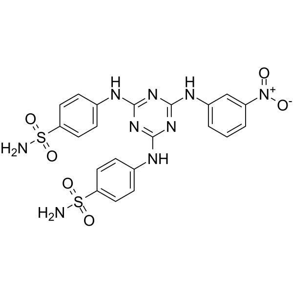
α-Glycosidase-IN-1 (compound MZ7) is a potent α-GLY (α-Glycosidase) inhibitor, with an IC50 of 44.72 nM and a KI of 41.74 nM. α-Glycosidase-IN-1 also shows inhibition profile against human carbonic anhydrase isoenzymes I and II (hCA I and hCA II), and acetylcholinesterase (AChE), with IC50 values of 104.87, 100.04, and 654.87 nM, respectively. α-Glycosidase-IN-1 can be used for the research of many diseases such as diabetes, Alzheimer’s disease, heart failure, ulcer, and epilepsy.
For research use only. We do not sell to patients.
- CAS No.: 2428389-66-4
- Formula: C21H19N9O6S2
- Molecular Weight:557.56
-
Storage:
Please store the product under the recommended conditions in the Certificate of Analysis.

To place orders, for customer services and technical support, please contact: MedChemExpress USA
Tel: 609-228-6898 E-mail: [email protected] [email protected]


-
 Biological Activity
-
Chemical Information
- Protocols
- Purity & Documentation
- References
-
 Help & FAQs




Biological Activity
Description
IC50 & Target
IC50: 44.72 ± 0.9964 nM (α-GLY), 100.04 ± 0.9688 nM (hCA II), 104.87 ± 0.9920 nM (hCA I), 654.87 ± 0.9383 nM (AChE); KI: 41.74 ± 8.08 nM (α-GLY), 114.78 ± 20.33 nM (hCA I), 114.78 ± 9.54 nM (hCA II), 597.33 ± 100.56 nM (AChE)[1]


Chemical Information
-
CAS No. 2428389-66-4
-
Molecular Weight 557.56
-
Formula C21H19N9O6S2
-
SMILES
O=[N+](C1=CC(NC2=NC(NC3=CC=C(S(=O)(N)=O)C=C3)=NC(NC4=CC=C(S(=O)(N)=O)C=C4)=N2)=CC=C1)[O-]
-
Shipping
Room temperature in continental US; may vary elsewhere.
-
Storage
Please store the product under the recommended conditions in the Certificate of Analysis.

Protocols
-
Cell Cytotoxicity Assay
Cytotoxicity assays are usually based on the assessment of cell membrane damage, which can also be indirectly detected by measuring cell viability. Detection methods include MTT assay, CKK-8 assay, LDH assay and ATP assay, etc.
-
Research Protocol for Cardiovascular Diseases
Cardiovascular disease can be modeled as maladaptive cardiac remodeling, where ischemic injury or pressure overload activates inflammatory signaling, fibroblast activation, extracellular-matrix deposition, cardiomyocyte hypertrophy, vascular remodeling, and progressive ventricular dysfunction. The TGF-β/SMAD axis is a central profibrotic pathway after myocardial injury and pressure overload, while innate immune and cytokine pathways regulate leukocyte recruitment, scar formation, and adverse remodeling. Key unresolved questions include which inflammatory signals are reparative versus harmful, when fibrosis is protective versus maladaptive, and whether pathway inhibition improves function without weakening necessary infarct healing or compensatory remodeling.
-
Research Protocol for Metabolic Diseases
AMP-activated protein kinase, AMPK, is a conserved cellular energy sensor that responds to reduced cellular energy status and coordinates metabolism by increasing ATP-generating catabolic pathways while suppressing ATP-consuming anabolic processes. In metabolic disease research, the AMPK pathway is experimentally relevant because it regulates hepatic lipid synthesis, fatty acid oxidation, glucose production, skeletal-muscle glucose disposal, mTORC1-linked biosynthesis, autophagy, mitochondrial homeostasis, and whole-body energy balance. The central pathway logic is that energy stress, metformin, exercise-like stimulation, or direct AMPK activators increase AMPKα Thr172 phosphorylation and downstream substrate phosphorylation, including ACC and RAPTOR. Phosphorylation of ACC suppresses lipogenesis and supports fatty acid oxidation, whereas phosphorylation of RAPTOR suppresses mTORC1 signaling and links cellular energy status to growth and protein synthesis control. The pathway is linked


Purity & Documentation
References

Calculators
Concentration (start) × Volume (start) = Concentration (final) × Volume (final)


α-Glycosidase-IN-1 Related Classifications
HY-147954 Related Classifications
 Powered by Bioz