



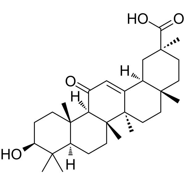
18α-Glycyrrhetinic acid
Based on 4 publication(s) in Google Scholar
18α-Glycyrrhetinic acid, a diet-derived compound, is an inhibitor of NF-kB and an activator of proteasome, which serves as pro-longevity and anti-aggregation factor in a multicellular organism. 18α-Glycyrrhetinic acid induces apoptosis.
For research use only. We do not sell to patients.
- Purity : 99.87%
- CAS No.: 1449-05-4
- Formula: C30H46O4
- Molecular Weight:470.68
-
Storage:Powder -20°C, 3 years , 4°C, 2 years ; In solvent -80°C, 6 months , -20°C, 1 month

To place orders, for customer services and technical support, please contact: MedChemExpress USA
Tel: 609-228-6898 E-mail: [email protected] [email protected]


-
 Biological Activity
-
Chemical Information
-
Solvent & Solubility
- Protocols
- Purity & Documentation
- References
-
 Help & FAQs


-
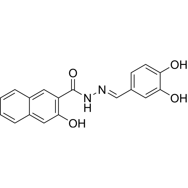
HY-15304
DynasoreAutophagy Inducer -
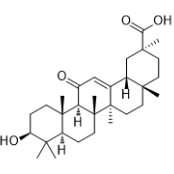
HY-N0180
18β-Glycyrrhetinic acidEndogenous Metabolite -
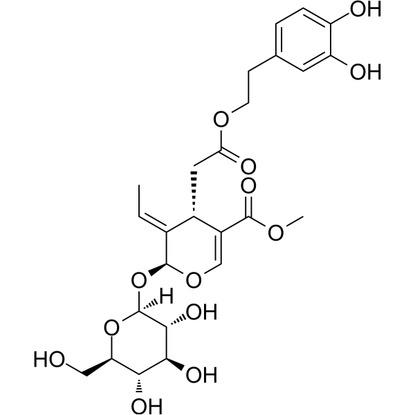
HY-N0292
OleuropeinApoptosis Inducer -
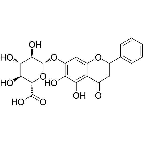
HY-N0197
BaicalinAutophagy Inducer -
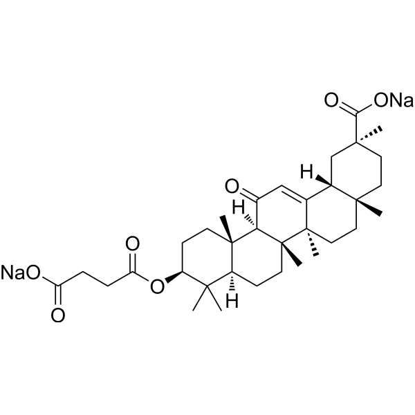
HY-B1367
Carbenoxolone disodiumGap Junction Inhibitor -
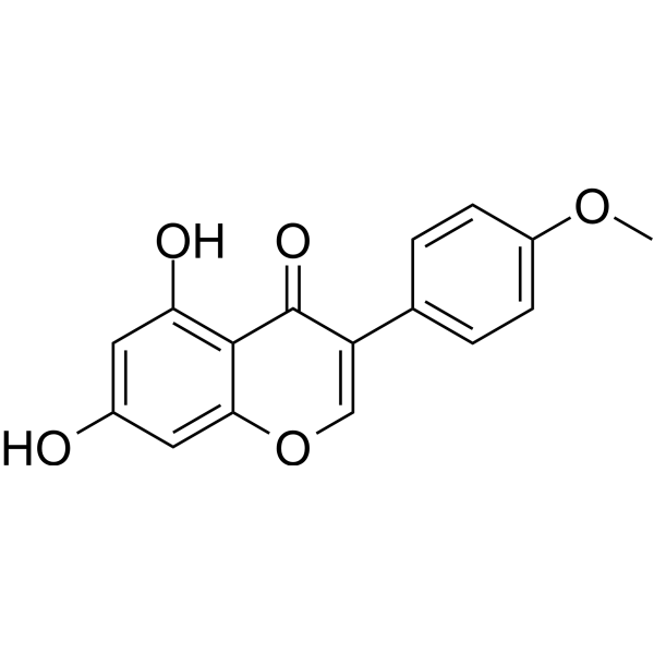
HY-14595
Biochanin AEndogenous Metabolite -
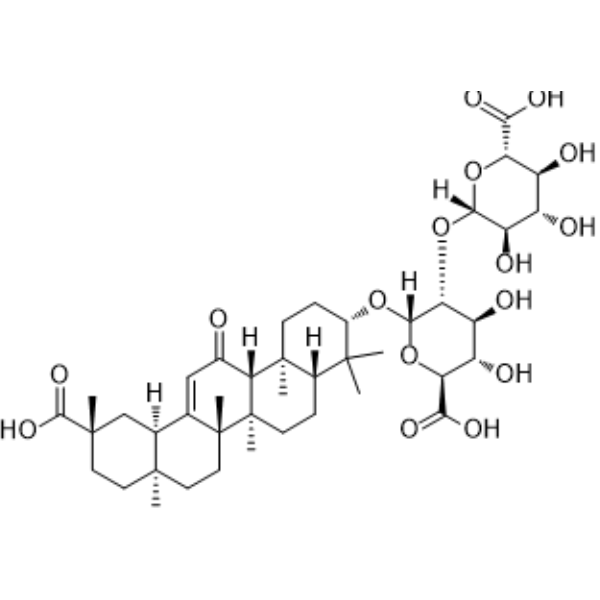
HY-N0184
Glycyrrhizic acidHMGB1 Antagonist -
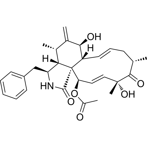
HY-N6682
Cytochalasin DActin Polymerization Inhibitor -
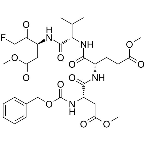
HY-12466
Z-DEVD-FMKCaspase-3 Inhibitor -
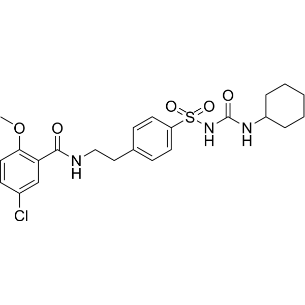
HY-15206
GlibenclamideKATP Inhibitor -
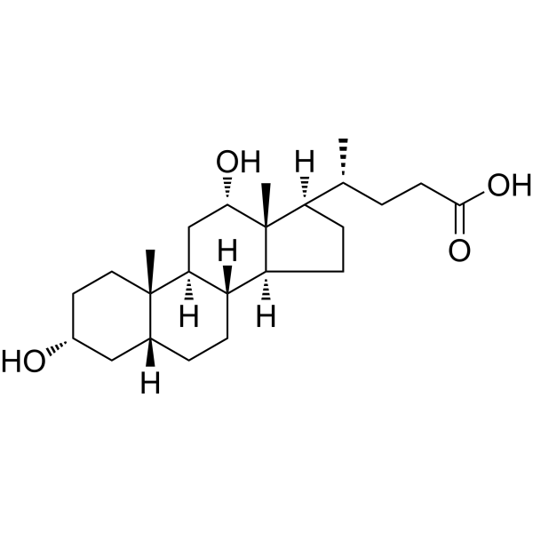
HY-N0593
Deoxycholic acidBile Acid Receptor Activator -
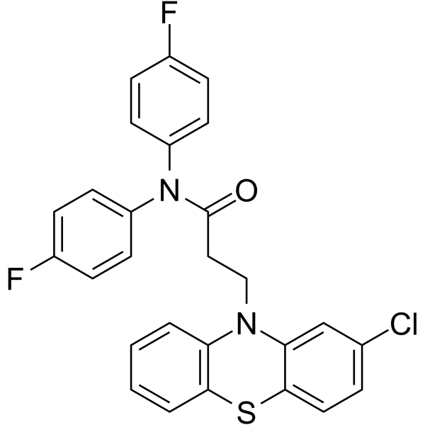
HY-150602
20S Proteasome activator 120S Proteasome Activator
Publications Citing Use of MedChemExpress (MCE) 18α-Glycyrrhetinic acid
More Customer Validation & Images

-
Cell Proliferation/Viability Assay

-
Cell Migration/Invasion Assay

-
Bio/Physico-chemical Assay

-
Bio/Physico-chemical Assay


Biological Activity
Description
|
Proteasome |
NF-κB |
Cellular Effect
|
Cell Line

|
Type | Value | Description | References |
|---|---|---|---|---|
| 4T1 | IC50 |
96.22 μM
Compound: 2; GA
|
Cytotoxicity in mouse 4T1 cells assessed as reduction in cell viability incubated for 96 hrs by SRB assay
Cytotoxicity in mouse 4T1 cells assessed as reduction in cell viability incubated for 96 hrs by SRB assay
|
[PMID: 28754470] |
| CT26 | IC50 |
70.48 μM
Compound: 2; GA
|
Cytotoxicity in mouse CT26 cells assessed as reduction in cell viability incubated for 96 hrs by SRB assay
Cytotoxicity in mouse CT26 cells assessed as reduction in cell viability incubated for 96 hrs by SRB assay
|
[PMID: 28754470] |
| DU-145 | IC50 |
136.4 μM
Compound: 2; GA
|
Cytotoxicity in human DU145 cells assessed as reduction in cell viability incubated for 96 hrs by SRB assay
Cytotoxicity in human DU145 cells assessed as reduction in cell viability incubated for 96 hrs by SRB assay
|
[PMID: 28754470] |
| EA.hy 926 | IC50 |
>100 μM
Compound: 2; GA
|
Cytotoxicity in human EAhy926 cells assessed as reduction in cell viability incubated for 72 hrs by MTT assay
Cytotoxicity in human EAhy926 cells assessed as reduction in cell viability incubated for 72 hrs by MTT assay
|
[PMID: 28754470] |
| H9c2 | IC50 |
>100 μM
Compound: 2; GA
|
Cytotoxicity in rat H9c2 cells assessed as reduction in cell viability incubated for 72 hrs by MTT assay
Cytotoxicity in rat H9c2 cells assessed as reduction in cell viability incubated for 72 hrs by MTT assay
|
[PMID: 28754470] |
| HCT-116 | IC50 |
75.08 μM
Compound: 2; GA
|
Cytotoxicity in human HCT116 cells assessed as reduction in cell viability incubated for 96 hrs by SRB assay
Cytotoxicity in human HCT116 cells assessed as reduction in cell viability incubated for 96 hrs by SRB assay
|
[PMID: 28754470] |
| HCT-8 | IC50 |
82.22 μM
Compound: 2; GA
|
Cytotoxicity in human HCT8 cells assessed as reduction in cell viability incubated for 96 hrs by SRB assay
Cytotoxicity in human HCT8 cells assessed as reduction in cell viability incubated for 96 hrs by SRB assay
|
[PMID: 28754470] |
| HeLa | IC50 |
>50 μM
Compound: 2; GA
|
Cytotoxicity in human HeLa cells assessed as reduction in cell viability incubated for 72 hrs by MTT assay
Cytotoxicity in human HeLa cells assessed as reduction in cell viability incubated for 72 hrs by MTT assay
|
[PMID: 28754470] |
| HepG2 | IC50 |
>50 μM
Compound: 2; GA
|
Cytotoxicity in human HepG2 cells assessed as reduction in cell viability incubated for 72 hrs by MTT assay
Cytotoxicity in human HepG2 cells assessed as reduction in cell viability incubated for 72 hrs by MTT assay
|
[PMID: 28754470] |
| HT-29 | IC50 |
>50 μM
Compound: 2; GA
|
Cytotoxicity in human HT-29 cells assessed as reduction in cell viability incubated for 72 hrs by MTT assay
Cytotoxicity in human HT-29 cells assessed as reduction in cell viability incubated for 72 hrs by MTT assay
|
[PMID: 28754470] |
| HT-29 | IC50 |
77.23 μM
Compound: 2; GA
|
Cytotoxicity in human HT-29 cells assessed as reduction in cell viability incubated for 96 hrs by SRB assay
Cytotoxicity in human HT-29 cells assessed as reduction in cell viability incubated for 96 hrs by SRB assay
|
[PMID: 28754470] |
| MCF7 | IC50 |
75.66 μM
Compound: 2; GA
|
Cytotoxicity in human MCF7 cells assessed as reduction in cell viability incubated for 24 hrs by MTT assay
Cytotoxicity in human MCF7 cells assessed as reduction in cell viability incubated for 24 hrs by MTT assay
|
[PMID: 28754470] |
| MDA-MB-231 | IC50 |
101.2 μM
Compound: 2; GA
|
Cytotoxicity in human MDA-MB-231 cells assessed as reduction in cell viability incubated for 96 hrs by SRB assay
Cytotoxicity in human MDA-MB-231 cells assessed as reduction in cell viability incubated for 96 hrs by SRB assay
|
[PMID: 28754470] |
| MDA-MB-231 | IC50 |
84.7 μM
Compound: 2; GA
|
Cytotoxicity in human MDA-MB-231 cells assessed as reduction in cell viability incubated for 24 hrs by MTT assay
Cytotoxicity in human MDA-MB-231 cells assessed as reduction in cell viability incubated for 24 hrs by MTT assay
|
[PMID: 28754470] |
| MDCK | IC50 |
>100 μM
Compound: 2; GA
|
Cytotoxicity in human MDCK cells assessed as reduction in cell viability incubated for 72 hrs by MTT assay
Cytotoxicity in human MDCK cells assessed as reduction in cell viability incubated for 72 hrs by MTT assay
|
[PMID: 28754470] |
| PC-3 | IC50 |
88.04 μM
Compound: 2; GA
|
Cytotoxicity in human PC3 cells assessed as reduction in cell viability incubated for 96 hrs by SRB assay
Cytotoxicity in human PC3 cells assessed as reduction in cell viability incubated for 96 hrs by SRB assay
|
[PMID: 28754470] |
| TERT-RPE1 | IC50 |
63.41 μM
Compound: 2; GA
|
Cytotoxicity in human hTERT-RPE1 cells assessed as reduction in cell viability incubated for 24 hrs by MTT assay
Cytotoxicity in human hTERT-RPE1 cells assessed as reduction in cell viability incubated for 24 hrs by MTT assay
|
[PMID: 28754470] |
In Vitro
18α-Glycyrrhetinic acid (18a-GA) markedly reduces LX-2 cell numbers by 14.8% and 31.2% after 48 h and 72 h of treatment, respectively (P< 0.05). 18α-Glycyrrhetinic acid also significantly increases the percentage of LX-2 cells in phase G0/G1 and decreases it in phase S after treated for 48 h and 72 h compare with the control group. 18α-Glycyrrhetinic acid increases apoptosis to 6.8% at 48 h, compare with control (2.5%), and at 72 h the percentages of apoptotic cells in control and the treatment groups are 3.1% and 15.6%, respectively, in LX-2 cells (P<0.01). Furthermore, 18α-Glycyrrhetinic acid induces expression of PPAR-γ and alters some cell cycle and apoptosis-related proteins. 18α-Glycyrrhetinic acid also inhibits NF-κB DNA-binding activity[1].
MedChemExpress (MCE) has not independently confirmed the accuracy of these methods. They are for reference only. Further protocols information, click here.
In Vivo
MedChemExpress (MCE) has not independently confirmed the accuracy of these methods. They are for reference only.


Chemical Information
-
CAS No. 1449-05-4
-
Appearance Solid
-
Molecular Weight 470.68
-
Formula C30H46O4
-
Color White to off-white
-
SMILES
CC1(C)[C@@H](O)CC[C@@]2(C)[C@@]1([H])CC[C@]([C@@](CC[C@]3(C)[C@]4([H])C[C@@](C)(C(O)=O)CC3)(C)C4=C5)(C)[C@]2([H])C5=O
-
Structure Classification
-
Initial Source
-
Shipping
Room temperature in continental US; may vary elsewhere.
-
Storage
Powder -20°C 3 years 4°C 2 years In solvent -80°C 6 months -20°C 1 month


Publications (4)
-
Journal Impact Factor
-
Most Recent
-
NPJ Precis Oncol
Calcium-modulated GJB6-GRHL3 positive feedback loop attenuates ESCC progression through AKT signaling pathway inhibition. [Abstract]2026 Jun 4. PMID: 42243308
18α-Glycyrrhetinic acid purchased from MedChemExpress. Usage Cited in: NPJ Precis Oncol. 2026 Jun 4. [Abstract]
CCK-8 growth-curve analysis of GJB6-knockdown KYSE30 cells after treatment with 18α-GA (18α-Glycyrrhetinic acid; 10 μM; 1 h). Cell proliferation was partly restored by 18α-GA (10 μM, 1 h) in the GJB6 knockdown group of KYSE30 cells.
18α-Glycyrrhetinic acid purchased from MedChemExpress. Usage Cited in: NPJ Precis Oncol. 2026 Jun 4. [Abstract]
Transwell migration assay with GJIC inhibitor 18α-GA (18α-Glycyrrhetinic acid; 10 μM; 1 h) in GJB6 RNA interfered KYSE30 cells.
-
Cell Calcium
Drug repurposing DMSO from an old solvent to a new candidate for the treatment of colitis and sepsis in male mice. [Abstract]2026 May 23:136:103155. PMID: 42235215
18α-Glycyrrhetinic acid purchased from MedChemExpress. Usage Cited in: Cell Calcium. 2026 May 23:136:103155. [Abstract]
Cumulative concentration-response curves, Rmax, AUC, and EC50 of DMSO-induced vasorelaxation in arterioles from mice after incubation with 18α-G (18α-Glycyrrhetinic acid; 10 μM; 20 min). 18α-G significantly attenuated DMSO-induced vasorelaxation.
-
Microvasc Res
Functional heterogeneity of endothelium-dependent vasorelaxation in different order branches of mesenteric artery in female/male mice. [Abstract]2025 Mar:158:104777. PMID: 39613183
18α-Glycyrrhetinic acid purchased from MedChemExpress. Usage Cited in: Microvasc Res. 2025 Mar:158:104777. [Abstract]
Representative tracings show ACh concentration-dependently induced vasorelaxation of the first-order branch of or second-order branch of male mice mesenteric arteries in the presence of 10 μmol/L 18α-Glycyrrhetinic acid (10 μM; 20 min). ACh-induced vasorelaxation was inhibited by 18α-glycyrrhetinic acid in second but not in first order branch.
-


Solvent & Solubility
In Vitro:
DMSO : 10 mg/mL (21.25 mM; ultrasonic and warming and heat to 60°C; Hygroscopic DMSO has a significant impact on the solubility of product, please use newly opened DMSO)
Please refer to the solubility information to select the appropriate solvent. Once prepared, please aliquot and store the solution to prevent product inactivation from repeated freeze-thaw cycles.
Storage method and period of stock solution: -80°C, 6 months; -20°C, 1 month. When stored at -80°C, please use it within 6 months. When stored at -20°C, please use it within 1 month.
Please refer to the solubility information to select the appropriate solvent. Once prepared, please aliquot and store the solution to prevent product inactivation from repeated freeze-thaw cycles.
Storage method and period of stock solution: -80°C, 6 months; -20°C, 1 month. When stored at -80°C, please use it within 6 months. When stored at -20°C, please use it within 1 month.
Concentration (start) × Volume (start) = Concentration (final) × Volume (final)
In Vivo:
Select the appropriate dissolution method based on your experimental animal and administration route.
- For the following dissolution methods, please ensure to first prepare a clear stock solution using an In Vitro approach and then sequentially add co-solvents:
- To ensure reliable experimental results, the clarified stock solution can be appropriately stored based on storage conditions. As for the working solution for In Vivo experiments, it is recommended to prepare freshly and use it on the same day.
- The percentages shown for the solvents indicate their volumetric ratio in the final prepared solution. If precipitation or phase separation occurs during preparation, heat and/or sonication can be used to aid dissolution.
Add each solvent one by one: 10% DMSO 40% PEG300 5% Tween-80 45% Saline
Solubility: 1.14 mg/mL (2.42 mM); Suspended solution; Need ultrasonic
This protocol yields a suspended solution of 1.14 mg/mL. Suspended solution can be used for oral and intraperitoneal injection.
Taking 1 mL working solution as an example, add 100 μL DMSO stock solution (11.4 mg/mL) to 400 μL PEG300, and mix evenly; then add 50 μL Tween-80 and mix evenly; then add 450 μL Saline to adjust the volume to 1 mL.
Preparation of Saline: Dissolve 0.9 g sodium chloride in ddH₂O and dilute to 100 mL to obtain a clear Saline solution.
In Vivo Dissolution Calculator
Please enter the basic information of animal experiments:
-
-
-
-
Recommended: Prepare an additional quantity of animals to account for potential losses during experiments.
Please enter your animal formula composition:
-
%DMSO +
Recommended: Keep the proportion of DMSO in working solution below 2% if your animal is weak.
-
%+
-
+%Tween-80 + +
-
%Saline +
The co-solvents required include: DMSO, . All of co-solvents are available by MedChemExpress (MCE). , Tween 80. All of co-solvents are available by MedChemExpress (MCE).
Working solution concentration: 0.22 mg/mL
Method for preparing stock solution: mg drug dissolved in μL DMSO. Stock solution concentration: mg/mL.
1. Take μL DMSO stock solution;
2. Add μL .
μL , mix evenly;
3. Then add μL Tween 80, mix evenly;
4. Then add μL
Please ensure that the stock solution in the first step is dissolved to a clear state, and add co-solvents in sequence. You can use ultrasonic heating (ultrasonic cleaner, recommended frequency 20-40 kHz), vortexing, etc. to assist dissolution.

Protocols
-
Apoptosis
Apoptosis, also called programmed cell death, is generally characterized by distinct morphological characteristics.
-
TUNEL staining for apoptotic DNA fragmentation
TUNEL staining detects DNA strand breaks by using terminal deoxynucleotidyl transferase to add labeled nucleotides to exposed 3′-OH DNA termini, generating either microscopic staining in fixed cells or tissue sections, or fluorescence/cytometric signal in cell suspensions. TUNEL positivity reflects DNA fragmentation but should not be interpreted alone as definitive apoptosis, because TUNEL can also label necrotic, autolytic, mechanically damaged, or DNA-repair-associated DNA breaks.
-
Annexin V plus membrane-impermeant dye apoptosis staining
Annexin V-based apoptosis assays rely on the detection of phosphatidylserine (PS) externalization from the inner leaflet of the plasma membrane to the outer leaflet, an early biochemical hallmark of apoptosis. Fluorescently labeled Annexin V binds PS in a calcium-dependent manner, enabling identification of early apoptotic cells by flow cytometry or fluorescence microscopy. When combined with a membrane-impermeant DNA-binding dye (e. g. , propidium iodide), this approach allows discrimination between viable (Annexin V−/dye−), early apoptotic (Annexin V+/dye−), and late apoptotic or necrotic (Annexin V+/dye+) cell populations by assessing membrane integrity and PS exposure.
-
Apoptosis Solutions
Apoptosis is a regulated, generally non-lytic cell-death pathway that removes unwanted, damaged, infected, or abnormal cells through coordinated morphological changes, caspase activation, DNA fragmentation, and membrane remodeling. The intrinsic apoptosis pathway is controlled mainly by mitochondrial outer membrane permeabilization, BCL-2 family proteins, cytochrome c release, apoptosome formation, caspase-9 activation, and downstream executioner caspase-3/7 activation. The extrinsic apoptosis pathway is initiated by death receptors such as Fas, TNFR, and TRAIL receptors, which recruit adaptor proteins and activate caspase-8 before engaging executioner caspases or mitochondrial amplification through BID cleavage. Apoptosis is linked to many phenotypes, including cancer cell killing, tissue homeostasis, immune regulation, neurodegeneration, infection response, and treatment-induced cytotoxicity; unresolved questions include how apoptosis interacts with necroptosis, pyroptosis, ferroptos


Purity & Documentation
-
Data Sheet (285 KB)
-
SDS (394 KB)
- English - EN (394 KB)
- Français - FR (394 KB)
- Deutsch - DE (394 KB)
- Norwegian - NO (394 KB)
- Español - ES (394 KB)
- Swedish - SV (394 KB)
- Italian - IT (394 KB)
- Korean - KR (394 KB)
- Portuguese - PT (394 KB)
-
Handling Instructions (2659 KB)
References
[1]. Zong L et al. 18α-glycyrrhetinic acid extracted from Glycyrrhiza radix inhibits proliferation and promotes apoptosis of the hepatic stellate cell line. J Dig Dis. 2013 Jun;14(6):328-36. [Content Brief]
[2]. Papaevgeniou N, et al. 18α-Glycyrrhetinic Acid Proteasome Activator Decelerates Aging and Alzheimer's Disease Progression in Caenorhabditis elegans and Neuronal Cultures. Antioxid Redox Signal. 2016 Dec 1;25(16):855-869. [Content Brief]


Complete Stock Solution Preparation Table
Please refer to the solubility information to select the appropriate solvent. Once prepared, please aliquot and store the solution to prevent product inactivation from repeated freeze-thaw cycles.
Storage method and period of stock solution: -80°C, 6 months; -20°C, 1 month. When stored at -80°C, please use it within 6 months. When stored at -20°C, please use it within 1 month.
| Optional Solvent | Concentration Solvent Mass | 1 mg | 5 mg | 10 mg | 25 mg |
|---|---|---|---|---|---|
| DMSO | 1 mM | 2.1246 mL | 10.6229 mL | 21.2459 mL | 53.1146 mL |
| 5 mM | 0.4249 mL | 2.1246 mL | 4.2492 mL | 10.6229 mL | |
| 10 mM | 0.2125 mL | 1.0623 mL | 2.1246 mL | 5.3115 mL | |
| 15 mM | 0.1416 mL | 0.7082 mL | 1.4164 mL | 3.5410 mL | |
| 20 mM | 0.1062 mL | 0.5311 mL | 1.0623 mL | 2.6557 mL |


18α-Glycyrrhetinic acid Related Classifications
HY-N0375 Related Classifications
 Powered by Bioz