



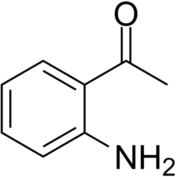
2'-Aminoacetophenone

Based on 1 Customer Validation
2'-Aminoacetophenone is an orally active inducer of apoptosis and respiratory biomarker. 2'-Aminoacetophenone can be used to detect Pseudomonas aeruginosa infections in the lungs of cystic fibrosis patients. 2'-Aminoacetophenone can inhibit the protein levels of LC3BII and p62 in macrophages infected with pqsA or mvfR and regulate autophagy. 2'-Aminoacetophenone can disrupt mitochondrial function by inducing oxidative stress and apoptosis signaling, leading to dysfunction in mouse skeletal muscle.
연구목적의 판매만을 진행합니다. 환자를 대상으로 한 판매는 하지 않습니다.
- Purity : 99.12%
- CAS No.: 551-93-9
- 화학식: C8H9NO
- 분자량:135.16
-
보관:
4°C, protect from light
* In solvent : -80°C, 6 months; -20°C, 1 month (protect from light)

To place orders, for customer services and technical support, please contact: MedChemExpress USA
Tel: 609-228-6898 E-mail: [email protected] [email protected]


-
 Biological Activity
-
Chemical Information
-
용액&용해도
- Protocol
- 순도&문서
- References
-
 Help & FAQs


-

HY-I0501R
2'-Aminoacetophenone (Standard)Apoptosis Inducer -
HY-W018791
BifendateAntioxidant, Anti-HBV Agent -
HY-W015740
DimedoneBiochemical Reagent -
HY-N8441
NeriifolinApoptosis Inhibitor -
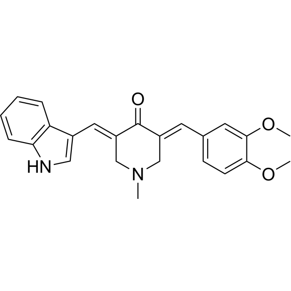
HY-112698
CA-5fLate-Stage Autophagy Inhibitor -
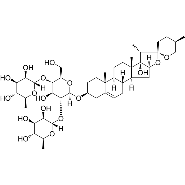
HY-N4180
Pennogenin 3-O-beta-chacotriosideAutophagy Inducer -
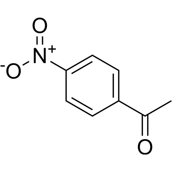
HY-Y0185
4'-NitroacetophenoneNitro-aromatic Compound -
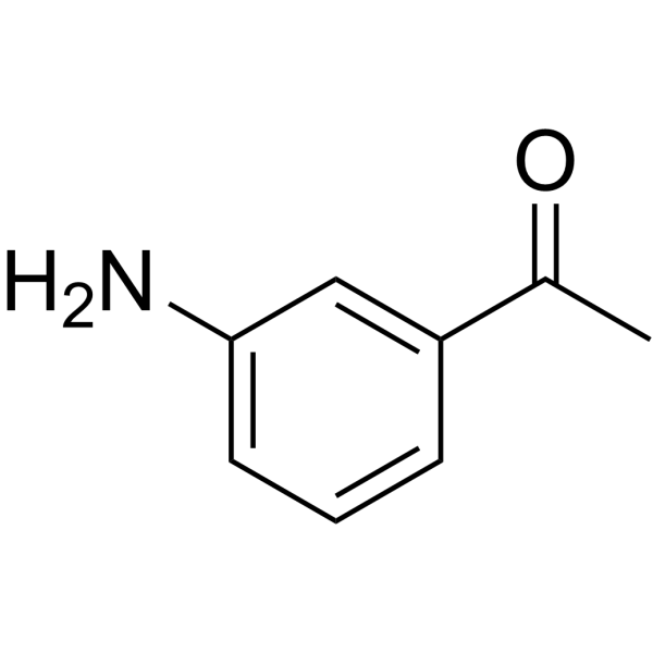
HY-W012637
3-AcetylanilineDrug Intermediate -

HY-W018791R
Bifendate (Standard)Antioxidant, Anti-HBV Agent -

HY-32144
1-(3-Nitrophenyl)ethanoneNatural Product -

HY-33040
2,2',4'-TrichloroacetophenoneAntibacterial Agent -
HY-79034
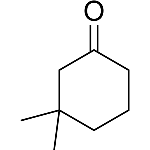
3,3-DimethylcyclohexanoneIntermediate For Boll Weevil Sex Attractant


Biological Activity
제품 설명
In Vitro
2'-Aminoacetophenone (200 μM, 3 h) can inhibit the protein levels of LC3BII and p62 in macrophages infected with pqsA or mvfR, and interfere with autophagy through epigenetic regulation of lipid biosynthesis, mediating the persistence of Pseudomonas aeruginosa in macrophages[1]. 2'-Aminoacetophenone can increase the generation of diallylmethylene fatty acyl protons and ethylene matrix subunits, inducing apoptosis of skeletal muscle cells[4].
MedChemExpress (MCE) has not independently confirmed the accuracy of these methods. They are for reference only.
-
Cell Line:pqsA- or mvfR-infected macrophages
-
Concentration:200 μM
-
Incubation Time:3 h
-
Result:Inhibited the protein expression of LC3BII and p62.
In Vivo
MedChemExpress (MCE) has not independently confirmed the accuracy of these methods. They are for reference only.
-
Animal Model:Female Oregon-R flies, 5-7-days-old[3].
-
Dosage:Sucrose solution (4%, 0.2 mL) with 2'-Aminoacetophenone (1.5 mM)
-
Administration:Oral gavage (p.o.); 2 days
-
Result:Increased the level of long-term colonization of the flies’ intestine by P. aeruginosa.
-
Animal Model:Six-week-old male CD1 mice weighing approximately 20-25 g[4].
-
Dosage:6.75 mg/kg
-
Administration:Intraperitoneal injection (i.p.); once
-
Result:Resulted in upregulation of 97% of genes involved in the apoptotic signaling pathway.
- Collagen: Sirius Red Staining
- Mitochondrial membrane-potential fluorescent assay
- Annexin V plus membrane-impermeant dye apoptosis staining
- ROS/oxidative-stress fluorescent staining
- Connective Tissue: Masson's Trichrome/Collagen Trichrome Staining
- Fibrosis/Collagen Morphometry
- Detection of Apoptosis
- TUNEL staining for apoptotic DNA fragmentation
- Autophagy
- Mitochondrial membrane-potential and mitochondrial mass staining
- Apoptosis Solutions
- Lysosome and acidic-vesicle live-cell staining
- Macroautophagy Solutions


Chemical Information
-
CAS No. 551-93-9
-
Appearance <20°C Solid, >20°C Liquid
-
분자량 135.16
-
화학식 C8H9NO
-
Color Light yellow to yellow
-
SMILES
CC(C1=CC=CC=C1N)=O
-
Synonyms
o-aminoacetophenone
-
Structure Classification
-
Initial Source
Pseudomonas aeruginosa
-
선적
Room temperature in continental US; may vary elsewhere.
-
보관
4°C, protect from light
* In solvent : -80°C, 6 months; -20°C, 1 month (protect from light)


용액&용해도
In Vitro:
DMSO : 100 mg/mL (739.86 mM; Need ultrasonic; Hygroscopic DMSO has a significant impact on the solubility of product, please use newly opened DMSO)
Please refer to the solubility information to select the appropriate solvent. Once prepared, please aliquot and store the solution to prevent product inactivation from repeated freeze-thaw cycles.
Storage method and period of stock solution: -80°C, 6 months; -20°C, 1 month (protect from light). When stored at -80°C, please use it within 6 months. When stored at -20°C, please use it within 1 month.
Please refer to the solubility information to select the appropriate solvent. Once prepared, please aliquot and store the solution to prevent product inactivation from repeated freeze-thaw cycles.
Storage method and period of stock solution: -80°C, 6 months; -20°C, 1 month (protect from light). When stored at -80°C, please use it within 6 months. When stored at -20°C, please use it within 1 month.
Concentration (start) × Volume (start) = Concentration (final) × Volume (final)
In Vivo:
Select the appropriate dissolution method based on your experimental animal and administration route.
- For the following dissolution methods, please ensure to first prepare a clear stock solution using an In Vitro approach and then sequentially add co-solvents:
- To ensure reliable experimental results, the clarified stock solution can be appropriately stored based on storage conditions. As for the working solution for In Vivo experiments, it is recommended to prepare freshly and use it on the same day.
- The percentages shown for the solvents indicate their volumetric ratio in the final prepared solution. If precipitation or phase separation occurs during preparation, heat and/or sonication can be used to aid dissolution.
Add each solvent one by one: 10% DMSO 40% PEG300 5% Tween-80 45% Saline
Solubility: ≥ 2.5 mg/mL (18.50 mM); Clear solution
This protocol yields a clear solution of ≥ 2.5 mg/mL (saturation unknown).
Taking 1 mL working solution as an example, add 100 μL DMSO stock solution (25.0 mg/mL) to 400 μL PEG300, and mix evenly; then add 50 μL Tween-80 and mix evenly; then add 450 μL Saline to adjust the volume to 1 mL.
Preparation of Saline: Dissolve 0.9 g sodium chloride in ddH₂O and dilute to 100 mL to obtain a clear Saline solution.
Add each solvent one by one: 10% DMSO 90% (20% SBE-β-CD in Saline)
Solubility: ≥ 2.5 mg/mL (18.50 mM); Clear solution
This protocol yields a clear solution of ≥ 2.5 mg/mL (saturation unknown).
Taking 1 mL working solution as an example, add 100 μL DMSO stock solution (25.0 mg/mL) to 900 μL 20% SBE-β-CD in Saline, and mix evenly.
Preparation of 20% SBE-β-CD in Saline (4°C, storage for one week): 2 g SBE-β-CD powder is dissolved in 10 mL Saline, completely dissolve until clear.
In Vivo Dissolution Calculator
Please enter the basic information of animal experiments:
-
-
-
-
Recommended: Prepare an additional quantity of animals to account for potential losses during experiments.
Please enter your animal formula composition:
-
%DMSO +
Recommended: Keep the proportion of DMSO in working solution below 2% if your animal is weak.
-
%+
-
+%Tween-80 + +
-
%Saline +
The co-solvents required include: DMSO, . All of co-solvents are available by MedChemExpress (MCE). , Tween 80. All of co-solvents are available by MedChemExpress (MCE).
Working solution concentration: 0.22 mg/mL
Method for preparing stock solution: mg drug dissolved in μL DMSO. Stock solution concentration: mg/mL. * In solvent : -80°C, 6 months; -20°C, 1 month (protect from light)
1. Take μL DMSO stock solution;
2. Add μL .
μL , mix evenly;
3. Then add μL Tween 80, mix evenly;
4. Then add μL
Please ensure that the stock solution in the first step is dissolved to a clear state, and add co-solvents in sequence. You can use ultrasonic heating (ultrasonic cleaner, recommended frequency 20-40 kHz), vortexing, etc. to assist dissolution.


순도&문서
-
Data Sheet (279 KB)
-
SDS (394 KB)
- English - EN (394 KB)
- Français - FR (394 KB)
- Deutsch - DE (394 KB)
- Norwegian - NO (394 KB)
- Español - ES (394 KB)
- Swedish - SV (394 KB)
- Italian - IT (394 KB)
- Korean - KR (394 KB)
- Portuguese - PT (394 KB)
-
Handling Instructions (2659 KB)
References
[1]. Chakraborty A, et al. Quorum-Sensing Signaling Molecule 2-Aminoacetophenone Mediates the Persistence of Pseudomonas aeruginosa in Macrophages by Interference with Autophagy through Epigenetic Regulation of Lipid Biosynthesis. mBio. 2023 Apr 25;14(2):e0015923. [Content Brief]
[2]. Scott-Thomas AJ, et al. 2-Aminoacetophenone as a potential breath biomarker for Pseudomonas aeruginosa in the cystic fibrosis lung. BMC Pulm Med. 2010 Nov 7;10:56. [Content Brief]
[3]. Kapsetaki SE, et al. The bacterial metabolite 2-aminoacetophenone promotes association of pathogenic bacteria with flies. Nat Commun. 2014 Jul 21;5:4401. [Content Brief]
[4]. Bandyopadhaya A, et al. Bacterial-excreted small volatile molecule 2-aminoacetophenone induces oxidative stress and apoptosis in murine skeletal muscle. Int J Mol Med. 2016 Apr;37(4):867-78. [Content Brief]


Complete Stock Solution Preparation Table
Please refer to the solubility information to select the appropriate solvent. Once prepared, please aliquot and store the solution to prevent product inactivation from repeated freeze-thaw cycles.
Storage method and period of stock solution: -80°C, 6 months; -20°C, 1 month (protect from light). When stored at -80°C, please use it within 6 months. When stored at -20°C, please use it within 1 month.
| Optional Solvent | Concentration Solvent Mass | 1 mg | 5 mg | 10 mg | 25 mg |
|---|---|---|---|---|---|
| DMSO | 1 mM | 7.3986 mL | 36.9932 mL | 73.9864 mL | 184.9660 mL |
| 5 mM | 1.4797 mL | 7.3986 mL | 14.7973 mL | 36.9932 mL | |
| 10 mM | 0.7399 mL | 3.6993 mL | 7.3986 mL | 18.4966 mL | |
| 15 mM | 0.4932 mL | 2.4662 mL | 4.9324 mL | 12.3311 mL | |
| 20 mM | 0.3699 mL | 1.8497 mL | 3.6993 mL | 9.2483 mL | |
| 25 mM | 0.2959 mL | 1.4797 mL | 2.9595 mL | 7.3986 mL | |
| 30 mM | 0.2466 mL | 1.2331 mL | 2.4662 mL | 6.1655 mL | |
| 40 mM | 0.1850 mL | 0.9248 mL | 1.8497 mL | 4.6241 mL | |
| 50 mM | 0.1480 mL | 0.7399 mL | 1.4797 mL | 3.6993 mL | |
| 60 mM | 0.1233 mL | 0.6166 mL | 1.2331 mL | 3.0828 mL | |
| 80 mM | 0.0925 mL | 0.4624 mL | 0.9248 mL | 2.3121 mL | |
| 100 mM | 0.0740 mL | 0.3699 mL | 0.7399 mL | 1.8497 mL |


2'-Aminoacetophenone Related Classifications
HY-I0501 Related Classifications
 Powered by Bioz