



3,4-Dicaffeoylquinic acid
Based on 10 publication(s) in Google Scholar
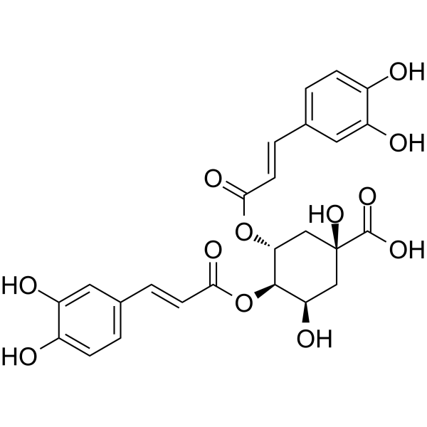
3,4-Dicaffeoylquinic acid (3,4-Di-O-caffeoylquinic acid), naturally isolated from Laggera alata, has antioxidative, DNA protective, neuroprotective and hepatoprotective properties. 3,4-Dicaffeoylquinic acid exerts apoptosis-mediated cytotoxicity and α-glucosidase inhibitory effects. 3,4-Dicaffeoylquinic acid possesses a unique mechanism of anti-influenza viral activity, that is, enhancing viral clearance by increasing TRAIL.
For research use only. We do not sell to patients.
- Purity : 98.15%
- CAS No.: 14534-61-3
- Formula: C25H24O12
- Molecular Weight:516.45
-
Storage:Powder -20°C, 3 years , 4°C, 2 years ; In solvent -80°C, 6 months , -20°C, 1 month

To place orders, for customer services and technical support, please contact: MedChemExpress USA
Tel: 609-228-6898 E-mail: [email protected] [email protected]


-
 Biological Activity
-
Chemical Information
-
Solvent & Solubility
- Protocols
- Purity & Documentation
- References
-
 Help & FAQs


-
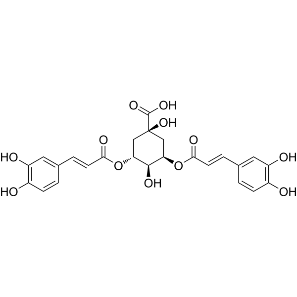
HY-N0056
Isochlorogenic acid AAntioxidant -
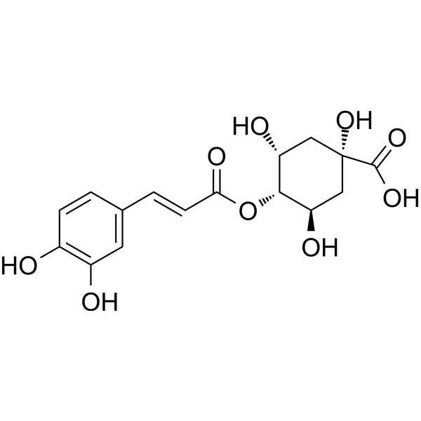
HY-N0787
Cryptochlorogenic acidphenolic Acid Compound -
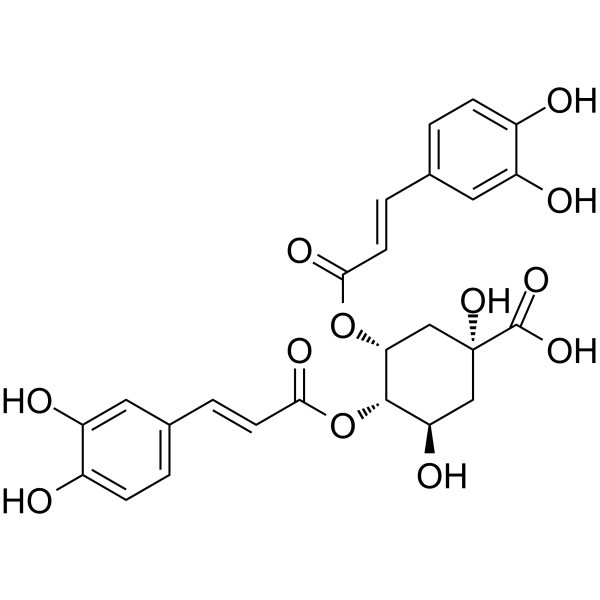
HY-N0058
4,5-Dicaffeoylquinic acidAntioxidant -
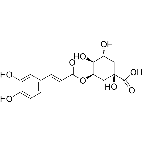
HY-N0722
Neochlorogenic acidCOX Inhibitor -
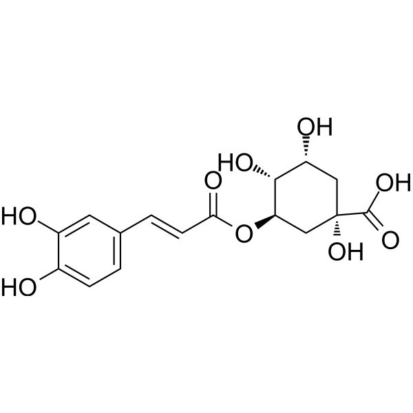
HY-N0055
Chlorogenic acidAntioxidant -
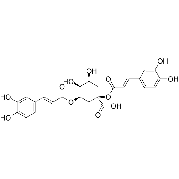
HY-N0359
CynarinAntioxidant -
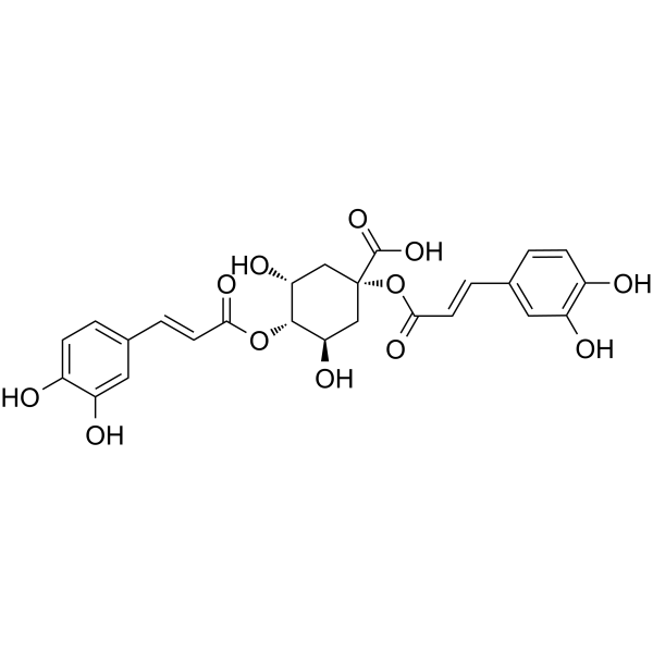
HY-N0358
1,4-Dicaffeoylquinic acidAnti-inflammatory Agent -

HY-N0057R
3,4-Dicaffeoylquinic acid (Standard)Antioxidant -
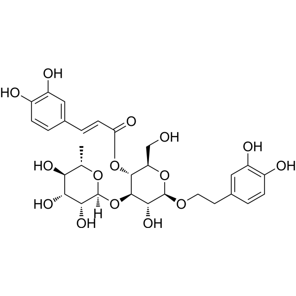
HY-N0021
VerbascosideBacterial Inhibitor -

HY-10583
Y-27632 dihydrochlorideROCK Inhibitor -
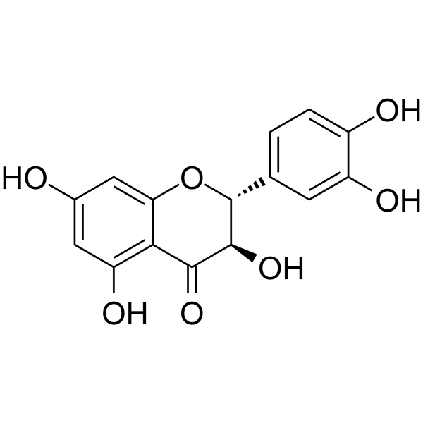
HY-N0136
TaxifolinAntifibrotic Agent -
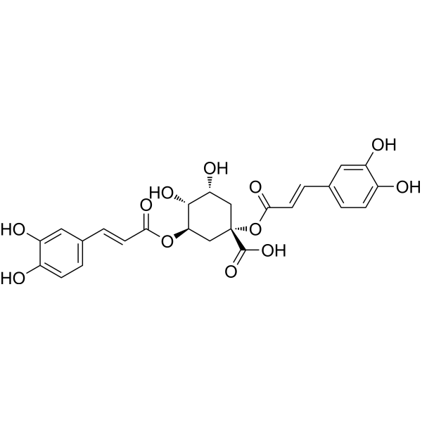
HY-N1412
1,3-Dicaffeoylquinic acidAntioxidant Agent
Publications Citing Use of MedChemExpress (MCE) 3,4-Dicaffeoylquinic acid
More- Nat Aging. 2024 Sep;4(9):1231-1248. [Abstract]
- Phytomedicine. 2026 Feb:151:157776. [Abstract]
- Cell Biosci. 2023 Nov 14;13(1):210. [Abstract]
- Cells. 2026 Feb 3;15(3):287. [Abstract]
- Foods. 2024 Apr 3;13(7):1101. [Abstract]
- ACS Omega. 2025 Sep 22;10(38):44260-44269. [Abstract]
- Vet Microbiol. 2026 May:316:110992. [Abstract]
- Chem Pharm Bull. 2024;72(1):93-97. [Abstract]
- SSRN. 2026 Apr 14.
- SSRN. 2026 Apr 8.
 Customer Validation & Images

-
Cell Proliferation/Viability Assay
-
Cell Proliferation/Viability Assay
-
IP
-
Bio/Physico-chemical Assay
-
WB
All Endogenous Metabolite Isoforms
More

Biological Activity
Description
Cellular Effect
|
Cell Line

|
Type | Value | Description | References |
|---|---|---|---|---|
| Jurkat | IC50 |
>35 mM
Compound: 3
|
Cytotoxicity against human Jurkat T cells
Cytotoxicity against human Jurkat T cells
|
[PMID: 15921434] |
| Jurkat | IC50 |
>35 μM/mL
Compound: 3
|
Cytotoxicity against human Jurkat T cells
Cytotoxicity against human Jurkat T cells
|
[PMID: 15921434] |
In Vitro
3,4-Dicaffeoylquinic acid acts as a greater primary antioxidant than its methanol extract, by having higher ferric reducing activity (EC50 2.18 μg/ml), β-carotene bleaching activity (EC50 23.85 μg/ml) and DPPH scavenging activity (EC50 68.91 μg/ml). 3,4-Dicaffeoylquinic acid exhibits a remarkable dose-dependent inhibitory effect on NCI-H23 (human lung adenocarcinoma) cell lines (EC50 3.26 μg/ml) and is found to be apoptotic in nature based on a clear indication of DNA fragmentation. 3,4-Dicaffeoylquinic acid also displays a concentration-dependent α-glucosidase inhibition with EC50 241.80 μg/ml[1].
MedChemExpress (MCE) has not independently confirmed the accuracy of these methods. They are for reference only. Further protocols information, click here.


Chemical Information
-
CAS No. 14534-61-3
-
Appearance Solid
-
Molecular Weight 516.45
-
Formula C25H24O12
-
Color Off-white to light yellow
-
SMILES
O=C([C@@]1(O)C[C@@H](OC(/C=C/C2=CC=C(O)C(O)=C2)=O)[C@H](OC(/C=C/C3=CC=C(O)C(O)=C3)=O)[C@H](O)C1)O
-
Synonyms
3,4-Di-O-caffeoylquinic acid; Isochlorogenic acid B
-
Structure Classification
-
Initial Source
-
Shipping
Room temperature in continental US; may vary elsewhere.
-
Storage
Powder -20°C 3 years 4°C 2 years In solvent -80°C 6 months -20°C 1 month


Publications (10)
-
Journal Impact Factor
-
Most Recent
-
Nat Aging
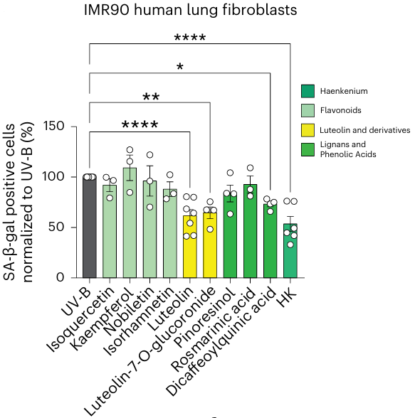
Targeting senescence induced by age or chemotherapy with a polyphenol-rich natural extract improves longevity and healthspan in mice. [Abstract]2024 Sep;4(9):1231-1248. PMID: 38951692
3,4-Dicaffeoylquinic acid purchased from MedChemExpress. Usage Cited in: Nat Aging. 2024 Sep;4(9):1231-1248. [Abstract]
Quantification of SA-β-Gal assay of UV-B-irradiated IMR90 fibroblasts, treated with the indicated compounds (3,4-Dicaffeoylquinic acid (Dicaffeoylquinic acid), etc.) at 1 μM concentration or 10 μg/mL for HK.
-
Phytomedicine
A novel mechanism of Reduning injection in sepsis treatment: Targeting inflammatory kinases TBK1 and IKKβ. [Abstract]2026 Feb:151:157776. PMID: 41512388 -
Cell Biosci
2023 Nov 14;13(1):210. PMID: 37964389
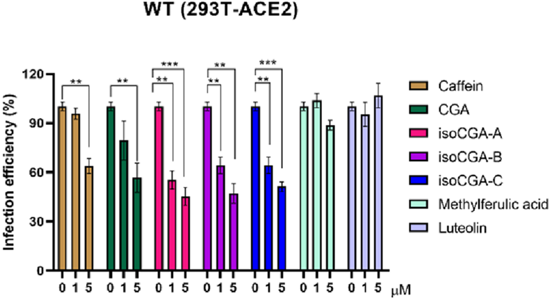
3,4-Dicaffeoylquinic acid purchased from MedChemExpress. Usage Cited in: Cell Biosci. 2023 Nov 14;13(1):210. [Abstract]
Examination of the inhibition capability of caffeine, CGA, isoCGA-A, isoCGA-B (3,4-Dicaffeoylquinic acid) (1-5 μM; 1 h), isoCGA-C, methylferulic acid and luteolin against the entry of wild-type SARS-CoV-2 on 293 T-ACE2 cells by Vpp (MOI = 0.1).
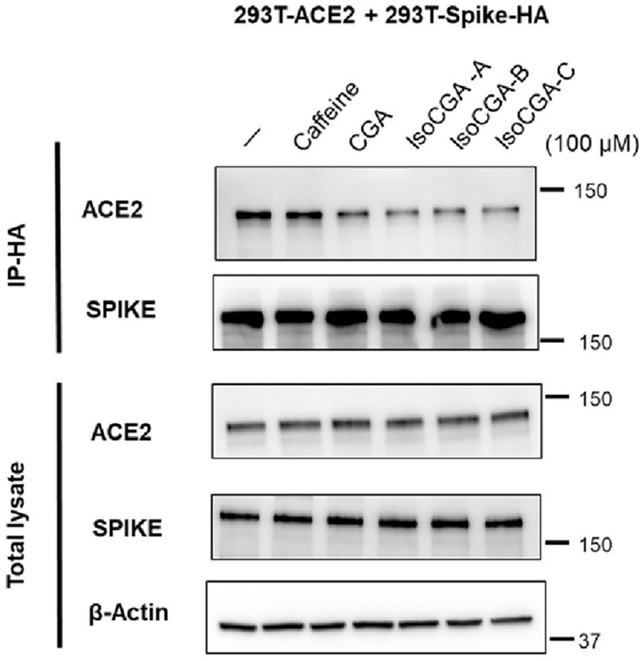
3,4-Dicaffeoylquinic acid purchased from MedChemExpress. Usage Cited in: Cell Biosci. 2023 Nov 14;13(1):210. [Abstract]
Co-immunoprecipitation using 293 T cells expressing SARS-CoV-2 spike-HA and ACE2 to verify that 5 compounds can inhibit the binding of ACE2 and spike. After anti-HA pulldown, adding 5 compounds (3,4-Dicaffeoylquinic acid (IsoCGA-B), etc.) (100 μM; 1 h), ACE2, and spike antibodies were used to detect by immunoblotting.
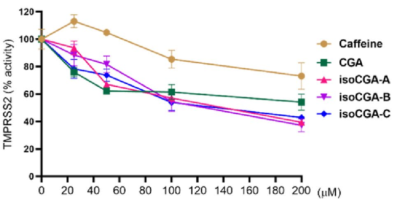
3,4-Dicaffeoylquinic acid purchased from MedChemExpress. Usage Cited in: Cell Biosci. 2023 Nov 14;13(1):210. [Abstract]
Validation of the ability of caffeine, CGA, isoCGA-A, isoCGA-B (3,4-Dicaffeoylquinic acid) (1h), and isoCGA-C at 0, 25, 50, 100, and 200 μM to repress TMPRSS2 activity using a cell-based TMPRSS2 enzyme activity assay.
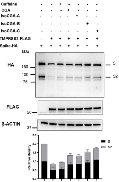
3,4-Dicaffeoylquinic acid purchased from MedChemExpress. Usage Cited in: Cell Biosci. 2023 Nov 14;13(1):210. [Abstract]
Through Western blot to test 5 compounds (3,4-Dicaffeoylquinic acid (IsoCGA-B) (1 h), etc.) at 100 μM impact cleavage of SARS-CoV-2 S by TMPRSS2.
-
Cells
Identification of Natural Compounds Triggering MRGPRX2-Mediated Calcium Flux and Degranulation in RBL-2H3 Cells. [Abstract]2026 Feb 3;15(3):287. PMID: 41677650 -
Foods
Effects of Cooking Methods on Caffeoylquinic Acids and Radical Scavenging Activity of Sweet Potato. [Abstract]2024 Apr 3;13(7):1101. PMID: 38611405 -
ACS Omega
Selective Enhancement of Caffeoylquinic Acid Derivative via UV Irradiation and Validation of Analytical Method in the Aerial Aster × chusanensis Y. S. Lim. [Abstract]2025 Sep 22;10(38):44260-44269. PMID: 41048756 -
Vet Microbiol
The Chinese medicine monomer Schisandrin C inhibits PRRSV infection by regulating the OGT-PI3K/AKT/mTOR signaling pathway. [Abstract]2026 May:316:110992. PMID: 41865607 -
Chem Pharm Bull
Chromatographic Evaluation and Characterization of Constituents of Sunflower Seed Extract Used as Food Additives. [Abstract]2024;72(1):93-97. PMID: 38233137 -
-


Solvent & Solubility
In Vitro:
DMSO : 50 mg/mL (96.81 mM; Need ultrasonic; Hygroscopic DMSO has a significant impact on the solubility of product, please use newly opened DMSO)
Please refer to the solubility information to select the appropriate solvent. Once prepared, please aliquot and store the solution to prevent product inactivation from repeated freeze-thaw cycles.
Storage method and period of stock solution: -80°C, 6 months; -20°C, 1 month. When stored at -80°C, please use it within 6 months. When stored at -20°C, please use it within 1 month.
Please refer to the solubility information to select the appropriate solvent. Once prepared, please aliquot and store the solution to prevent product inactivation from repeated freeze-thaw cycles.
Storage method and period of stock solution: -80°C, 6 months; -20°C, 1 month. When stored at -80°C, please use it within 6 months. When stored at -20°C, please use it within 1 month.
Concentration (start) × Volume (start) = Concentration (final) × Volume (final)

Protocols
-
Apoptosis
Apoptosis, also called programmed cell death, is generally characterized by distinct morphological characteristics.
-
TUNEL staining for apoptotic DNA fragmentation
TUNEL staining detects DNA strand breaks by using terminal deoxynucleotidyl transferase to add labeled nucleotides to exposed 3′-OH DNA termini, generating either microscopic staining in fixed cells or tissue sections, or fluorescence/cytometric signal in cell suspensions. TUNEL positivity reflects DNA fragmentation but should not be interpreted alone as definitive apoptosis, because TUNEL can also label necrotic, autolytic, mechanically damaged, or DNA-repair-associated DNA breaks.
-
Cell Cytotoxicity Assay
Cytotoxicity assays are usually based on the assessment of cell membrane damage, which can also be indirectly detected by measuring cell viability. Detection methods include MTT assay, CKK-8 assay, LDH assay and ATP assay, etc.
-
Mammalian live/dead viability and cytotoxicity staining
Live/dead viability and cytotoxicity staining assays are based on the simultaneous detection of intracellular esterase activity in metabolically active (viable) cells and membrane integrity loss in non-viable cells. In commonly used dual-staining approaches, membrane-permeant fluorogenic substrates are converted by intracellular esterases into fluorescent products in live cells, while impermeant DNA-binding dyes selectively enter cells with compromised plasma membranes and label nucleic acids in dead or dying cells, enabling discrimination between viable and non-viable populations by fluorescence microscopy or flow cytometry.
-
Annexin V plus membrane-impermeant dye apoptosis staining
Annexin V-based apoptosis assays rely on the detection of phosphatidylserine (PS) externalization from the inner leaflet of the plasma membrane to the outer leaflet, an early biochemical hallmark of apoptosis. Fluorescently labeled Annexin V binds PS in a calcium-dependent manner, enabling identification of early apoptotic cells by flow cytometry or fluorescence microscopy. When combined with a membrane-impermeant DNA-binding dye (e. g. , propidium iodide), this approach allows discrimination between viable (Annexin V−/dye−), early apoptotic (Annexin V+/dye−), and late apoptotic or necrotic (Annexin V+/dye+) cell populations by assessing membrane integrity and PS exposure.
-
Protocol for Pharmacokinetic Study
Pharmacokinetic studies quantify how an organism handles a drug over time through absorption, distribution, metabolism, and excretion, and the core experimental readout is the concentration-time profile of parent drug and, when relevant, metabolites in biological matrices such as plasma, whole blood, urine, bile, or tissue. Pharmacokinetic analysis links dose, route, exposure, clearance, half-life, distribution, bioavailability, and systemic exposure to drug efficacy and toxicity hypotheses rather than measuring a signaling pathway directly. The literature links pharmacokinetics to drug-development phenotypes by showing that drug metabolism and pharmacokinetics influence compound progression, exposure-response interpretation, safety margins, dosing strategy, and failure risk during discovery and development. DMPK science contributes to compound optimization by integrating physicochemical properties, in vitro metabolism, transporter behavior, in vivo exposure, and pharmacodynamic contex
-
Apoptosis Solutions
Apoptosis is a regulated, generally non-lytic cell-death pathway that removes unwanted, damaged, infected, or abnormal cells through coordinated morphological changes, caspase activation, DNA fragmentation, and membrane remodeling. The intrinsic apoptosis pathway is controlled mainly by mitochondrial outer membrane permeabilization, BCL-2 family proteins, cytochrome c release, apoptosome formation, caspase-9 activation, and downstream executioner caspase-3/7 activation. The extrinsic apoptosis pathway is initiated by death receptors such as Fas, TNFR, and TRAIL receptors, which recruit adaptor proteins and activate caspase-8 before engaging executioner caspases or mitochondrial amplification through BID cleavage. Apoptosis is linked to many phenotypes, including cancer cell killing, tissue homeostasis, immune regulation, neurodegeneration, infection response, and treatment-induced cytotoxicity; unresolved questions include how apoptosis interacts with necroptosis, pyroptosis, ferroptos
-
Cell Viability Determination by MTT Colorimetric Assay
The following protocol uses the MTT colorimetric assay as a classic literature-established method for assessing cell viability/metabolic activity in cultured mammalian cells. MTT[3-(4,5-dimethylthiazol-2-yl)-2,5-diphenyltetrazolium bromide] is reduced by metabolically active cells to a colored formazan product; the amount of formazan is quantified spectrophotometrically and provides an indirect measure of metabolically active viable cells. Importantly, MTT reduction reflects cellular oxidoreductase/metabolic activity rather than an absolute direct count of living cells, so changes in cellular metabolism can alter the signal independently of cell number.


Purity & Documentation
-
Data Sheet (278 KB)
-
SDS (396 KB)
- English - EN (396 KB)
- Français - FR (396 KB)
- Deutsch - DE (396 KB)
- Norwegian - NO (396 KB)
- Español - ES (396 KB)
- Swedish - SV (396 KB)
- Italian - IT (396 KB)
- Korean - KR (396 KB)
- Portuguese - PT (396 KB)
-
Handling Instructions (2659 KB)
References
[1]. Takemura T, et al. 3,4-Dicaffeoylquinic Acid, a Major Constituent of Brazilian Propolis, Increases TRAIL Expression and Extends the Lifetimes of Mice Infected with the Influenza A Virus. Evid Based Complement Alternat Med. 2012;2012:946867. [Content Brief]
[2]. Liu X, et al. Protective effect of isochlorogenic acid B on liver fibrosis in non-alcoholic steatohepatitis of mice. Basic Clin Pharmacol Toxicol. 2019;124(2):144-153. [Content Brief]
[3]. Ooi KL, et al. Cytotoxic, apoptotic and anti-α-glucosidase activities of 3,4-di-O-caffeoyl quinic acid, an antioxidant isolated from the polyphenolic-rich extract of Elephantopus mollis Kunth. J Ethnopharmacol. 2011;135(3):685-695. [Content Brief]


Complete Stock Solution Preparation Table
Please refer to the solubility information to select the appropriate solvent. Once prepared, please aliquot and store the solution to prevent product inactivation from repeated freeze-thaw cycles.
Storage method and period of stock solution: -80°C, 6 months; -20°C, 1 month. When stored at -80°C, please use it within 6 months. When stored at -20°C, please use it within 1 month.
| Optional Solvent | Concentration Solvent Mass | 1 mg | 5 mg | 10 mg | 25 mg |
|---|---|---|---|---|---|
| DMSO | 1 mM | 1.9363 mL | 9.6815 mL | 19.3630 mL | 48.4074 mL |
| 5 mM | 0.3873 mL | 1.9363 mL | 3.8726 mL | 9.6815 mL | |
| 10 mM | 0.1936 mL | 0.9681 mL | 1.9363 mL | 4.8407 mL | |
| 15 mM | 0.1291 mL | 0.6454 mL | 1.2909 mL | 3.2272 mL | |
| 20 mM | 0.0968 mL | 0.4841 mL | 0.9681 mL | 2.4204 mL | |
| 25 mM | 0.0775 mL | 0.3873 mL | 0.7745 mL | 1.9363 mL | |
| 30 mM | 0.0645 mL | 0.3227 mL | 0.6454 mL | 1.6136 mL | |
| 40 mM | 0.0484 mL | 0.2420 mL | 0.4841 mL | 1.2102 mL | |
| 50 mM | 0.0387 mL | 0.1936 mL | 0.3873 mL | 0.9681 mL | |
| 60 mM | 0.0323 mL | 0.1614 mL | 0.3227 mL | 0.8068 mL | |
| 80 mM | 0.0242 mL | 0.1210 mL | 0.2420 mL | 0.6051 mL |


3,4-Dicaffeoylquinic acid Related Classifications
HY-N0057 Related Classifications
 Powered by Bioz