



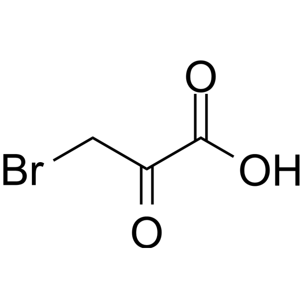
3-Bromopyruvic acid
Based on 36 publication(s) in Google Scholar
3-Bromopyruvate (Bromopyruvic acid) is an analogue of pyruvate and a potent hexokinase (HK)-II inhibitor with high tumor selectivity. 3-Bromopyruvate inhibits cell growth and induces apoptosis through interfering with glycolysis. 3-Bromopyruvate induces autophagy by stimulating ROS formation in breast cancer cells. Antimicrobial activities.
For research use only. We do not sell to patients.
- Purity : 98.0%
- CAS No.: 1113-59-3
- Formula: C3H3BrO3
- Molecular Weight:166.96
-
Storage:
4°C, sealed storage, away from moisture
* In solvent : -80°C, 6 months; -20°C, 1 month (sealed storage, away from moisture)

To place orders, for customer services and technical support, please contact: MedChemExpress USA
Tel: 609-228-6898 E-mail: [email protected] [email protected]


-
 Biological Activity
-
Chemical Information
-
Solvent & Solubility
- Protocols
- Purity & Documentation
- References
-
 Help & FAQs


-
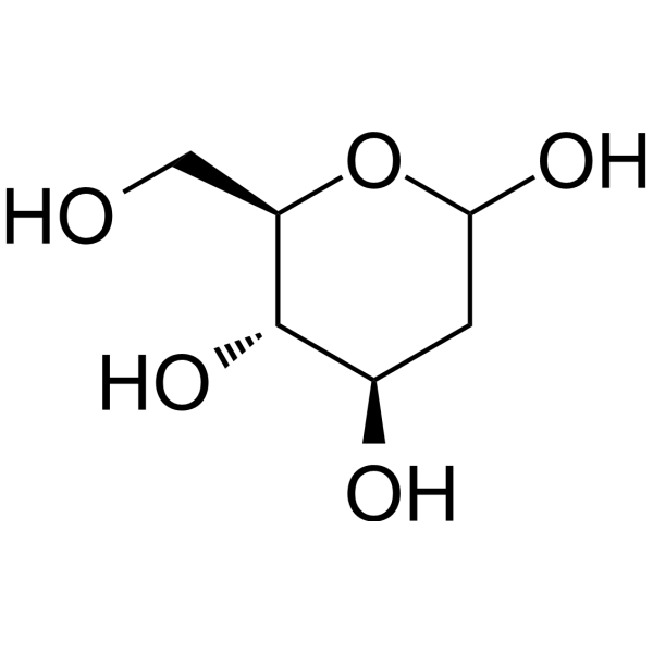
HY-13966
2-Deoxy-D-glucoseGlycolysis Inhibitor -
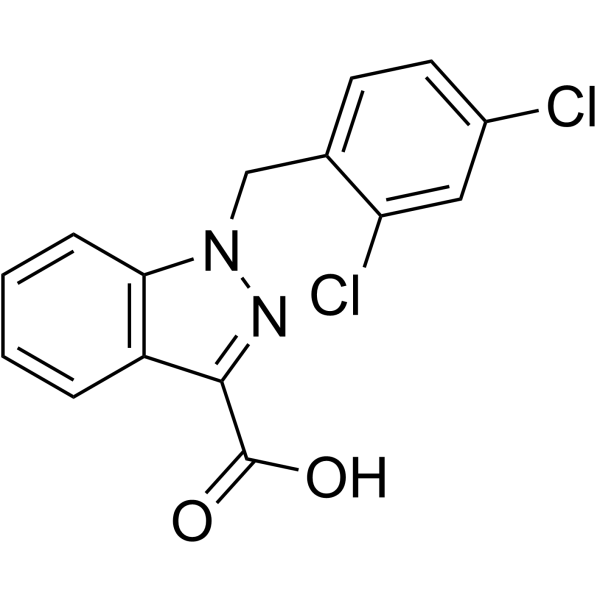
HY-B0486
LonidamineHexokinase Inhibitor -
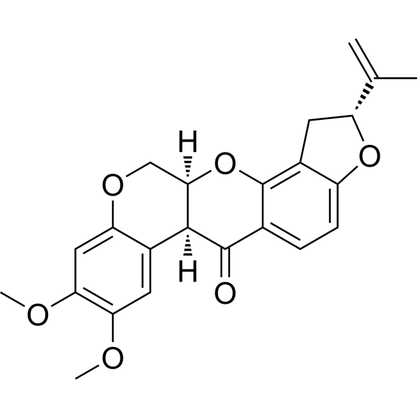
HY-B1756
RotenoneMitochondrial Complex I Inhibitor -
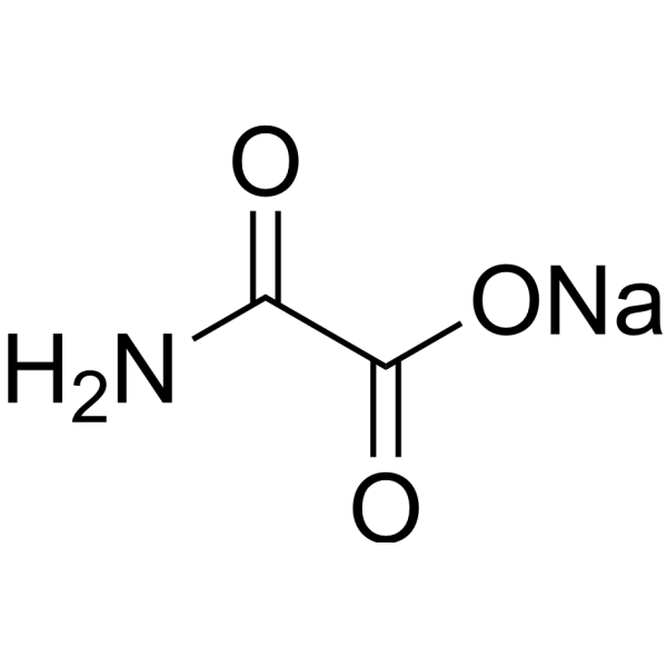
HY-W013032A
Oxamic acid sodiumLDH-A Inhibitor -

HY-10583
Y-27632 dihydrochlorideROCK Inhibitor -
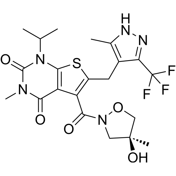
HY-12750
AZD3965MCT1 Inhibitor -
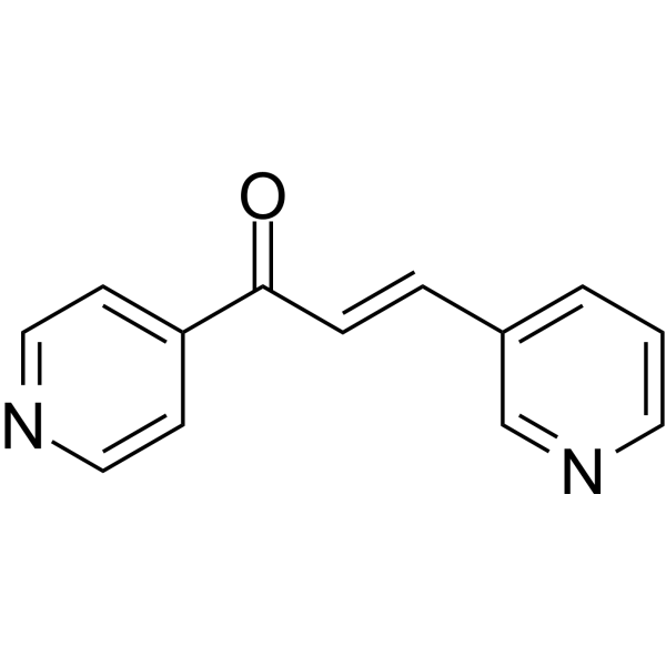
HY-19824
3POPFKFB3 Blocker -
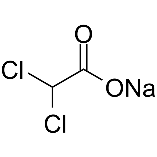
HY-Y0445A
Sodium dichloroacetatePDK Inhibitor -
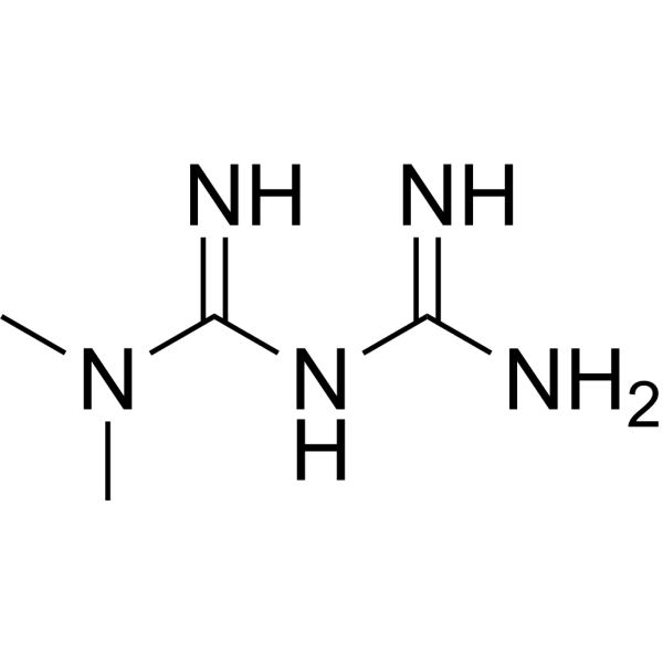
HY-B0627
MetforminAntihyperglycemic Agent -
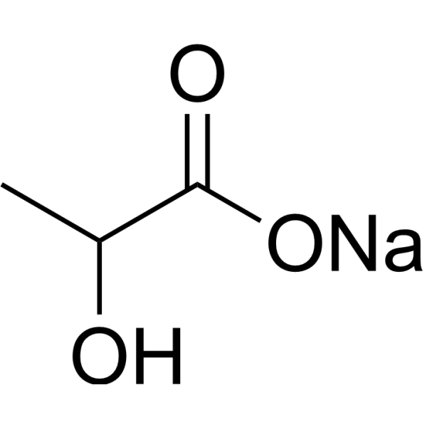
HY-B2227B
Lactic acid sodium (60% w/w in water)Glycolysis Production -
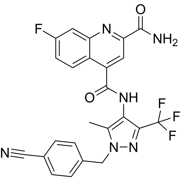
HY-100017
BAY-876GLUT1 Inhibitor -
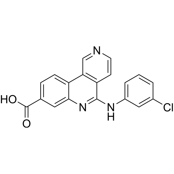
HY-50855
SilmitasertibCasein Kinase 2 Inhibitor
Publications Citing Use of MedChemExpress (MCE) 3-Bromopyruvic acid
More- Cell Stem Cell. 2023 Apr 6;30(4):450-459.e9. [Abstract]
- Bone Res. 2024 Aug 28;12(1):49. [Abstract]
- Mol Cell. 2025 Jul 17;85(14):2733-2748.e7. [Abstract]
- J Clin Invest. 2026 Mar 2;136(5):e172380. [Abstract]
- Chem Eng J. 2025 Mar 1.
- Cell Death Dis. 2025 Mar 17;16(1):182. [Abstract]
- Cell Death Dis. 2022 Sep 20;13(9):803. [Abstract]
- Mater Today Bio. 2025 Apr 10:32:101732. [Abstract]
- Arch Toxicol. 2022 Nov;96(11):2913-2926. [Abstract]
- Arch Toxicol. 2022 Jul;96(7):2113-2122. [Abstract]
- Cell Death Discov. 2025 Oct 31;11(1):493. [Abstract]
- Int J Biol Macromol. 2024 Dec;282(Pt 4):137070. [Abstract]
- J Agric Food Chem. 2021 Nov 3;69(43):12862-12869. [Abstract]
- Neurobiol Dis. 2022 Feb:163:105605. [Abstract]
- Eur J Pharmacol. 2026 May 10:1023:178866. [Abstract]
- Int Immunopharmacol. 2026 Sep 1:184:116961. [Abstract]
- Arthritis Res Ther. 2025 Sep 26;27(1):180. [Abstract]
- Molecules. 2022 Apr 11;27(8):2447. [Abstract]
- Sci Rep. 2025 Mar 28;15(1):10772. [Abstract]
- Sci Rep. 2024 Jul 17;14(1):16561. [Abstract]
- Cell Signal. 2024 Jun:118:111126. [Abstract]
- Poult Sci. 2024 Jun;103(6):103717. [Abstract]
- Fish Shellfish Immunol. 2022 Oct:129:22-29. [Abstract]
- Cell Stress Chaperones. 2022 May;27(3):273-283. [Abstract]
- Exp Cell Res. 2020 Mar 15;388(2):111876. [Abstract]
- Food Chem Toxicol. 2021 Feb:148:111926. [Abstract]
- Exp Eye Res. 2022 Apr 28;220:109095. [Abstract]
- J Inorg Biochem. 2022 Apr 20;233:111838. [Abstract]
- J Inorg Biochem. 2022 Apr:229:111725. [Abstract]
- Vet Parasitol. 2025 Jul:337:110471. [Abstract]
- Clin Exp Pharmacol Physiol. 2026 Jan;53(1):e70096. [Abstract]
- Res Sq. 2026 Jun 15.
- bioRxiv. 2026 May 29.
- SSRN. 2025 Dec 2.
- Research Square Print. posted 01 Nov, 2022
- Research Square Preprint. 2021 Jun.
 Customer Validation & Images

-
IHC
-
IP
-
WB
-
IHC
-
WB


Biological Activity
Description
Cellular Effect
|
Cell Line

|
Type | Value | Description | References |
|---|---|---|---|---|
| A549 | GI50 |
25 μM
Compound: 3-BP
|
Growth inhibition of human A549 cells expressing MCT1 incubated for 72 hrs by MTT assay
Growth inhibition of human A549 cells expressing MCT1 incubated for 72 hrs by MTT assay
|
[PMID: 36584238] |
In Vitro
3-Bromopyruvate enhances TRAIL-induced apoptosis in breast cancer cells[2].
3-Bromopyruvate (Bromopyruvic acid), a hexokinase II inhibitor, can induce apoptosis in hepatocellular carcinoma cells by inducing endoplasmic reticulum (ER) stress[2].
3-Bromopyruvate inhibits ATP generation and upregulates the expression of DR5. 3-Bromopyruvate upregulates CHOP, GRP78 and the phosphorylation of AMPK and augments TRAIL-induced Bax and caspase-3 levels[2].
MedChemExpress (MCE) has not independently confirmed the accuracy of these methods. They are for reference only. Further protocols information, click here.
-
Cell Line:MCF-7 and MDA-MB-231 cells
-
Concentration:40, 80, 160 or 320 µM
-
Incubation Time:24 hours
-
Result:3-Bromopyruvate (80 and 160 µmol/l) and TRAIL (400 ng/ml) significantly inhibited cell viability.
In Vivo
MedChemExpress (MCE) has not independently confirmed the accuracy of these methods. They are for reference only.
-
Animal Model:Female nude mice (BALB/c; 4-5-weeks old and 18-20 g)[2]
-
Dosage:8 mg/kg
-
Administration:I.p.; every 4 days for 28 days
-
Result:Showed antitumor efficacy in tumor xenografts.


Chemical Information
-
CAS No. 1113-59-3
-
Appearance Solid
-
Molecular Weight 166.96
-
Formula C3H3BrO3
-
Color Off-white to light yellow
-
SMILES
O=C(O)C(CBr)=O
-
Synonyms
Bromopyruvic acid; 3-BrPA; Hexokinase II Inhibitor II, 3-BP
-
Shipping
Room temperature in continental US; may vary elsewhere.
-
Storage
4°C, sealed storage, away from moisture
* In solvent : -80°C, 6 months; -20°C, 1 month (sealed storage, away from moisture)


Publications (36)
-
Journal Impact Factor
-
Most Recent
-
Cell Stem Cell
Highly efficient and rapid generation of human pluripotent stem cells by chemical reprogramming. [Abstract]2023 Apr 6;30(4):450-459.e9. PMID: 36944335 -
Bone Res
2024 Aug 28;12(1):49. PMID: 39198395
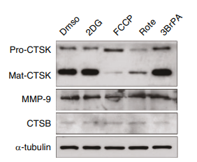
3-Bromopyruvic acid purchased from MedChemExpress. Usage Cited in: Bone Res. 2024 Aug 28;12(1):49. [Abstract]
Immunoblots of the pre-proenzyme and active CTSK levels in osteoclasts treated with 2DG (1 μM), FCCP (1 μM), Rotenone (100 nmol/L) or 3-BrPA (3-Bromopyruvic acid, 1 μM) for 10 h. The MMP-9 and CTSB expression levels were also detected. DMSO-treated osteoclasts were used as controls.
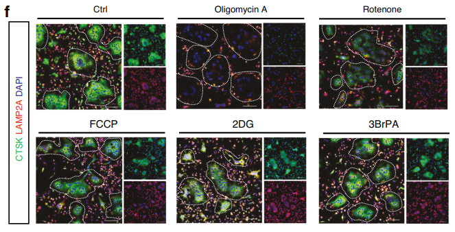
3-Bromopyruvic acid purchased from MedChemExpress. Usage Cited in: Bone Res. 2024 Aug 28;12(1):49. [Abstract]
Immunofluorescence staining to show the CTSK expression level in osteoclasts that were treated with Oligomycin A (10 nM), Rotenone (100 nM), FCCP (1 μM), 2DG (1 μM) or 3BrPA (3-Bromopyruvic acid, 1 μM) for 6 h.
-
Mol Cell
YTHDC1 lactylation regulates its phase separation to enhance target mRNA stability and promote RCC progression. [Abstract]2025 Jul 17;85(14):2733-2748.e7. PMID: 40680722 -
J Clin Invest
α-Ketoglutarate protects against cartilage damage via epigenetically driven metabolic reprogramming in osteoarthritis models. [Abstract]2026 Mar 2;136(5):e172380. PMID: 41766657 -
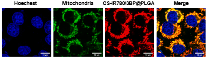
3-Bromopyruvic acid purchased from MedChemExpress. Usage Cited in: Chem Eng J. 2025 Mar 1.
Representative TEM images showed that IR780/3BP@PLGA and CSIR780/3BP@PLGA (Groups 4 and 6, 1 mg/mL PLGA concentration) nanoparticles presented a spherical structure.
-
Cell Death Dis
High glucose levels promote glycolysis and cholesterol synthesis via ERRα and suppress the autophagy-lysosomal pathway in endometrial cancer. [Abstract]2025 Mar 17;16(1):182. PMID: 40097416
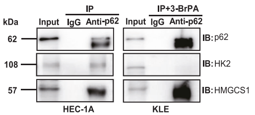
3-Bromopyruvic acid purchased from MedChemExpress. Usage Cited in: Cell Death Dis. 2025 Mar 17;16(1):182. [Abstract]
Co-IP was performed to clarify the relationship between HK2 and HMGCS1 binding with p62 when added in 10 μM 3-BrPA (3-Bromopyruvic acid) in KLE cells.
-
Cell Death Dis
Hnrnpk maintains chondrocytes survival and function during growth plate development via regulating Hif1α-glycolysis axis. [Abstract]2022 Sep 20;13(9):803. PMID: 36127325 -
Mater Today Bio
An exosome-based nanoplatform for siRNA delivery combined with starvation therapy promotes tumor cell death through autophagy, overcoming refractory KRAS-mutated tumors and restoring cetuximab chemosensitivity. [Abstract]2025 Apr 10:32:101732. PMID: 40290881 -
Arch Toxicol
CuO-NPs-triggered heterophil extracellular traps exacerbate liver injury in chicks by promoting oxidative stress and inflammatory responses. [Abstract]2022 Nov;96(11):2913-2926. PMID: 35962800 -
Arch Toxicol
Chicken heterophils extracellular traps act as early effectors against cyclopiazonic acid dependent upon NADPH oxidase, ROS and glycolysis. [Abstract]2022 Jul;96(7):2113-2122. PMID: 35508807 -
Cell Death Discov
Tanshinone IIA inhibits heat-induced growth of p53-mutant Huh-7 hepatocellular carcinoma by modulating osmotic homeostasis and glycolysis through targeting ALDH7A1. [Abstract]2025 Oct 31;11(1):493. PMID: 41173836 -
Int J Biol Macromol
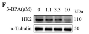
Cepharanthine inhibits African swine fever virus replication by suppressing AKT-associated pathways through disrupting Hsp90-Cdc37 complex. [Abstract]2024 Dec;282(Pt 4):137070. PMID: 39486740
3-Bromopyruvic acid purchased from MedChemExpress. Usage Cited in: Int J Biol Macromol. 2024 Dec;282(Pt 4):137070. [Abstract]
3-BPA (3-Bromopyruvic acid) at concentrations from 1.1 to 10 μM and GSK at concentrations from 3.3 to 30 μM inhibited ASFV replication in a dose-dependent manner, indicating that glycolysis inhibition leads to suppression of ASFV replication in PAMs.
-
J Agric Food Chem
Glycolysis and Reactive Oxygen Species Production Participate in T-2 Toxin-Stimulated Chicken Heterophil Extracellular Traps. [Abstract]2021 Nov 3;69(43):12862-12869. PMID: 34694797 -
Neurobiol Dis
Upregulated hexokinase 2 expression induces the apoptosis of dopaminergic neurons by promoting lactate production in Parkinson's disease. [Abstract]2022 Feb:163:105605. PMID: 34973450 -
Eur J Pharmacol
Inhibition of PKM2 lactylation by geniposide ameliorates synovial hyperplasia in experimental arthritis. [Abstract]2026 May 10:1023:178866. PMID: 41990904 -
Int Immunopharmacol
Rheumatoid arthritis synovial fibroblasts promote the glycolysis of myeloid-derived suppressor cells via TREM1/mTOR axis. [Abstract]2026 Sep 1:184:116961. PMID: 42242136 -
Arthritis Res Ther
2025 Sep 26;27(1):180. PMID: 41013704 -
Molecules
2022 Apr 11;27(8):2447. PMID: 35458645 -
Sci Rep
PKM2 accelerated the progression of chronic fatigue syndrome via promoting the H4K12la/ NF-κB induced neuroinflammation and mitochondrial damage. [Abstract]2025 Mar 28;15(1):10772. PMID: 40155479 -
Sci Rep
Controlling glycolysis to generate characteristic volatile organic compounds of lung cancer cells. [Abstract]2024 Jul 17;14(1):16561. PMID: 39020066 -
Cell Signal
HK2 promotes migration and invasion of intrahepatic cholangiocarcinoma via enhancing cancer stem-like cells' resistance to anoikis. [Abstract]2024 Jun:118:111126. PMID: 38453126 -
Poult Sci
2024 Jun;103(6):103717. PMID: 38643746 -
Fish Shellfish Immunol
Zinc oxide nanoparticles (ZnO-NPs) exhibit immune toxicity to crucian carp (Carassius carassius) by neutrophil extracellular traps (NETs) release and oxidative stress. [Abstract]2022 Oct:129:22-29. PMID: 35932984 -
Cell Stress Chaperones
Role of endoplasmic reticulum stress in apoptosis induced by HK2 inhibitor and its potential as a new drug combination strategy. [Abstract]2022 May;27(3):273-283. PMID: 35355227 -
Exp Cell Res
CSN5 upregulates glycolysis to promote hepatocellular carcinoma metastasis via stabilizing the HK2 protein. [Abstract]2020 Mar 15;388(2):111876. PMID: 31991125 -
Food Chem Toxicol
Diacetoxyscirpenol-induced heterophil extracellular traps contribute to the immune toxicity of liver injury in chickens. [Abstract]2021 Feb:148:111926. PMID: 33352262 -
Exp Eye Res
Inhibition of Drp1 ameliorates diabetic retinopathy by regulating mitochondrial homeostasis. [Abstract]2022 Apr 28;220:109095. PMID: 35490835 -
J Inorg Biochem
Nanosilver-stimulated heterophil extracellular traps promoted liver and kidney injury in chicken. [Abstract]2022 Apr 20;233:111838. PMID: 35504082 -
J Inorg Biochem
Alumina nanoparticles-induced heterophil extracellular traps exacerbate liver injury by regulating oxidative stress and inflammation in chickens. [Abstract]2022 Apr:229:111725. PMID: 35063926 -
Vet Parasitol
2025 Jul:337:110471. PMID: 40245810 -
Clin Exp Pharmacol Physiol
Esculin Facilitates Aerobic Glycolysis via the Wnt/β-Catenin/HIF-1α Pathway to Reduce the Progression of Gastric Cancer. [Abstract]2026 Jan;53(1):e70096. PMID: 41340325 -
-
-
-
-


Solvent & Solubility
In Vitro:
H2O : 175 mg/mL (1048.16 mM; Need ultrasonic)
DMSO : 100 mg/mL (598.95 mM; Need ultrasonic; Hygroscopic DMSO has a significant impact on the solubility of product, please use newly opened DMSO)
Please refer to the solubility information to select the appropriate solvent. Once prepared, please aliquot and store the solution to prevent product inactivation from repeated freeze-thaw cycles.
Storage method and period of stock solution: -80°C, 6 months; -20°C, 1 month (sealed storage, away from moisture). When stored at -80°C, please use it within 6 months. When stored at -20°C, please use it within 1 month.
* Note: If you choose water as the stock solution, please dilute it to the working solution, then filter and sterilize it with a 0.22 μm filter before use.
Please refer to the solubility information to select the appropriate solvent. Once prepared, please aliquot and store the solution to prevent product inactivation from repeated freeze-thaw cycles.
Storage method and period of stock solution: -80°C, 6 months; -20°C, 1 month (sealed storage, away from moisture). When stored at -80°C, please use it within 6 months. When stored at -20°C, please use it within 1 month.
* Note: If you choose water as the stock solution, please dilute it to the working solution, then filter and sterilize it with a 0.22 μm filter before use.
Concentration (start) × Volume (start) = Concentration (final) × Volume (final)
In Vivo:
Select the appropriate dissolution method based on your experimental animal and administration route.
- For the following dissolution methods, please ensure to first prepare a clear stock solution using an In Vitro approach and then sequentially add co-solvents:
- To ensure reliable experimental results, the clarified stock solution can be appropriately stored based on storage conditions. As for the working solution for In Vivo experiments, it is recommended to prepare freshly and use it on the same day.
- The percentages shown for the solvents indicate their volumetric ratio in the final prepared solution. If precipitation or phase separation occurs during preparation, heat and/or sonication can be used to aid dissolution.
Add each solvent one by one: 10% DMSO 40% PEG300 5% Tween-80 45% Saline
Solubility: ≥ 2.5 mg/mL (14.97 mM); Clear solution
This protocol yields a clear solution of ≥ 2.5 mg/mL (saturation unknown).
Taking 1 mL working solution as an example, add 100 μL DMSO stock solution (25.0 mg/mL) to 400 μL PEG300, and mix evenly; then add 50 μL Tween-80 and mix evenly; then add 450 μL Saline to adjust the volume to 1 mL.
Preparation of Saline: Dissolve 0.9 g sodium chloride in ddH₂O and dilute to 100 mL to obtain a clear Saline solution.
Add each solvent one by one: 10% DMSO 90% (20% SBE-β-CD in Saline)
Solubility: ≥ 2.5 mg/mL (14.97 mM); Clear solution
This protocol yields a clear solution of ≥ 2.5 mg/mL (saturation unknown).
Taking 1 mL working solution as an example, add 100 μL DMSO stock solution (25.0 mg/mL) to 900 μL 20% SBE-β-CD in Saline, and mix evenly.
Preparation of 20% SBE-β-CD in Saline (4°C, storage for one week): 2 g SBE-β-CD powder is dissolved in 10 mL Saline, completely dissolve until clear.
For the following dissolution methods, please prepare the working solution directly:
It is recommended to prepare fresh solutions and use them promptly within a short period of time.
The percentages shown for the solvents indicate their volumetric ratio in the final prepared solution. If precipitation or phase separation occurs during preparation, heat and/or sonication can be used to aid dissolution.
Add each solvent one by one: PBS
Solubility: 100 mg/mL (598.95 mM); Clear solution; Need ultrasonic
In Vivo Dissolution Calculator
Please enter the basic information of animal experiments:
-
-
-
-
Recommended: Prepare an additional quantity of animals to account for potential losses during experiments.
Working solution concentration: 0.22 mg/mL
This product has good water solubility, please refer to the measured solubility data in water/PBS/Saline for details.

Protocols
-
Research Protocol for Infectious Diseases
Infectious-disease experiments test how pathogens interact with host barriers, innate immune receptors, inflammatory signaling, pathogen replication, and tissue injury; pattern-recognition receptors such as TLRs, RIG-I-like receptors, NOD-like receptors, and inflammasomes detect microbial molecules and activate NF-κB, interferon, and cytokine responses. The central hypothesis is that infection severity reflects the balance between pathogen burden and host response: protective inflammation restricts pathogen growth, whereas excessive or mislocalized inflammation contributes to tissue damage and disease phenotype. Unresolved questions include which host pathways are protective versus pathogenic, why some infection models fail to translate to human disease, and which combined readouts best predict clinically relevant infection outcomes.
-
Annexin V plus membrane-impermeant dye apoptosis staining
Annexin V-based apoptosis assays rely on the detection of phosphatidylserine (PS) externalization from the inner leaflet of the plasma membrane to the outer leaflet, an early biochemical hallmark of apoptosis. Fluorescently labeled Annexin V binds PS in a calcium-dependent manner, enabling identification of early apoptotic cells by flow cytometry or fluorescence microscopy. When combined with a membrane-impermeant DNA-binding dye (e. g. , propidium iodide), this approach allows discrimination between viable (Annexin V−/dye−), early apoptotic (Annexin V+/dye−), and late apoptotic or necrotic (Annexin V+/dye+) cell populations by assessing membrane integrity and PS exposure.
-
ROS/oxidative-stress fluorescent staining
ROS/oxidative-stress fluorescent staining uses cell-permeant fluorogenic probes that become fluorescent after oxidation inside cells or tissues; commonly used examples include DCFH-DA/DCFDA for broad cellular oxidant detection, DHE for superoxide-related signal detection, MitoSOX for mitochondrial superoxide-related signal detection, and CellROX probes for oxidative-stress-associated fluorescence readouts. The assay detects probe oxidation rather than a single ROS species unless the probe and analysis method have been chemically validated for that species. DCFH-DA enters cells, is deacetylated by intracellular esterases to DCFH, and produces fluorescent DCF after oxidation, so the readout is used as an operational measure of total cellular oxidative stress rather than a species-specific ROS measurement. DHE and MitoSOX can report superoxide-related oxidation, but red fluorescence alone can include non-specific ethidium-like oxidation products; HPLC or optimized spectral approaches are
-
Somatic Cell Culture
A method of simulating the in vivo environment in vitro to maintain the cell growth, differentation and main functions.
-
CCK-8/WST-8 Cell Proliferation Assay
The CCK-8/WST-8 assay is based on the reduction of the water-soluble tetrazolium salt WST-8 to a water-soluble formazan product by cellular dehydrogenases in metabolically active cells, where the generated formazan amount is proportional to the number of living cells and is quantified by measuring absorbance in the visible range, providing a colorimetric readout for cell viability and proliferation assessment. This class of tetrazolium-based assays improves upon earlier MTT-based systems by producing a water-soluble formazan, eliminating the need for organic solubilization steps and enabling direct spectrophotometric measurement in culture medium.
-
Cell Counting-Based Growth Curve Assay
Cell counting-based growth curve assays quantify cell proliferation by directly measuring changes in viable cell number over time using manual or automated counting methods such as hemocytometer-based counting or instrument-assisted cell enumeration, enabling construction of growth curves that reflect population expansion dynamics in response to culture conditions. A widely used approach is trypan blue exclusion with hemocytometer counting, where membrane-compromised (non-viable) cells take up the dye, allowing discrimination between viable and non-viable cells while simultaneously enabling total cell number quantification. Repeated sampling across time points allows estimation of proliferation rate, growth phases, and comparative growth kinetics between experimental conditions.
-
Breast Cancer Modeling
Breast cancer is a heterogeneous cancer, and it has been distinguished into four subtypes: luminal A, luminal B, HER2-positive and basal-like. Molecular mutations, epigenetic alterations, hormone exposure and immune microenvironment are related to the progression of breast cancer.
-
Cell Viability Determination by MTT Colorimetric Assay
The following protocol uses the MTT colorimetric assay as a classic literature-established method for assessing cell viability/metabolic activity in cultured mammalian cells. MTT[3-(4,5-dimethylthiazol-2-yl)-2,5-diphenyltetrazolium bromide] is reduced by metabolically active cells to a colored formazan product; the amount of formazan is quantified spectrophotometrically and provides an indirect measure of metabolically active viable cells. Importantly, MTT reduction reflects cellular oxidoreductase/metabolic activity rather than an absolute direct count of living cells, so changes in cellular metabolism can alter the signal independently of cell number.
-
Apoptosis
Apoptosis, also called programmed cell death, is generally characterized by distinct morphological characteristics.
-
TUNEL staining for apoptotic DNA fragmentation
TUNEL staining detects DNA strand breaks by using terminal deoxynucleotidyl transferase to add labeled nucleotides to exposed 3′-OH DNA termini, generating either microscopic staining in fixed cells or tissue sections, or fluorescence/cytometric signal in cell suspensions. TUNEL positivity reflects DNA fragmentation but should not be interpreted alone as definitive apoptosis, because TUNEL can also label necrotic, autolytic, mechanically damaged, or DNA-repair-associated DNA breaks.
-
Protocol for Cell Counting and Cell Density Analysis
Cell counting and cell-density analysis estimate the number of cells in a known volume or field area. Manual hemocytometer counting uses a chamber of defined geometry to convert counted cells into cells/mL, while automated counters and image-analysis workflows detect cell objects from optical, brightfield, fluorescence, impedance, or digital-image features. Trypan blue viability counting is based on dye exclusion: viable cells with intact membranes exclude dye, while non-viable cells with compromised membranes stain blue. The readout is total cell density, viable-cell density, dead-cell density, and percent viability. Cell density can also be estimated from microscopy images by counting objects per image area, from flow cytometry using calibrated volume or reference particles, or from in situ microscopy in bioreactors after calibration against reference methods such as hemocytometer or flow cytometry.
-
Autophagy
Autophagy is a process in which eukaryotic cells use lysosomes to degrade their own cytoplasmic proteins and damaged organelles under the regulation of autophagy related gene (Atg). Microtubule-associated proteins light chain 3 (LC3) is recognized as autophagy marker, which transfers from cytoplasmic LC3 (LC3-I) to membrane type (LC3-II). LC3-II/I ratio could be detected by Western Blot and fluorescence microscopy.
-
Apoptosis Solutions
Apoptosis is a regulated, generally non-lytic cell-death pathway that removes unwanted, damaged, infected, or abnormal cells through coordinated morphological changes, caspase activation, DNA fragmentation, and membrane remodeling. The intrinsic apoptosis pathway is controlled mainly by mitochondrial outer membrane permeabilization, BCL-2 family proteins, cytochrome c release, apoptosome formation, caspase-9 activation, and downstream executioner caspase-3/7 activation. The extrinsic apoptosis pathway is initiated by death receptors such as Fas, TNFR, and TRAIL receptors, which recruit adaptor proteins and activate caspase-8 before engaging executioner caspases or mitochondrial amplification through BID cleavage. Apoptosis is linked to many phenotypes, including cancer cell killing, tissue homeostasis, immune regulation, neurodegeneration, infection response, and treatment-induced cytotoxicity; unresolved questions include how apoptosis interacts with necroptosis, pyroptosis, ferroptos
-
Lysosome and acidic-vesicle live-cell staining
Lysosome and acidic-vesicle live-cell staining detects acidic intracellular compartments by using membrane-permeant acidotropic probes that accumulate in low-pH vesicles, including lysosomes, late endosomes, autolysosomes, and acidic phagosomes. LysoTracker staining is commonly used as an intensity-based readout of acidic lysosomal compartment abundance or enlargement, while acridine orange produces green fluorescence in less concentrated compartments and red fluorescence after concentration-dependent accumulation in acidic vesicular organelles. Loss or reduction of acridine-orange red signal can be used as a readout of lysosomal membrane permeabilization or reduced acidic-vesicle integrity. This protocol is designed for live cultured cells and can be adapted for fluorescence microscopy, high-content imaging, plate-reader readout, or flow cytometry when the selected literature supports the readout. Because these dyes report acidotropic accumulation rather than lysosome identity alone,
-
MTT Cell Proliferation Assay
The MTT assay is a colorimetric endpoint assay for estimating viable cell number, cell growth, cytotoxicity, or cell activation in cultured mammalian cells. Living cells reduce the yellow tetrazolium salt MTT into purple/blue formazan, while dead cells do not generate the same signal; the resulting color can be quantified with a multiwell spectrophotometer. MTT reduction is commonly interpreted as a readout of metabolic activity that often correlates with viable cell number, but it should not be treated as a direct cell-counting method unless the assay is optimized for the cell type and experimental condition. Studies show that MTT reduction can involve mitochondrial and non-mitochondrial reducing systems, and formazan may accumulate in intracellular lipid droplets rather than simply marking mitochondria.
-
Macroautophagy Solutions
Macroautophagy is a conserved lysosome-dependent degradation pathway in which cytoplasmic material is sequestered into double-membrane autophagosomes and delivered to lysosomes for degradation and recycling. The pathway supports cellular homeostasis during nutrient limitation, organelle stress, protein-aggregate accumulation, infection, differentiation, and tissue remodeling by coupling cargo sequestration, autophagosome maturation, lysosomal fusion, and degradation of cargo-derived macromolecules. The core molecular sequence includes initiation by nutrient- and stress-regulated autophagy machinery, autophagosome nucleation, LC3/ATG8-family conjugation to autophagosomal membranes, cargo selection through receptors such as SQSTM1/p62, autophagosome-lysosome fusion, and lysosomal degradation. LC3 was identified as a mammalian homolog of yeast Atg8 that localizes to autophagosomal membranes after processing, and p62/SQSTM1 was shown to connect ubiquitinated cargo with autophagic degradati


Purity & Documentation
-
Data Sheet (276 KB)
-
SDS (477 KB)
- English - EN (477 KB)
- Français - FR (477 KB)
- Deutsch - DE (477 KB)
- Norwegian - NO (477 KB)
- Español - ES (477 KB)
- Swedish - SV (477 KB)
- Italian - IT (477 KB)
- Korean - KR (477 KB)
- Portuguese - PT (477 KB)
-
Handling Instructions (2659 KB)
References
[1]. Gan L, et al. Synergistic Effect of 3-Bromopyruvate in Combination with Rapamycin Impacted Neuroblastoma Metabolism by Inhibiting Autophagy. Onco Targets Ther. 2020;13:11125-11137. Published 2020 Oct 29. [Content Brief]
[2]. Chen Y, et al. 3 Bromopyruvate sensitizes human breast cancer cells to TRAIL induced apoptosis via the phosphorylated AMPK mediated upregulation of DR5. Oncol Rep. 2018;40(5):2435-2444. [Content Brief]
[3]. Zhang Q, et al. Hexokinase II inhibitor, 3-BrPA induced autophagy by stimulating ROS formation in human breast cancer cells. Genes Cancer. 2014;5(3-4):100-112. [Content Brief]


Complete Stock Solution Preparation Table
Please refer to the solubility information to select the appropriate solvent. Once prepared, please aliquot and store the solution to prevent product inactivation from repeated freeze-thaw cycles.
Storage method and period of stock solution: -80°C, 6 months; -20°C, 1 month (sealed storage, away from moisture). When stored at -80°C, please use it within 6 months. When stored at -20°C, please use it within 1 month.
| Optional Solvent | Concentration Solvent Mass | 1 mg | 5 mg | 10 mg | 25 mg |
|---|---|---|---|---|---|
| DMSO / H2O | 1 mM | 5.9895 mL | 29.9473 mL | 59.8946 mL | 149.7365 mL |
| 5 mM | 1.1979 mL | 5.9895 mL | 11.9789 mL | 29.9473 mL | |
| 10 mM | 0.5989 mL | 2.9947 mL | 5.9895 mL | 14.9736 mL | |
| 15 mM | 0.3993 mL | 1.9965 mL | 3.9930 mL | 9.9824 mL | |
| 20 mM | 0.2995 mL | 1.4974 mL | 2.9947 mL | 7.4868 mL | |
| 25 mM | 0.2396 mL | 1.1979 mL | 2.3958 mL | 5.9895 mL | |
| 30 mM | 0.1996 mL | 0.9982 mL | 1.9965 mL | 4.9912 mL | |
| 40 mM | 0.1497 mL | 0.7487 mL | 1.4974 mL | 3.7434 mL | |
| 50 mM | 0.1198 mL | 0.5989 mL | 1.1979 mL | 2.9947 mL | |
| 60 mM | 0.0998 mL | 0.4991 mL | 0.9982 mL | 2.4956 mL | |
| 80 mM | 0.0749 mL | 0.3743 mL | 0.7487 mL | 1.8717 mL | |
| 100 mM | 0.0599 mL | 0.2995 mL | 0.5989 mL | 1.4974 mL |
* Note: If you choose water as the stock solution, please dilute it to the working solution, then filter and sterilize it with a 0.22 μm filter before use.


3-Bromopyruvic acid Related Classifications
HY-19992 Related Classifications
 Powered by Bioz