



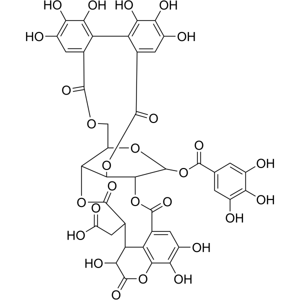
Chebulagic acid
Based on 5 publication(s) in Google Scholar
Chebulagic acid is a COX-LOX dual inhibitor isolated from the fruits of Terminalia chebula Retz, on angiogenesis. Chebulagic acid is a M2 serine to asparagine 31 mutation (S31N) inhibitor and influenza antiviral. Chebulagic acid also against SARS-CoV-2 viral replication with an EC50 of 9.76 μM.
For research use only. We do not sell to patients.
- Purity : 99.66%
- CAS No.: 23094-71-5
- Formula: C41H30O27
- Molecular Weight:954.66
-
Storage:Powder -20°C, 3 years , 4°C, 2 years ; In solvent -80°C, 6 months , -20°C, 1 month

To place orders, for customer services and technical support, please contact: MedChemExpress USA
Tel: 609-228-6898 E-mail: [email protected] [email protected]


-
 Biological Activity
-
Chemical Information
-
Solvent & Solubility
- Protocols
- Purity & Documentation
- References
-
 Help & FAQs


-
HY-N2033
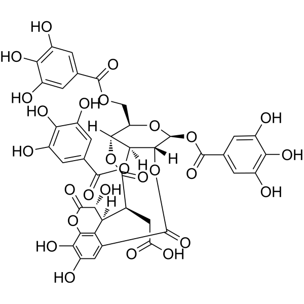
Chebulinic acidDNA Gyrase Inhibitor -
HY-N0527
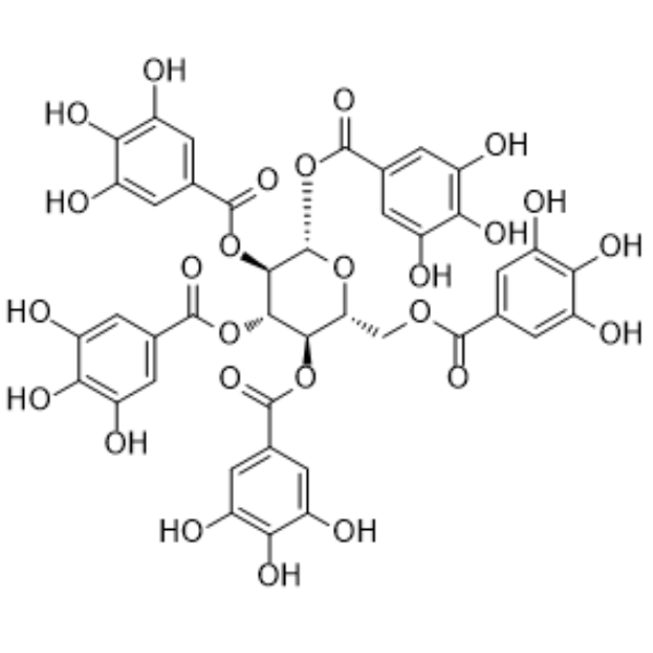
PentagalloylglucoseAntioxidant -
HY-N1992
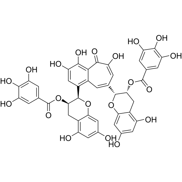
Theaflavin 3,3'-digallateVirus Protease Inhibitor -
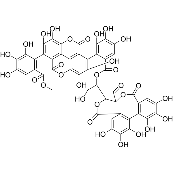
HY-N0063
Punicalagin3CLpro Inhibitor -
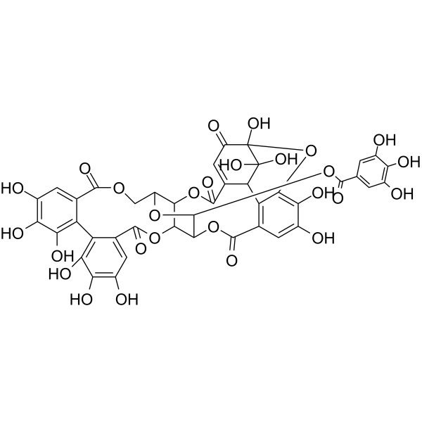
HY-N0472
GeraniinTNF Receptor Inhibitor -
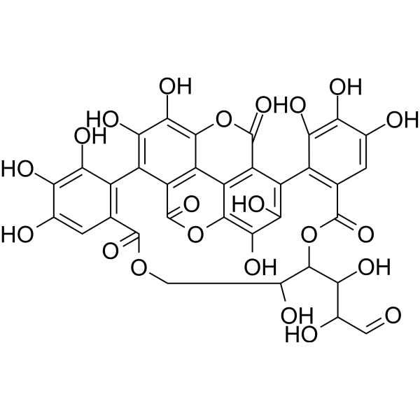
HY-N0639
PunicalinAnti-HBV Agent -
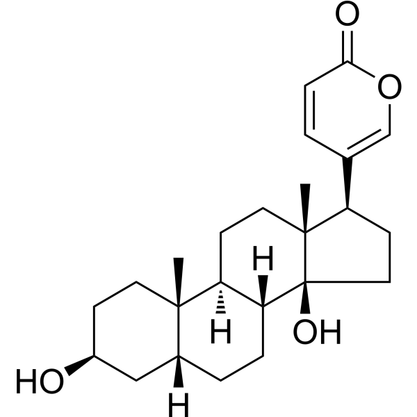
HY-N0877
BufalinNa+/K+ ATPase Inhibitor -

HY-N1996R
Chebulagic acid (Standard)COX Inhibitor -
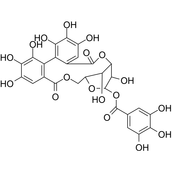
HY-N0462
CorilaginAnti-tumor Agent -
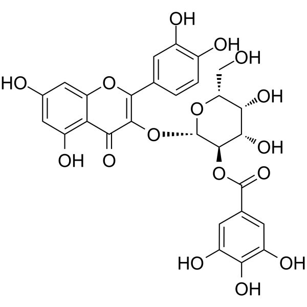
HY-N0526
2"-O-GalloylhyperinNatural Product -
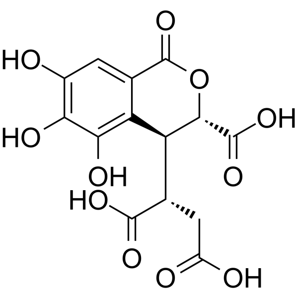
HY-N4170
Chebulic acidAntioxidant Agent -
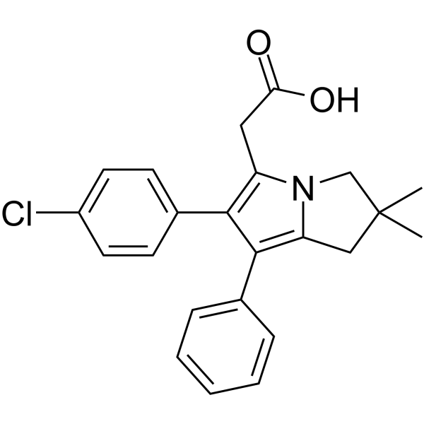
HY-B1452
LicofeloneCOX/5-LOX Inhibitor
Publications Citing Use of MedChemExpress (MCE) Chebulagic acid
More Customer Validation & Images

-
ELISA
-
RT-PCR
-
WB
-
Cell Proliferation/Viability Assay
-
Cell Proliferation/Viability Assay


Biological Activity
Description
In Vitro
Chebulagic acid can enhance the autophagy. Chebulagic acid exert anti-inflammatory and anti-infective effects. Chebulagic acid also shows a protective effect against 1-methyl-4-phenylpyridinium (MPP+)-induce cytotoxicity which mimics the pathological symptom of Parkinson's disease. Chebulagic acid inhibits the LPS-induced upregulation of TNF-α and IL-1β in a dose- and time-dependent manner. Furthermore, LPS-activated MAPK signaling is inhibited by Chebulagic acid treatment in the EA.hy926 cells.
MedChemExpress (MCE) has not independently confirmed the accuracy of these methods. They are for reference only. Further protocols information, click here.


Chemical Information
-
CAS No. 23094-71-5
-
Appearance Solid
-
Molecular Weight 954.66
-
Formula C41H30O27
-
Color White to light yellow
-
SMILES
OC(CC(C(O[C@H]([C@@H](COC(C1=CC(O)=C2O)=O)O[C@@H]3OC(C4=CC(O)=C(O)C(O)=C4)=O)[C@H](OC(C5=CC(O)=C(O)C(O)=C5C1=C2O)=O)[C@H]3OC6=O)=O)C(C7O)C(C6=CC(O)=C8O)=C8OC7=O)=O
-
Structure Classification
-
Initial Source
-
Shipping
Room temperature in continental US; may vary elsewhere.
-
Storage
Powder -20°C 3 years 4°C 2 years In solvent -80°C 6 months -20°C 1 month


Publications (5)
-
Journal Impact Factor
-
Most Recent
-
Cell Commun Signal
Nesfatin-1 enhances vascular smooth muscle calcification through facilitating BMP-2 osteogenic signaling. [Abstract]2024 Oct 11;22(1):488. PMID: 39394127 -
Pharmaceutics
Inhibition of SARS-CoV-2-Induced NLRP3 Inflammasome-Mediated Lung Cell Inflammation by Triphala-Loaded Nanoparticle Targeting Spike Glycoprotein S1. [Abstract]2024 Jun 2;16(6):751. PMID: 38931873
Chebulagic acid purchased from MedChemExpress. Usage Cited in: Pharmaceutics. 2024 Jun 2;16(6):751. [Abstract]
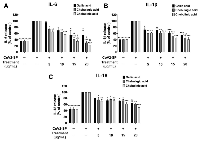
A549 cells were pre-treated with active compounds (Gallic acid, Chebulagic acid, and Chebulinic acid) (0–20 µg/mL) for 24 h. Then, the cells were exposed to CoV2-SP (100 ng/mL) for 3 h. The IL-6 (A), IL-1β (B), and IL-18 secretions (C) in the culture supernatant were examined by ELISA.
Chebulagic acid purchased from MedChemExpress. Usage Cited in: Pharmaceutics. 2024 Jun 2;16(6):751. [Abstract]
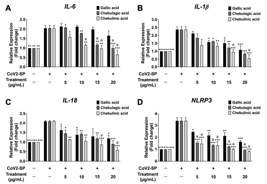
Inhibitory effects of active compounds (Gallic acid, Chebulagic acid and Chebulinic acid) on the IL-6 (A), IL-1β (B), IL-18 (C), and NLRP3 gene expressions (D) in CoV2-SP-induced A549 cells. A549 cells were pre-treated with active compounds (gallic acid, chebulagic acid and chebulinic acid) (0–20 µg/mL) for 24 h. Then, the cells were exposed to CoV2-SP (100 ng/mL) for 3 h. The mRNA expressions were determined using RT-qPCR.
Chebulagic acid purchased from MedChemExpress. Usage Cited in: Pharmaceutics. 2024 Jun 2;16(6):751. [Abstract]
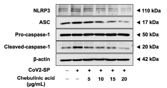
The effects of Chebulagic acid and chebulinic acid inhibited the NLRP3 inflammasome pathway in CoV2-SP-induced A549 cells. A549 cells were pre-treated with chebulagic acid and Chebulinic acid (0–20 µg/mL) for 24 h. Then, the cells were exposed to CoV2-SP (100 ng/mL) for 3 h. The inhibitory effects of chebulagic acid and chebulinic acid on the expression of NLRP3, ASC, and pro-caspase-1 (p50) and cleaved-caspase-1 (p20) proteins in A549 cells are displayed in western blot.
Chebulagic acid purchased from MedChemExpress. Usage Cited in: Pharmaceutics. 2024 Jun 2;16(6):751. [Abstract]
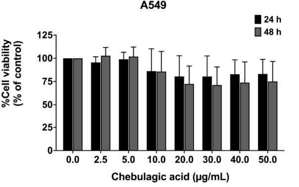
Chebulagic acid (0-50 μg/mL; 24, 48 h) showed no cytotoxicity towards A549 cells.
-
Molecules
Virtual Screening Identifies Chebulagic Acid as an Inhibitor of the M2(S31N) Viral Ion Channel and Influenza A Virus. [Abstract]2020 Jun 24;25(12):2903. PMID: 32599753 -
Antiviral Res
Discovery of chebulagic acid and punicalagin as novel allosteric inhibitors of SARS-CoV-2 3CLpro. [Abstract]2021 Jun:190:105075. PMID: 33872675
Chebulagic acid purchased from MedChemExpress. Usage Cited in: Antiviral Res. 2021 Jun:190:105075. [Abstract]
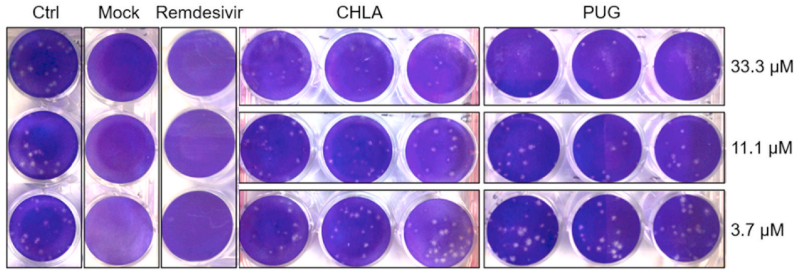
Chebulagic acid (CHLA; 3.7, 11.1, 33.3 μM; 48 h) inhibited the plaque formation of SARS-CoV-2 in a dose-dependent manner (Vero-E6 monolayers grown in 12-well plates were pre-treated with increasing concentrations of test compound for 1 h, followed by infection with SARS-CoV-2 (MOI of 0.0001) in the presence of test compounds).
-
Vet Microbiol
The Chinese medicine monomer Schisandrin C inhibits PRRSV infection by regulating the OGT-PI3K/AKT/mTOR signaling pathway. [Abstract]2026 May:316:110992. PMID: 41865607


Solvent & Solubility
In Vitro:
DMSO : 100 mg/mL (104.75 mM; Need ultrasonic; Hygroscopic DMSO has a significant impact on the solubility of product, please use newly opened DMSO)
Please refer to the solubility information to select the appropriate solvent. Once prepared, please aliquot and store the solution to prevent product inactivation from repeated freeze-thaw cycles.
Storage method and period of stock solution: -80°C, 6 months; -20°C, 1 month. When stored at -80°C, please use it within 6 months. When stored at -20°C, please use it within 1 month.
Please refer to the solubility information to select the appropriate solvent. Once prepared, please aliquot and store the solution to prevent product inactivation from repeated freeze-thaw cycles.
Storage method and period of stock solution: -80°C, 6 months; -20°C, 1 month. When stored at -80°C, please use it within 6 months. When stored at -20°C, please use it within 1 month.
Concentration (start) × Volume (start) = Concentration (final) × Volume (final)
In Vivo:
Select the appropriate dissolution method based on your experimental animal and administration route.
- For the following dissolution methods, please ensure to first prepare a clear stock solution using an In Vitro approach and then sequentially add co-solvents:
- To ensure reliable experimental results, the clarified stock solution can be appropriately stored based on storage conditions. As for the working solution for In Vivo experiments, it is recommended to prepare freshly and use it on the same day.
- The percentages shown for the solvents indicate their volumetric ratio in the final prepared solution. If precipitation or phase separation occurs during preparation, heat and/or sonication can be used to aid dissolution.
Add each solvent one by one: 10% DMSO 40% PEG300 5% Tween-80 45% Saline
Solubility: ≥ 0.83 mg/mL (0.87 mM); Clear solution
This protocol yields a clear solution of ≥ 0.83 mg/mL (saturation unknown).
Taking 1 mL working solution as an example, add 100 μL DMSO stock solution (8.3 mg/mL) to 400 μL PEG300, and mix evenly; then add 50 μL Tween-80 and mix evenly; then add 450 μL Saline to adjust the volume to 1 mL.
Preparation of Saline: Dissolve 0.9 g sodium chloride in ddH₂O and dilute to 100 mL to obtain a clear Saline solution.
Add each solvent one by one: 10% DMSO 90% (20% SBE-β-CD in Saline)
Solubility: ≥ 0.83 mg/mL (0.87 mM); Clear solution
This protocol yields a clear solution of ≥ 0.83 mg/mL (saturation unknown).
Taking 1 mL working solution as an example, add 100 μL DMSO stock solution (8.3 mg/mL) to 900 μL 20% SBE-β-CD in Saline, and mix evenly.
Preparation of 20% SBE-β-CD in Saline (4°C, storage for one week): 2 g SBE-β-CD powder is dissolved in 10 mL Saline, completely dissolve until clear.
In Vivo Dissolution Calculator
Please enter the basic information of animal experiments:
-
-
-
-
Recommended: Prepare an additional quantity of animals to account for potential losses during experiments.
Please enter your animal formula composition:
-
%DMSO +
Recommended: Keep the proportion of DMSO in working solution below 2% if your animal is weak.
-
%+
-
+%Tween-80 + +
-
%Saline +
The co-solvents required include: DMSO, . All of co-solvents are available by MedChemExpress (MCE). , Tween 80. All of co-solvents are available by MedChemExpress (MCE).
Working solution concentration: 0.22 mg/mL
Method for preparing stock solution: mg drug dissolved in μL DMSO. Stock solution concentration: mg/mL.
1. Take μL DMSO stock solution;
2. Add μL .
μL , mix evenly;
3. Then add μL Tween 80, mix evenly;
4. Then add μL
Please ensure that the stock solution in the first step is dissolved to a clear state, and add co-solvents in sequence. You can use ultrasonic heating (ultrasonic cleaner, recommended frequency 20-40 kHz), vortexing, etc. to assist dissolution.

Protocols
-
Research Protocol for Infectious Diseases
Infectious-disease experiments test how pathogens interact with host barriers, innate immune receptors, inflammatory signaling, pathogen replication, and tissue injury; pattern-recognition receptors such as TLRs, RIG-I-like receptors, NOD-like receptors, and inflammasomes detect microbial molecules and activate NF-κB, interferon, and cytokine responses. The central hypothesis is that infection severity reflects the balance between pathogen burden and host response: protective inflammation restricts pathogen growth, whereas excessive or mislocalized inflammation contributes to tissue damage and disease phenotype. Unresolved questions include which host pathways are protective versus pathogenic, why some infection models fail to translate to human disease, and which combined readouts best predict clinically relevant infection outcomes.
-
Human pluripotent stem cell endothelial-cell differentiation
Human pluripotent stem cell endothelial differentiation is based on stepwise developmental patterning: early activation of WNT/GSK3β inhibition promotes mesodermal or vascular progenitor entry, followed by endothelial specification using VEGF-related signaling, BMP4, FGF2, Notch modulation, or cAMP depending on the published protocol. Endothelial differentiation is read out by acquisition of CD31, CD34, VE-cadherin/CD144, KDR/VEGFR2, vWF, Tie2, NOS3, acetylated LDL uptake, tube/network formation, barrier function, and in vivo vessel-forming capacity where tested.
-
Vascular/Branching Fractal Analysis
Vascular/branching fractal analysis quantifies the geometric complexity of vessel trees or vascular networks from segmented 2D images, commonly by converting vessels into binary and/or skeletonized maps and estimating fractal dimension using box-counting or related approaches. Fractal dimension is interpreted as an image-derived readout of vascular branching complexity, space filling, or density, and has been applied to retinal photographs, fluorescein angiography, OCT angiography, capillary perfusion maps, and in vitro Matrigel angiogenesis networks. The assay readout is generated from vessel-positive pixels after image preprocessing, vessel segmentation, binarization, and optional skeletonization; reported outputs include fractal dimension, vessel density, branchpoint density, endpoint density, vessel length density, tortuosity, and generation-based branching metrics when VESGEN-style analysis is used. The biological interpretation is limited to quantitative vascular patterning and s
-
Endothelial Tube Formation Assay
Endothelial tube formation assay evaluates the ability of endothelial cells to attach, migrate, align, and organize into capillary-like networks when cultured on gelled basement membrane extract or Matrigel; the readout is the morphology and quantity of tube-like networks, which reflects an in vitro endothelial morphogenesis step related to angiogenesis. Basement membrane extract/Matrigel provides laminin-rich extracellular matrix cues that support endothelial differentiation into capillary-like structures, but it can contain biologically active growth factors, so growth-factor-reduced matrix is preferred when testing defined angiogenic stimulators or inhibitors.
-
Endothelial Cell Migration/Angiogenic Sprouting Assay
Endothelial cell migration and angiogenic sprouting assays are in vitro (and partially ex vivo-adapted) functional models that quantify the ability of endothelial cells to undergo coordinated migration, extracellular matrix invasion, and multicellular organization into capillary-like sprouts in response to pro-angiogenic stimuli such as VEGF, bFGF, or conditioned microenvironments. These assays are used to model early angiogenic events including tip-cell formation, directional migration, and lumen-like sprout extension, which collectively reflect angiogenic activation and vascular morphogenesis processes observed in vivo.
-
Genotoxicity/Mutagenicity Study
The bacterial reverse mutation assay detects point mutations that restore amino-acid prototrophy in auxotrophic Salmonella typhimurium or Escherichia coli tester strains; after exposure to a test article, mutagenic activity is read out as an increased number of revertant colonies on minimal agar compared with the vehicle control. The assay uses tester strains with different mutation targets so that base-substitution and frameshift mutagens can be detected, and testing is performed with and without exogenous mammalian metabolic activation because some chemicals require biotransformation to become mutagenic.


Purity & Documentation
-
Data Sheet (284 KB)
-
SDS (252 KB)
- English - EN (252 KB)
- Français - FR (252 KB)
- Deutsch - DE (252 KB)
- Norwegian - NO (252 KB)
- Español - ES (252 KB)
- Swedish - SV (252 KB)
- Italian - IT (252 KB)
- Korean - KR (252 KB)
- Portuguese - PT (252 KB)
-
Handling Instructions (2659 KB)
References
[1]. Kim HJ et al. Neuroprotective Effect of Chebulagic Acid via Autophagy Induction in SH-SY5Y Cells. Biomol Ther (Seoul). 2014 Jul;22(4):275-81. [Content Brief]
[2]. Liu Y et al. Chebulagic acid inhibits the LPS-induced expression of TNF-α and IL-1β in endothelial cells by suppressing MAPK activation. Exp Ther Med. 2015 Jul;10(1):263-268. [Content Brief]
[3]. Athira AP et al. Inhibition of Angiogenesis In Vitro by Chebulagic Acid: A COX-LOX Dual Inhibitor. Int J Vasc Med. 2013;2013:843897. [Content Brief]


Complete Stock Solution Preparation Table
Please refer to the solubility information to select the appropriate solvent. Once prepared, please aliquot and store the solution to prevent product inactivation from repeated freeze-thaw cycles.
Storage method and period of stock solution: -80°C, 6 months; -20°C, 1 month. When stored at -80°C, please use it within 6 months. When stored at -20°C, please use it within 1 month.
| Optional Solvent | Concentration Solvent Mass | 1 mg | 5 mg | 10 mg | 25 mg |
|---|---|---|---|---|---|
| DMSO | 1 mM | 1.0475 mL | 5.2375 mL | 10.4749 mL | 26.1873 mL |
| 5 mM | 0.2095 mL | 1.0475 mL | 2.0950 mL | 5.2375 mL | |
| 10 mM | 0.1047 mL | 0.5237 mL | 1.0475 mL | 2.6187 mL | |
| 15 mM | 0.0698 mL | 0.3492 mL | 0.6983 mL | 1.7458 mL | |
| 20 mM | 0.0524 mL | 0.2619 mL | 0.5237 mL | 1.3094 mL | |
| 25 mM | 0.0419 mL | 0.2095 mL | 0.4190 mL | 1.0475 mL | |
| 30 mM | 0.0349 mL | 0.1746 mL | 0.3492 mL | 0.8729 mL | |
| 40 mM | 0.0262 mL | 0.1309 mL | 0.2619 mL | 0.6547 mL | |
| 50 mM | 0.0209 mL | 0.1047 mL | 0.2095 mL | 0.5237 mL | |
| 60 mM | 0.0175 mL | 0.0873 mL | 0.1746 mL | 0.4365 mL | |
| 80 mM | 0.0131 mL | 0.0655 mL | 0.1309 mL | 0.3273 mL | |
| 100 mM | 0.0105 mL | 0.0524 mL | 0.1047 mL | 0.2619 mL |


Chebulagic acid Related Classifications
HY-N1996 Related Classifications
 Powered by Bioz