



PDE5-IN-2
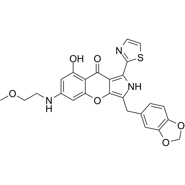
PDE5-IN-2 is a potent, highly selective, and orally active PDE5 inhibitor, with an IC50 of 0.31 nM, less potently inhibits PDE2A, PDE10A, PDE4D2, and PDE6C, with IC50s of 106, 46, 43, 1.2 nM, respectively. Anti-pulmonary arterial hypertension activity.
For research use only. We do not sell to patients.
- CAS No.: 2244517-61-9
- Formula: C25H21N3O6S
- Molecular Weight:491.52
-
Storage:
Please store the product under the recommended conditions in the Certificate of Analysis.

To place orders, for customer services and technical support, please contact: MedChemExpress USA
Tel: 609-228-6898 E-mail: [email protected] [email protected]


-
 Biological Activity
-
Chemical Information
- Protocols
- Purity & Documentation
- References
-
 Help & FAQs




Biological Activity
Description
IC50 & Target
[1]|
PDE5A1 0.31 nM (IC50) |
PDE6C 1.2 nM (IC50) |
PDE4D2 43 nM (IC50) |
PDE10A 46 nM (IC50) |
PDE2A 106 nM (IC50) |
In Vitro
PDE5-IN-2 (Compound 3) has at least 1000-fold selectivity over PDE1B, PDE3A, PDE7A1, PDE8A1, and PDE9A2 (IC50s, >32 000 nM)[1].
MedChemExpress (MCE) has not independently confirmed the accuracy of these methods. They are for reference only. Further protocols information, click here.
In Vivo
MedChemExpress (MCE) has not independently confirmed the accuracy of these methods. They are for reference only.


Chemical Information
-
CAS No. 2244517-61-9
-
Molecular Weight 491.52
-
Formula C25H21N3O6S
-
SMILES
O=C1C2=C(C3=NC=CS3)NC(CC4=CC(OCO5)=C5C=C4)=C2OC6=CC(NCCOC)=CC(O)=C61
-
Shipping
Room temperature in continental US; may vary elsewhere.
-
Storage
Please store the product under the recommended conditions in the Certificate of Analysis.

Protocols
-
Research Protocol for Cardiovascular Diseases
Cardiovascular disease can be modeled as maladaptive cardiac remodeling, where ischemic injury or pressure overload activates inflammatory signaling, fibroblast activation, extracellular-matrix deposition, cardiomyocyte hypertrophy, vascular remodeling, and progressive ventricular dysfunction. The TGF-β/SMAD axis is a central profibrotic pathway after myocardial injury and pressure overload, while innate immune and cytokine pathways regulate leukocyte recruitment, scar formation, and adverse remodeling. Key unresolved questions include which inflammatory signals are reparative versus harmful, when fibrosis is protective versus maladaptive, and whether pathway inhibition improves function without weakening necessary infarct healing or compensatory remodeling.


Purity & Documentation
References

Calculators
Concentration (start) × Volume (start) = Concentration (final) × Volume (final)


PDE5-IN-2 Related Classifications
HY-112704 Related Classifications
 Powered by Bioz