



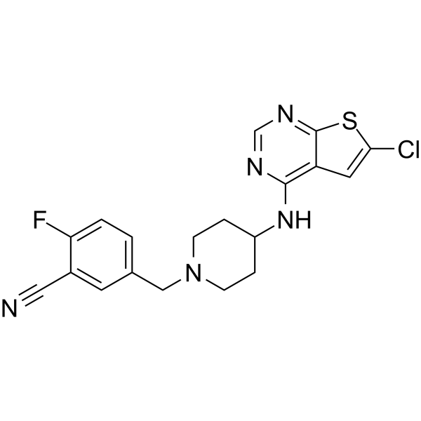
PRX-08066

Based on 1 Customer Validation
PRX-08066 is a selective and orally active 5-hydroxytryptamine receptor 2B (5-HT2BR) antagonist with a Ki of 3.4 nM. PRX-08066 inhibits the MAPK pathway, 5-HT release and fibrotic factor (TGFβ1, CTGF and FGF2) expression. PRX-08066 inhibits the proliferation of KRJ-I cells and induces apoptosis (caspase-3 activation). PRX-08066 inhibits pulmonary vascular remodeling. PRX-08066 can be used of pulmonary Arterial Hypertension (PAH) and neuroendocrine tumor (NET).
For research use only. We do not sell to patients.
- Purity : 97.46%
- CAS No.: 866206-54-4
- Formula: C19H17ClFN5S
- Molecular Weight:401.89
-
Storage:Powder -20°C, 3 years , 4°C, 2 years ; In solvent -80°C, 2 years , -20°C, 1 year

To place orders, for customer services and technical support, please contact: MedChemExpress USA
Tel: 609-228-6898 E-mail: [email protected] [email protected]


-
 Biological Activity
-
Chemical Information
-
Solvent & Solubility
- Protocols
- Purity & Documentation
- References
-
 Help & FAQs




Biological Activity
Description
IC50 & Target
|
5-HT2B Receptor 3.4 nM (IC50) |
Cellular Effect
|
Cell Line

|
Type | Value | Description | References |
|---|---|---|---|---|
| CHO-K1 | IC50 |
270 nM
Compound: A
|
Antagonist activity at 5-HT2B receptor expressed in CHOK1 cells assessed as inhibition of serotonin-induced intracellular Ca2+ flux by aequorin luminescence assay in presence of 4% human serum albumin
Antagonist activity at 5-HT2B receptor expressed in CHOK1 cells assessed as inhibition of serotonin-induced intracellular Ca2+ flux by aequorin luminescence assay in presence of 4% human serum albumin
|
[PMID: 19307114] |
| CHO-K1 | IC50 |
3.5 nM
Compound: A
|
Antagonist activity at 5-HT2B receptor expressed in CHOK1 cells assessed as inhibition of serotonin-induced intracellular Ca2+ flux by aequorin luminescence assay
Antagonist activity at 5-HT2B receptor expressed in CHOK1 cells assessed as inhibition of serotonin-induced intracellular Ca2+ flux by aequorin luminescence assay
|
[PMID: 19307114] |
In Vitro
PRX-08066 inhibits 5-HT induced mitogen-activated protein kinase activation (IC50 = 12 nM) and markedly reduced thymidine incorporation (IC50 = 3 nM) in hamster ovary cells expressing the human 5-HT2BR[1].
.
PRX-08066 (0.01 pM-100 pM, 24 h) significantly inhibits the proliferation of KRJ-I cells with an IC50 of 4.6 pM and has no significant effect on NCI-H720 cells (with no expression of 5-HT2B receptors)[2].
PRX-08066 (0.01 pM-100 pM) inhibits basal and Isoproterenol (HY-B0468)-stimulated 5-HT secretion with IC50s of 6.9 pM and 1.25 pM in KRJ-I cells[2].
PRX-08066 (0.5 pM) significantly reduces the phosphorylation level of ERK in KRJ-I cells[2].
PRX-08066 (1-2 h) reduces fibrotic factors (TGFβ1, CTGF, FGF2) in KRJ-I cells[2].
PRX-08066 (1 pM, 24 h) significantly increases the proportion of apoptotic cells by activating Caspase-3 in KRJ-I cells and HEK293 cells established in a coculture system[2].
MedChemExpress (MCE) has not independently confirmed the accuracy of these methods. They are for reference only.
-
Cell Line:KRJ-I cells and HEK293 cells established in a coculture system
-
Concentration:1 pM
-
Incubation Time:24 h
-
Result:Significantly increased the proportion of dead cells in KRJ-I, and the proportion of Caspase-3 positive cells among the dead cells also rose.
Significantly increased the proportion of positive cells for Caspase-3 in HEK293 cells.
In Vivo
MedChemExpress (MCE) has not independently confirmed the accuracy of these methods. They are for reference only.
-
Animal Model:MCT induced PAH model established in male Sprague-Dawley rats (200-225 g)[1]
-
Dosage:50 and 100 mg/kg
-
Administration:Oral administration (p.o.), once daily for 5 weeks
-
Result:Selectively reduced pulmonary artery pressure without affecting systemic blood pressure.
Reversed right ventricular hypertrophy.
Reduced the thickness of the media layer of pulmonary small arteries, improves the closure of the lumen, and reduces tissue pathological damage.
Inhibited the proliferation and contraction of vascular smooth muscle mediated by 5-HT.
No significant gastrointestinal side effects or systemic toxicity.
Clinical Trial
| NCT Number | Sponsor | Condition | Start Date |
Phase
|
|---|---|---|---|---|
| NCT01329991 | 2011-05 | PHASE1 |


Chemical Information
-
CAS No. 866206-54-4
-
Appearance Solid
-
Molecular Weight 401.89
-
Formula C19H17ClFN5S
-
Color White to off-white
-
SMILES
N#CC1=CC(CN2CCC(NC3=C(C=C(Cl)S4)C4=NC=N3)CC2)=CC=C1F
-
Shipping
Room temperature in continental US; may vary elsewhere.
-
Storage
Powder -20°C 3 years 4°C 2 years In solvent -80°C 2 years -20°C 1 year


Solvent & Solubility
In Vitro:
DMSO : 7 mg/mL (17.42 mM; Need ultrasonic; Hygroscopic DMSO has a significant impact on the solubility of product, please use newly opened DMSO)
Please refer to the solubility information to select the appropriate solvent. Once prepared, please aliquot and store the solution to prevent product inactivation from repeated freeze-thaw cycles.
Storage method and period of stock solution: -80°C, 2 years; -20°C, 1 year. When stored at -80°C, please use it within 2 years. When stored at -20°C, please use it within 1 year.
Please refer to the solubility information to select the appropriate solvent. Once prepared, please aliquot and store the solution to prevent product inactivation from repeated freeze-thaw cycles.
Storage method and period of stock solution: -80°C, 2 years; -20°C, 1 year. When stored at -80°C, please use it within 2 years. When stored at -20°C, please use it within 1 year.
Concentration (start) × Volume (start) = Concentration (final) × Volume (final)


Purity & Documentation
-
Data Sheet (274 KB)
-
SDS (392 KB)
- English - EN (392 KB)
- Français - FR (392 KB)
- Deutsch - DE (392 KB)
- Norwegian - NO (392 KB)
- Español - ES (392 KB)
- Swedish - SV (392 KB)
- Italian - IT (392 KB)
- Korean - KR (392 KB)
- Portuguese - PT (392 KB)
-
Handling Instructions (2659 KB)
References
[1]. Porvasnik SL, et al. PRX-08066, a novel 5-hydroxytryptamine receptor 2B antagonist, reduces monocrotaline-induced pulmonary arterial hypertension and right ventricular hypertrophy in rats. J Pharmacol Exp Ther. 2010 Aug;334(2):364-72. [Content Brief]
[2]. Svejda B, et al. The 5-HT(2B) receptor plays a key regulatory role in both neuroendocrine tumor cell proliferation and the modulation of the fibroblast component of the neoplastic microenvironment. Cancer. 2010 Jun 15;116(12):2902-12. [Content Brief]


Complete Stock Solution Preparation Table
Please refer to the solubility information to select the appropriate solvent. Once prepared, please aliquot and store the solution to prevent product inactivation from repeated freeze-thaw cycles.
Storage method and period of stock solution: -80°C, 2 years; -20°C, 1 year. When stored at -80°C, please use it within 2 years. When stored at -20°C, please use it within 1 year.
| Optional Solvent | Concentration Solvent Mass | 1 mg | 5 mg | 10 mg | 25 mg |
|---|---|---|---|---|---|
| DMSO | 1 mM | 2.4882 mL | 12.4412 mL | 24.8824 mL | 62.2061 mL |
| 5 mM | 0.4976 mL | 2.4882 mL | 4.9765 mL | 12.4412 mL | |
| 10 mM | 0.2488 mL | 1.2441 mL | 2.4882 mL | 6.2206 mL | |
| 15 mM | 0.1659 mL | 0.8294 mL | 1.6588 mL | 4.1471 mL |


PRX-08066 Related Classifications
HY-15472 Related Classifications
 Powered by Bioz