



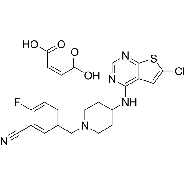
PRX-08066 maleate
PRX-08066 maleate is a selective and orally active 5-hydroxytryptamine receptor 2B (5-HT2BR) antagonist with a Ki of 3.4 nM. PRX-08066 maleate inhibits the MAPK pathway, 5-HT release and fibrotic factor (TGFβ1, CTGF and FGF2) expression. PRX-08066 maleate inhibits the proliferation of KRJ-I cells and induces apoptosis (caspase-3 activation). PRX-08066 maleate inhibits pulmonary vascular remodeling. PRX-08066 maleate can be used of pulmonary Arterial Hypertension (PAH) and neuroendocrine tumor (NET).
Nur für Forschungszwecke. Wir verkaufen nicht an Patienten.
- CAS. Nr.: 866206-55-5
- Formel: C23H21ClFN5O4S
- Molecular Weight:517.96
-
Speicherung:
Please store the product under the recommended conditions in the Certificate of Analysis.

To place orders, for customer services and technical support, please contact: MedChemExpress USA
Tel: 609-228-6898 E-mail: [email protected] [email protected]


-
 Biologische Aktivität
-
Chemical Information
- Protokoll
- Reinheit & Dokumentation
-
 Help & FAQs


Alle 5-HT Receptor Isoform-spezifische Produkte anzeigen
MoreAlle Caspase Isoform-spezifische Produkte anzeigen
More

Biologische Aktivität
Beschreibung
IC50 & Target
[1]|
5-HT2B Receptor 3.4 nM (Ki) |
In Vitro
PRX-08066 maleate inhibits 5-HT induced mitogen-activated protein kinase activation (IC50 = 12 nM) and markedly reduced thymidine incorporation (IC50 = 3 nM) in hamster ovary cells expressing the human 5-HT2BR[1].
.
PRX-08066 (0.01 pM-100 pM, 24 h) maleate significantly inhibits the proliferation of KRJ-I cells with an IC50 of 4.6 pM and has no significant effect on NCI-H720 cells (with no expression of 5-HT2B receptors)[2].
PRX-08066 (0.01 pM-100 pM) maleate inhibits basal and Isoproterenol (HY-B0468)-stimulated 5-HT secretion with IC50s of 6.9 pM and 1.25 pM in KRJ-I cells[2].
PRX-08066 (0.5 pM) maleate significantly reduces the phosphorylation level of ERK in KRJ-I cells[2].
PRX-08066 (1-2 h) maleate reduces fibrotic factors (TGFβ1, CTGF, FGF2) in KRJ-I cells[2].
PRX-08066 (1 pM, 24 h) maleate significantly increases the proportion of apoptotic cells by activating Caspase-3 in KRJ-I cells and HEK293 cells established in a coculture system[2].
MedChemExpress (MCE) has not independently confirmed the accuracy of these methods. They are for reference only. Further protocols information, click here.
-
Cell Line:KRJ-I cells and HEK293 cells established in a coculture system
-
Concentration:1 pM
-
Incubation Time:24 h
-
Result:Significantly increased the proportion of dead cells in KRJ-I, and the proportion of Caspase-3 positive cells among the dead cells also rose.
Significantly increased the proportion of positive cells for Caspase-3 in HEK293 cells.
In Vivo
MedChemExpress (MCE) has not independently confirmed the accuracy of these methods. They are for reference only.
-
Animal Model:MCT induced PAH model established in male Sprague-Dawley rats (200-225 g)[1]
-
Dosage:50 and 100 mg/kg
-
Administration:Oral administration (p.o.), once daily for 5 weeks
-
Result:Selectively reduced pulmonary artery pressure without affecting systemic blood pressure.
Reversed right ventricular hypertrophy.
Reduced the thickness of the media layer of pulmonary small arteries, improves the closure of the lumen, and reduces tissue pathological damage.
Inhibited the proliferation and contraction of vascular smooth muscle mediated by 5-HT.
No significant gastrointestinal side effects or systemic toxicity.


Chemical Information
-
CAS. Nr. 866206-55-5
-
Molecular Weight 517.96
-
Formel C23H21ClFN5O4S
-
SMILES
O=C(O)/C=C\C(O)=O.N#CC1=CC(CN2CCC(NC3=C(C=C(Cl)S4)C4=NC=N3)CC2)=CC=C1F
-
Versand
Room temperature in continental US; may vary elsewhere.
-
Speicherung
Please store the product under the recommended conditions in the Certificate of Analysis.

Protokoll
-
RNA extraction experimental
By lysing cells, releasing RNA, and removing impurities such as proteins and DNA, high-purity RNA products are finally obtained. The commonly used traditional method is the guanidine isothiocyanate/phenol/chloroform method (Trizol), which is suitable for a variety of animal materials including animal tissues, microorganisms, cultured cells, etc., and most plant materials.
-
Apoptosis
Apoptosis, also called programmed cell death, is generally characterized by distinct morphological characteristics.
-
TUNEL staining for apoptotic DNA fragmentation
TUNEL staining detects DNA strand breaks by using terminal deoxynucleotidyl transferase to add labeled nucleotides to exposed 3′-OH DNA termini, generating either microscopic staining in fixed cells or tissue sections, or fluorescence/cytometric signal in cell suspensions. TUNEL positivity reflects DNA fragmentation but should not be interpreted alone as definitive apoptosis, because TUNEL can also label necrotic, autolytic, mechanically damaged, or DNA-repair-associated DNA breaks.
-
Annexin V plus membrane-impermeant dye apoptosis staining
Annexin V-based apoptosis assays rely on the detection of phosphatidylserine (PS) externalization from the inner leaflet of the plasma membrane to the outer leaflet, an early biochemical hallmark of apoptosis. Fluorescently labeled Annexin V binds PS in a calcium-dependent manner, enabling identification of early apoptotic cells by flow cytometry or fluorescence microscopy. When combined with a membrane-impermeant DNA-binding dye (e. g. , propidium iodide), this approach allows discrimination between viable (Annexin V−/dye−), early apoptotic (Annexin V+/dye−), and late apoptotic or necrotic (Annexin V+/dye+) cell populations by assessing membrane integrity and PS exposure.
-
Apoptosis Solutions
Apoptosis is a regulated, generally non-lytic cell-death pathway that removes unwanted, damaged, infected, or abnormal cells through coordinated morphological changes, caspase activation, DNA fragmentation, and membrane remodeling. The intrinsic apoptosis pathway is controlled mainly by mitochondrial outer membrane permeabilization, BCL-2 family proteins, cytochrome c release, apoptosome formation, caspase-9 activation, and downstream executioner caspase-3/7 activation. The extrinsic apoptosis pathway is initiated by death receptors such as Fas, TNFR, and TRAIL receptors, which recruit adaptor proteins and activate caspase-8 before engaging executioner caspases or mitochondrial amplification through BID cleavage. Apoptosis is linked to many phenotypes, including cancer cell killing, tissue homeostasis, immune regulation, neurodegeneration, infection response, and treatment-induced cytotoxicity; unresolved questions include how apoptosis interacts with necroptosis, pyroptosis, ferroptos
-
Research Protocol for Cardiovascular Diseases
Cardiovascular disease can be modeled as maladaptive cardiac remodeling, where ischemic injury or pressure overload activates inflammatory signaling, fibroblast activation, extracellular-matrix deposition, cardiomyocyte hypertrophy, vascular remodeling, and progressive ventricular dysfunction. The TGF-β/SMAD axis is a central profibrotic pathway after myocardial injury and pressure overload, while innate immune and cytokine pathways regulate leukocyte recruitment, scar formation, and adverse remodeling. Key unresolved questions include which inflammatory signals are reparative versus harmful, when fibrosis is protective versus maladaptive, and whether pathway inhibition improves function without weakening necessary infarct healing or compensatory remodeling.
-
Cell Viability Determination by MTT Colorimetric Assay
The following protocol uses the MTT colorimetric assay as a classic literature-established method for assessing cell viability/metabolic activity in cultured mammalian cells. MTT[3-(4,5-dimethylthiazol-2-yl)-2,5-diphenyltetrazolium bromide] is reduced by metabolically active cells to a colored formazan product; the amount of formazan is quantified spectrophotometrically and provides an indirect measure of metabolically active viable cells. Importantly, MTT reduction reflects cellular oxidoreductase/metabolic activity rather than an absolute direct count of living cells, so changes in cellular metabolism can alter the signal independently of cell number.


Reinheit & Dokumentation

Calculators
Konzentration (Stammlösung) × Volumen (Stammlösung) = Konzentration (Ziellösung) × Volumen (Ziellösung)


PRX-08066 maleate Related Classifications
HY-15472A Related Classifications
 Powered by Bioz