



Fluorescent Dye
Fluorescence is a phenomenon of photoluminescence in luminescence. When an atom is irradiated by light, some electrons around the nucleus jump from their original orbitals to higher energy orbitals under the influence of light energy, i.e., from the ground state to the first or second excited single-line state, etc. Due to the instability of the first or second excited singlet state, the electron returns from the excited singlet state to the ground state accompanied by released energy, resulting in fluorescence.
Fluorescent dyes can be covalently bound or physically adsorbed in the structure of the substance. The fluorescence properties allow for the characterization, localization and quantitative analysis of the labeled object. Fluorescent dyes have shown great potential for the detection of proteins, nucleic acids, cells, and immunoassays, attributed to the advantages in non-radioactive contamination, simple experimental operation and easy observation.
Fluorescent dyes have penetrated many fields such as pharmacology, physiology, environmental science, and information science, as well as functional studies of proteins and drug screening.
All Product Categories
-
Fluorescent Dye Related Products (4914)
Related Products (4914)

-
Antibodies (4)
-
BG-SS-SulfoCy5
 0 ImagesCat. No.: HY-D2930Synonyms: SNAP-SS-SulfoCy5BG-SS-SulfoCy5 (SNAP-SS-SulfoCy5) is a SulfoCy5-labeled SNAP tag fluorescent probe, linked by a disulfide bond. BG-SS-SulfoCy5 combines the specific recognition of SNAP-tag, the cleavability of disulfide bond, and the excellent optical properties of SulfoCy5. BG-SS-SulfoCy5 can be used for tracking the internalization of cell surface proteins and multiple labeling experiments.
-
MitoLumina 750
 0 ImagesCat. No.: HY-D3513MitoLumina 750 is a mitochondria-targeting fluorescent probe used to label mitochondria in live cells. MitoLumina 750 contains a chloromethyl group that allows for covalent binding to mitochondrial thiol-containing proteins. MitoLumina 750 is primarily used for mitochondrial imaging, live-cell mitochondrial tracking, and multicolor fluorescence microscopy (Ex/Em = 745/775 nm).
-
Phazr-N3
 0 ImagesCat. No.: HY-D3439Phazr-N3 is a near-infrared (NIR) environment-sensitive fluorescent probe used to monitor the entire aggregation process of Aβ42 (amyloid-β42) from monomers/early aggregates to mature fibrils and plaques. Phazr-N3 binds Aβ42 in a structure-independent manner, exhibits low micromolar affinity (Kd = 6.6 μM) for free, disordered Aβ42 under non-aggregating conditions, and produces a solvatochromic fluorescence shift driven by local polarity changes during aggregation. Phazr-N3 uses a 580/680 nm excitation/emission setting in Aβ aggregation[1].
-
Janelia Fluor® 549, Tetrazine
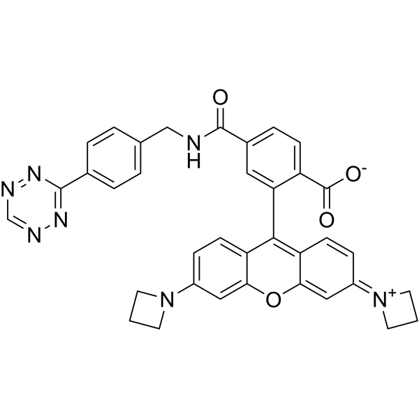 0 ImagesCat. No.: HY-131024Synonyms: JF549, TetrazineJanelia Fluor 549, Tetrazine (JF549, Tetrazine) is a fluorescent dye with the absorption maximum (λab (max)) of 549 nm and emission maximum (λem (max)) of 571 nm. Janelia Fluor fluorescent dyes can be chemically conjugated to the specific HaloTag substrate (a chloroalkane ligand) to form a fluorescent ligand, rather than binding directly to the HaloTag protein itself. Janelia Fluor products are licensed under U.S. Pat. Nos. 9,933,417, 10,018,624 and 10,161,932 and other patents from Howard Hughes Medical Institute.
-
Thiazole Orange (solution)
 0 ImagesCat. No.: HY-DY1097CAS No.: 107091-89-4Thiazole Orange (solution) is an asymmetric anthocyanin dye that can be coupled with oligonucleotides (ONs) to prepare fluorescent hybridization probes. Thiazole Orange has been widely used in biomolecular detection and staining of DNA/ RNA in gels and can be used for reticulocyte analysis. Thiazole orange generates a significant fluorescence enhancement and high quantum yield when it binds with nucleic acids, especially RNA. Thiazole orange can permeate living cell membranes. Thiazole orange can use UV light for detection, but can also be detected with blue light. The excitation and emission of Thiazole orange are λex = 510 nm (488 nm and 470 nm also show strong excitation) and λem = 527 nm, respectively.
Solvent and concentration: DMSO: 10 mM
The 1 mL volume is defined as the base specification. All larger sizes correspond to incremental volumes of this base.
-
Cy3-PEG10000-N3
 0 ImagesCat. No.: HY-D2540Cy3-PEG10000-N3 is a Cy3 (HY-D0822)-azide fluorescent dye used to label for protein and nucleic acid. Cy3-PEG10000-N3 is a click chemistry reagent, it contains an Azide group and can undergo copper-catalyzed azide-alkyne cycloaddition reaction (CuAAc) with molecules containing Alkyne groups. It can also undergo strain-promoted alkyne-azide cycloaddition (SPAAC) reactions with molecules containing DBCO or BCN groups.
-
FITC-PEG1000-N3
 0 ImagesCat. No.: HY-D2802AFITC-PEG1000-N3 is an azide fluorescent dye containing polyethylene glycol (PEG) and FITC (HY-66019). FITC-PEG1000-N3 is a click chemistry reagent, it contains an Azide group and can undergo copper-catalyzed azide-alkyne cycloaddition reaction (CuAAc) with molecules containing Alkyne groups. It can also undergo strain-promoted alkyne-azide cycloaddition (SPAAC) reactions with molecules containing DBCO or BCN groups.
-
Toluidine Blue Staining Solution (1%, Phosphate Buffer)
 0 ImagesCat. No.: HY-DY2019Toluidine Blue Staining Solution (1%, Phosphate Buffer) is an alkaline quinonimine dye (vivo dye) with high affinity for acidic tissue components, staining nuclei blue and polysaccharides purple. Toluidine Blue Staining Solution (1%, Phosphate Buffer) shows heterostaining properties for mast cells, mucins and chondrocytes. Toluidine Blue Staining Solution (1%, Phosphate Buffer) can stain different components of plant tissues and cells in different colours. Toluidine Blue Staining Solution (1%, Phosphate Buffer) is also used as a diagnostic aid to identify malignant lesions, such as cancer.
-
Squarefluor 631/646 free acid
 0 ImagesCat. No.: HY-N16327CAS No.: 3092242-67-3Squarefluor 631/646 free acid is a squaraine probe (Ex/Em = 631/646 nm).
-
Solanum Tuberosum (Potato) Lectin
 0 ImagesCat. No.: HY-NP081Synonyms: STLSolanum Tuberosum (Potato) Lectin (STL) is a plant lectin that can be used as a probe to specifically bind biomolecules (such as polysaccharides, peptides, etc.).Solanum Tuberosum (Potato) Lectin (STL) is a biological material or organic compound that can be used in life science research.
-
Cy 3 (Non-Sulfonated) triethylamine
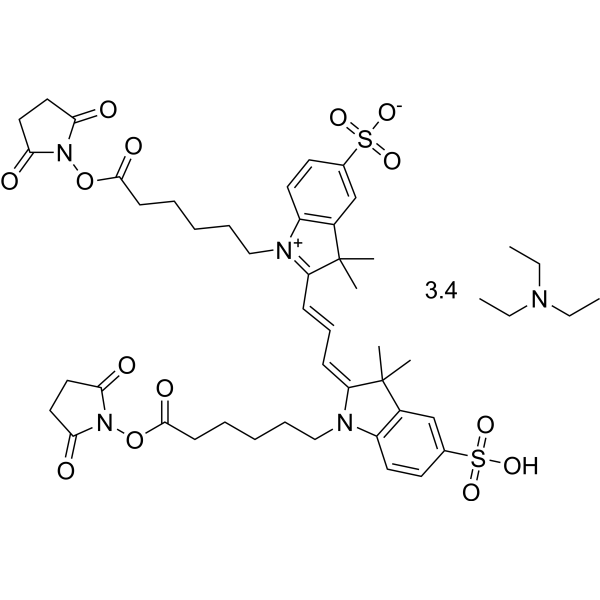 0 ImagesCat. No.: HY-D0968BSynonyms: Cyanine3 triethylamineCy 3 Non-Sulfonated (Cyanine3) triethylamine is a cyanine (Cy) dye, and a fluorescent label with green channel for protein and nucleic acid. Cy 3 Non-Sulfonated triethylamine is a fluorescent photoproduct of Cyanine5 via photoconversion upon photoexcitation. Cy 3 Non-Sulfonated triethylamine can be used to high-density single-particle tracking in a living cell without using UV illumination and cell-toxic additives (Ex=470 nm; Em=515 nm and 565 nm nm).
-
TAMRA-PEG7-N3
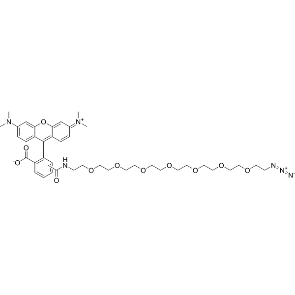 0 ImagesCat. No.: HY-D2112TAMRA-PEG7-N3 is a TAMRA (HY-135640) dye derivative containing 7 PEG units. TAMRA-PEG7-N3 undergoes copper-catalyzed azide-alkyne cycloaddition (CuAAc) with molecules containing an alkynyl group (N3). It also undergoes strain-promoted alkyne-azide cycloaddition (SPAAC) with molecules containing DBCO or BCN groups. It contains an azide group and can undergo copper-catalyzed azide-alkyne cycloaddition (CuAAc) with molecules containing alkyne groups. It can also undergo ring strain-promoted alkyne-azide cycloaddition (SPAAC) with molecules containing DBCO or BCN groups.
-
Griffonia (Bandeiraea) Simplicifolia Lectin I (Fluorescein)
 0 ImagesCat. No.: HY-NP0131Synonyms: GSL I (Fluorescein)Griffonia (Bandeiraea) Simplicifolia Lectin I (GSL I) Fluorescein is a plant lectin that can be used as a probe to specifically bind biomolecules (such as polysaccharides, peptides, etc.).Griffonia (Bandeiraea) Simplicifolia Lectin I (GSL I) Fluorescein is a biological material or organic compound that can be used in life science research.
-
Concanavalin A-Cy5
 0 ImagesCat. No.: HY-NP164OSynonyms: Con A-Cy5Concanavalin A-Cy5 (Con A-Cy5) is a Cy5 (HY-D0821)-labled Concanavalin A (HY-P2149). Concanavalin A is a Ca2+/Mn2+-dependent and mannose/glucose-binding plant lectin. Concanavalin A binds the N- and O-glycosylated proteins of head and neck carcinoma cells glycoproteins (Ex/Em = 630/670 nm).
-
Sambucus Nigra Lectin-AF568
 0 ImagesCat. No.: HY-NP080DSynonyms: SNA-AF568Sambucus Nigra Lectin-AF568 (SNA-AF568) is a phytohemagglutinin labeled with AF568, which can be used as a probe to specifically bind to biomolecules (such as polysaccharides and peptides). Sambucus Nigra Lectin-AF568 precisely locates the distribution of glycan chains on the cell surface through stable fluorescence signals, and can be used for glycobiology research (Ex/Em = 579/603 nm).
-
Soybean Agglutinin
 0 ImagesCat. No.: HY-NP089Synonyms: SBASoybean Agglutinin (SBA) is a plant lectin that can be used as a probe to specifically bind biomolecules (such as polysaccharides, peptides, etc.).Soybean Agglutinin (SBA) is a biological material or organic compound that can be used in life science research.
-
Violamine R
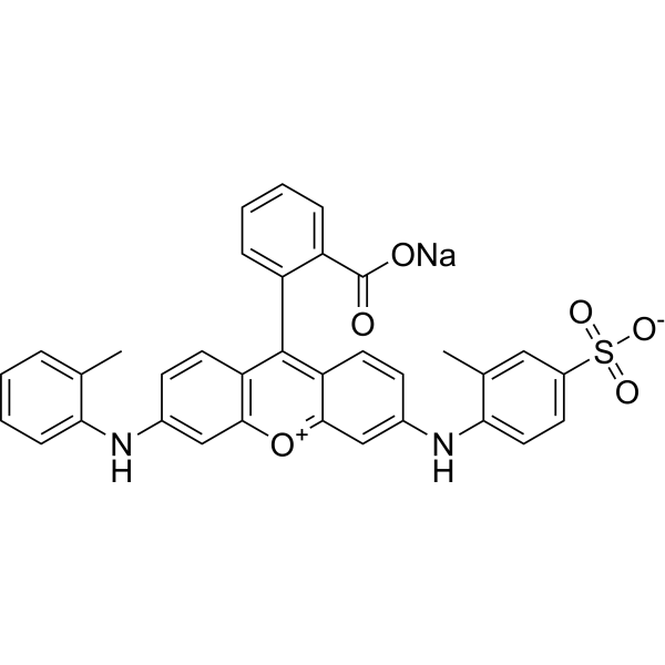 0 ImagesViolamine R (Acid Violet 9) is a potent fluorophore. Violamine R shows environment and temperature-dependent photoluminescence (PL) intermittency in poly(vinyl alcohol) (PVOH) and potassium acid phthalate (KAP). Violamine R can be used to measure the temperatures spanning the glass-transition temperature (Tg) of the polymer host by the fluorescence intermittency or blinking in poly(vinyl alcohol) (PVA).
-
RIM-1
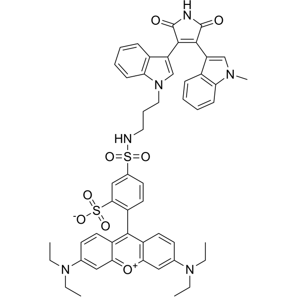 0 ImagesCat. No.: HY-D1751CAS No.: 150206-04-5

-
LHA930
 0 ImagesCat. No.: HY-D3403CAS No.: 3121511-19-8LHA930 is a fluorescent probe for in vivo detection of early-stage ferroptosis. By introducing a 4-phenyl-3-methylbut-2-enyl reactive group responsive to lipid hydrogen abstraction, LHA930 exhibits fluorescence turn-on upon the action of oxidative radicals associated with lipid peroxidation, enabling detection of lipid peroxidation during the early stage of ferroptosis. The detection wavelength of LHA930 is Ex/Em = 860/930 nm. LHA930 can be used for near-infrared fluorescence imaging studies of ferroptosis in APAP-induced mouse liver injury models.




