



Abituzumab

Based on 1 Customer Validation
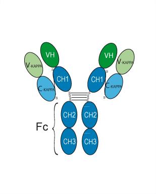
Abituzumab (DI17E6) is a humanised anti-integrin αV monoclonal antibody (IgG2 type). Abituzumab effectively reduces the phosphorylation of FAK, Akt and ERK. Abituzumab can be used in cancer research, particularly in prostate cancer.
For research use only. We do not sell to patients.
- Purity : 99.38%
- CAS No.: 1105038-73-0
- Molecular Weight:144.58 kDa
-
Storage:
Please store the product under the recommended conditions in the Certificate of Analysis.


To place orders, for customer services and technical support, please contact: MedChemExpress USA
Tel: 609-228-6898 E-mail: [email protected] [email protected]


-
 Biological Activity
-
Chemical Information
- Protocols
- Purity & Documentation
- References
-
 Help & FAQs


-

HY-P99296
IntetumumabAnti-αv Integrin Monoclonal Antibody -
HY-16141
CilengitideIntegrin Inhibitor -

HY-P99291
Etaracizumabαvβ3 Integrin mAb -

HY-P99473
BersanlimabAnti-ICAM-1 Antibody -

HY-P9911
Vedolizumab -
HY-100506
GLPG0187Integrin Receptor Antagonist -

HY-P9911A
Vedolizumab (anti-α4β7-integrin) -
HY-15102
MK-0429αvβ3 Integrin Inhibitor -

HY-P9981
TafasitamabCD19 Monoclonal Antibody -

HY-P99192
EmibetuzumabBivalent MET Antibody -

HY-P99002
Human IgG2 kappa, Isotype ControlIgG2κ Isotype Control -

HY-P990673
VandortuzumabSTEAP1 ADC Antibody


Biological Activity
Description
Isotype
Human IgG2 kappa
Recommend Isotype Controls
Species Reactivity
Human
IC50 & Target
Integrin aV/ITGAV/CD51
In Vitro
Abituzumab (DI17E6) (0.01, 0.1, 1, 10, 30,100 μg/mL; 24 h) inhibits adhesion of PCa cells to multiple extracellular matrix proteins but not collagen I[1]
Abituzumab (100 μg/mL; 12, 18 h) inhibits inhibits motility and invasion of PCa cells[1].
Abituzumab (0.01, 0.1, 1, 10, 30,100 μg/mL; 24 h) inhibits the ability of PCa cells to adhere to osteoblast and bone stromal cell lines[1].
Abituzumab (100 μg/mL; 24 h) blocks integrin-mediated cell signaling in PCa cancer cell lines[1].
MedChemExpress (MCE) has not independently confirmed the accuracy of these methods. They are for reference only. Further protocols information, click here.
-
Cell Line:PCa cells
-
Concentration:0.01, 0.1, 1, 10, 30,100 µg/mL
-
Incubation Time:24 h
-
Result:Promoted detachment of PCa cells from vitronectin (up to approximately 20% cells detached at 100 μg/mL), osteopontin (up to approximately 10% cells detached at 100 μg/mL) and fibronectin (up to approximately 10% cells detached at 100 μg/mL) but not from collagen I.
-
Cell Line:PCa cells
-
Concentration:100 µg/mL
-
Incubation Time:12, 18 h
-
Result:Inhibited invasive ability by approximately 25 to 30% compared to vehicle, and inhibited motility by approximately 30 to 40%.
-
Cell Line:PCa, hFOB, Saos2, HS-5, HDMEC cells
-
Concentration:0.01, 0.1, 1, 10, 30,100 µg/mL
-
Incubation Time:24 h
-
Result:Inhibited PCa cells adhesion to human osteoblast cell lines hFOB and Saos2 and a bone marrow stromal cell line, HS-5.
Promoted detachment of PCa cells from to a known expressor of αv integrins, HDMEC cells.
-
Cell Line:PC3, DU145, C4-2B, LNCaP, ARCaP and VCaP cells
-
Concentration:100 µg/mL
-
Incubation Time:24 h
-
Result:Inhibited FAK phosphorylation starting at 12 h and 6 h in C4-2B and LNCaP, respectively; AKT phosphorylation starting at 3 h and 2 h in C4-2B and LNCaP, respectively; and ERK phosphorylation at starting at 1 h and 1.5 h in C4-2B and LNCaP, respectively.
Clinical Trial
| NCT Number | Sponsor | Condition | Start Date |
Phase

|
|---|---|---|---|---|
| NCT01329991 | Plexxikon| | 2011-05 | PHASE1 |
Gene ID
Accession
Conjugated
Unconjugated
Reconsititution
The product can be reconstituted/diluted with sterile PBS or saline.
Format
-
Human IgG2 kappa
Application
ELISA, FACS, Functional assay
Verified Bioactivity
-
 Immobilized Integrin alpha V beta 3 Protein, Human (HEK293, His, solution, HY-P73868) can bind Abituzumab. The EC50 for this effect is 4.5 ng/mL.
 -
 Flow cytometric analysis of 1X10⁶ A549 cells with Abituzumab (HY-P99183, red). Cells were fixed with 4% paraformaldehyde. Then stained with the primary antibody at 1/200 dilution for an hour at 4℃. Alexa Fluor 488-conjugated AffiniPure Goat Anti-Human IgG H&L (AF488) (HY-P83776) was used as the secondary antibody at 1/1,000 dilution for 30 minutes at 4℃. Human IgG1 kappa (HY-P99001, blue) was used as the isotype control, cells without incubation with primary antibody were used as the unlabeled control (black).


Chemical Information
-
CAS No. 1105038-73-0
-
Appearance Liquid
-
Molecular Weight 144.58 kDa
-
Color Colorless to light yellow
-
SMILES
[Abituzumab]
-
Synonyms
EMD 525797; DI17E6
-
Shipping
Shipping with dry ice.
-
Formulation
Please refer to the lot-specific COA for specific buffer information.
-
Storage
Please store the product under the recommended conditions in the Certificate of Analysis.

Protocols
-
Kinase activity and phosphorylation assays
Kinase activity assays measure the ability of kinases to transfer phosphate groups from ATP to specific substrates, while phosphorylation assays detect the presence and levels of phosphorylated proteins. Common methods include radiolabeled ATP incorporation (e. g. ,), ADP release detection via bioluminescence (e. g. ,[3]), enzyme-linked immunosorbent assays (ELISA) for phospho-specific epitopes (e. g. ,[6]), and microtiter-based formats for high-throughput screening (e. g. ,[8]). The ADP-Glo assay quantifies kinase activity by measuring ADP produced during phosphorylation using a luciferase-based system. Radiometric assays involve autoradiography or scintillation counting after incorporation of 32P-labeled ATP into substrate proteins. ELISA-based approaches rely on phospho-specific antibodies to detect activated kinases in cell lysates or purified samples.
-
Western Blot
Western blotting (WB) is a commonly used experimental method in molecular biology, biochemistry, and immunogenetics for identifying and quantifying target proteins. It combines gel electrophoresis with immunoassay, enabling researchers to analyze protein expression, post-translational modifications, and molecular weight.
-
Protocol for Kinase activity and phosphorylation assays
Kinase activity assays measure transfer of phosphate from ATP to a protein or peptide substrate, generating phosphorylated substrate, ADP, or incorporated radiolabeled phosphate as the readout; phosphorylation assays measure site-specific phosphorylation in cells or tissues as a proxy for kinase-pathway activation, inhibition, or substrate regulation. Phosphorylation can be detected by phospho-specific Western blot, immunoprecipitation kinase assay, phospho-immunofluorescence, phospho-flow cytometry, luminescent ADP detection, radiolabeled ATP incorporation, or reporter-based pathway assays, and these readouts can be applied to cancer cells, primary neurons, mouse tumors, organoids, inflammatory macrophages, ferroptosis studies, and mitophagy studies when the kinase target is biologically relevant.


Purity & Documentation
-
Data Sheet (261 KB)
-
SDS (252 KB)
- English - EN (252 KB)
- Français - FR (252 KB)
- Deutsch - DE (252 KB)
- Norwegian - NO (252 KB)
- Español - ES (252 KB)
- Swedish - SV (252 KB)
- Italian - IT (252 KB)
- Korean - KR (252 KB)
- Portuguese - PT (252 KB)
-
Inhibitory Antibodies User Guide (603 KB)
References

Calculators
Concentration (start) × Volume (start) = Concentration (final) × Volume (final)


Abituzumab Related Classifications
HY-P99183 Related Classifications
 Powered by Bioz