



Albofungin

Based on 1 Customer Validation
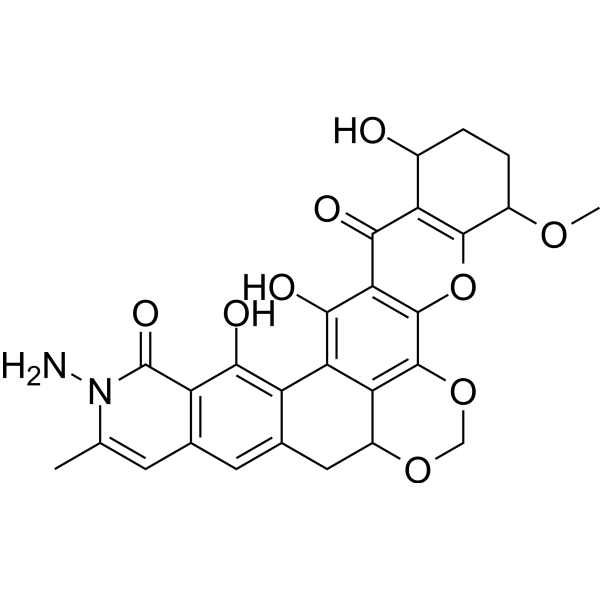
Albofungin (Antibiotic P-42-1) is isolated from the culture filtrate of Actinomyces tumemacerans strain INMI.P-42. Albofungin shows highly active on a wide variety of gram-positive bacteria and fungi. Albofungin shows cytotoxic to HeLa cell cultures and exhibited antitumor activity on EHRLICH ascites tumor in mice.
For research use only. We do not sell to patients.
- CAS No.: 37895-35-5
- Formula: C27H24N2O9
- Molecular Weight:520.49
-
Storage:Powder -20°C, 3 years ; In solvent -80°C, 6 months , -20°C, 1 month

To place orders, for customer services and technical support, please contact: MedChemExpress USA
Tel: 609-228-6898 E-mail: [email protected] [email protected]


-
 Biological Activity
-
Chemical Information
- Protocols
- Purity & Documentation
- References
-
 Help & FAQs


-
HY-116213
Sparstolonin BTLR2/TLR4 Antagonist -
HY-17021
EsomeprazoleH+, K+-ATPase Inhibitor -

HY-124493
AluminonComplexometric Titration Indicator -

HY-17021R
Esomeprazole (Standard)H+, K+-ATPase Inhibitor -
HY-122482
β-RubromycinHIV Reverse Transcriptase Inhibitor -

HY-165145
Carbazomycin C -
HY-13502S
Mitoxantrone-d8Stable Isotope -

HY-W739930
Desmethyl ofloxacin hydrochloride
All Antibiotic Isoforms
More

Biological Activity
Description


Chemical Information
-
CAS No. 37895-35-5
-
Appearance Solid
-
Molecular Weight 520.49
-
Formula C27H24N2O9
-
Color Off-white to light yellow
-
SMILES
NN1C(C)=CC2=C(C1=O)C(O)=C3C(CC4C5=C(OCO4)C(OC6=C(C7=O)C(O)CCC6OC)=C7C(O)=C53)=C2
-
Structure Classification
-
Initial Source
Actinomyces tumecerans
-
Shipping
Room temperature in continental US; may vary elsewhere.
-
Storage
Powder -20°C 3 years In solvent -80°C 6 months -20°C 1 month


Purity & Documentation
-
Data Sheet (270 KB)
-
SDS (252 KB)
- English - EN (252 KB)
- Français - FR (252 KB)
- Deutsch - DE (252 KB)
- Norwegian - NO (252 KB)
- Español - ES (252 KB)
- Swedish - SV (252 KB)
- Italian - IT (252 KB)
- Korean - KR (252 KB)
- Portuguese - PT (252 KB)
-
Handling Instructions (2659 KB)
References

Calculators
Concentration (start) × Volume (start) = Concentration (final) × Volume (final)


Albofungin Related Classifications
HY-119789 Related Classifications
 Powered by Bioz