



AMPK-IN-5
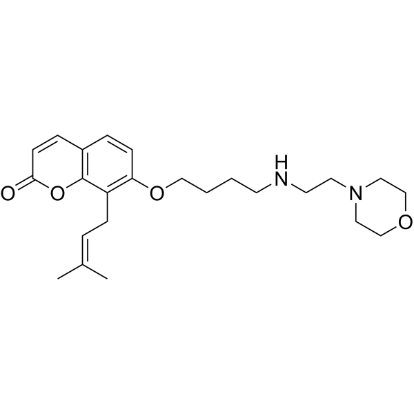
AMPK-IN-5 (compound 7m) is a Osthole (HY-N0054) derivative, and blocks MAPK signal transduction by inhibiting the phosphorylation of JNK and p38, thereby inhibiting the release of inflammatory cytokines. AMPK-IN-5 reduce DSS-induced ulcerative colitis and LPS (HY-D1056)-induced acute lung injury.
Nur für Forschungszwecke. Wir verkaufen nicht an Patienten.
- Formel: C24H34N2O4
- Molecular Weight:414.54
-
Speicherung:
Please store the product under the recommended conditions in the Certificate of Analysis.

To place orders, for customer services and technical support, please contact: MedChemExpress USA
Tel: 609-228-6898 E-mail: [email protected] [email protected]


-
 Biologische Aktivität
-
Chemical Information
- Protokoll
- Reinheit & Dokumentation
- Verweise
-
 Help & FAQs




Biologische Aktivität
Beschreibung
Cellular Effect
|
Cell Line

|
Type | Value | Description | References |
|---|---|---|---|---|
| J774.A1 | IC50 |
18.14 μM
Compound: 7m
|
Antiinflammatory activity against LPS-induced mouse J774.A1 cells assessed as inhibition of TNF-alpha release pretreated for 30 mins followed by LPS stimulation and measured after 24 hrs by ELISA
Antiinflammatory activity against LPS-induced mouse J774.A1 cells assessed as inhibition of TNF-alpha release pretreated for 30 mins followed by LPS stimulation and measured after 24 hrs by ELISA
|
[PMID: 38422703] |
| J774.A1 | IC50 |
4.23 μM
Compound: 7m
|
Antiinflammatory activity against LPS-induced mouse J774.A1 cells assessed as inhibition of IL-6 release pretreated for 30 mins followed by LPS stimulation and measured after 24 hrs by ELISA
Antiinflammatory activity against LPS-induced mouse J774.A1 cells assessed as inhibition of IL-6 release pretreated for 30 mins followed by LPS stimulation and measured after 24 hrs by ELISA
|
[PMID: 38422703] |
| RAW264.7 | IC50 |
29.72 μM
Compound: 7m
|
Antiinflammatory activity against LPS-induced mouse RAW264.7 cells assessed as inhibition of TNF-alpha release pretreated for 30 mins followed by LPS stimulation and measured after 24 hrs by ELISA
Antiinflammatory activity against LPS-induced mouse RAW264.7 cells assessed as inhibition of TNF-alpha release pretreated for 30 mins followed by LPS stimulation and measured after 24 hrs by ELISA
|
[PMID: 38422703] |
| RAW264.7 | IC50 |
4.571 μM
Compound: 7m
|
Antiinflammatory activity against LPS-induced mouse RAW264.7 cells assessed as inhibition of IL-6 release pretreated for 30 mins followed by LPS stimulation and measured after 24 hrs by ELISA
Antiinflammatory activity against LPS-induced mouse RAW264.7 cells assessed as inhibition of IL-6 release pretreated for 30 mins followed by LPS stimulation and measured after 24 hrs by ELISA
|
[PMID: 38422703] |
| THP-1 | IC50 |
10.61 μM
Compound: 7m
|
Antiinflammatory activity against LPS-induced PMA-differentiated human THP-1 cells assessed as inhibition of IL-6 release pretreated for 30 mins followed by LPS stimulation and measured after 24 hrs by ELISA
Antiinflammatory activity against LPS-induced PMA-differentiated human THP-1 cells assessed as inhibition of IL-6 release pretreated for 30 mins followed by LPS stimulation and measured after 24 hrs by ELISA
|
[PMID: 38422703] |
| THP-1 | IC50 |
27.45 μM
Compound: 7m
|
Antiinflammatory activity against LPS-induced PMA-differentiated human THP-1 cells assessed as inhibition of TNF-alpha release pretreated for 30 mins followed by LPS stimulation and measured after 24 hrs by ELISA
Antiinflammatory activity against LPS-induced PMA-differentiated human THP-1 cells assessed as inhibition of TNF-alpha release pretreated for 30 mins followed by LPS stimulation and measured after 24 hrs by ELISA
|
[PMID: 38422703] |


Chemical Information
-
Molecular Weight 414.54
-
Formel C24H34N2O4
-
SMILES
O=C1C=CC2=C(C(C/C=C(C)/C)=C(OCCCCNCCN3CCOCC3)C=C2)O1
-
Versand
Room temperature in continental US; may vary elsewhere.
-
Speicherung
Please store the product under the recommended conditions in the Certificate of Analysis.

Protokoll
-
Kinase activity and phosphorylation assays
Kinase activity assays measure the ability of kinases to transfer phosphate groups from ATP to specific substrates, while phosphorylation assays detect the presence and levels of phosphorylated proteins. Common methods include radiolabeled ATP incorporation (e. g. ,), ADP release detection via bioluminescence (e. g. ,[3]), enzyme-linked immunosorbent assays (ELISA) for phospho-specific epitopes (e. g. ,[6]), and microtiter-based formats for high-throughput screening (e. g. ,[8]). The ADP-Glo assay quantifies kinase activity by measuring ADP produced during phosphorylation using a luciferase-based system. Radiometric assays involve autoradiography or scintillation counting after incorporation of 32P-labeled ATP into substrate proteins. ELISA-based approaches rely on phospho-specific antibodies to detect activated kinases in cell lysates or purified samples.
-
Western Blot
Western blotting (WB) is a commonly used experimental method in molecular biology, biochemistry, and immunogenetics for identifying and quantifying target proteins. It combines gel electrophoresis with immunoassay, enabling researchers to analyze protein expression, post-translational modifications, and molecular weight.
-
LPS-Induced Endotoxemia/Systemic Inflammation
Lipopolysaccharide (LPS)-induced endotoxemia is a widely used in vivo model of acute systemic inflammation in which LPS, a Gram-negative bacterial endotoxin, activates innate immune signaling primarily through TLR4, leading to rapid and transient induction of pro-inflammatory cytokines such as TNF-α, IL-6, and IL-1β in circulation and tissues. This cytokine surge is commonly used as a measurable readout of systemic inflammatory activation and immune dysregulation, and is typically assessed within hours after intraperitoneal LPS administration in mouse models of endotoxemia. The model captures key features of systemic inflammatory response syndrome, including cytokine release, immune cell activation, and downstream tissue responses, and has been used to evaluate anti-inflammatory interventions such as cytokine modulation, lipid mediators, and immune cell-targeting therapies.
-
DSS-Induced Colitis
Dextran sulfate sodium (DSS)-induced colitis is generated by administering DSS in mouse drinking water, producing epithelial injury, barrier disruption, weight loss, diarrhea, fecal blood, colon shortening, histologic mucosal damage, and inflammatory mediator changes; the model is mainly used to study acute or chronic intestinal inflammation resembling selected features of ulcerative colitis. DSS injury is interpreted through clinical and tissue readouts rather than a single molecular endpoint: daily body weight, stool consistency, and bleeding are combined into a disease activity index, while colon length, histology, cytokines, myeloperoxidase activity, intestinal permeability, and tight-junction markers provide complementary measures of inflammation and barrier damage.
-
TNBS-Induced Colitis
TNBS-induced colitis is produced by intrarectal delivery of 2,4,6-trinitrobenzene sulfonic acid in ethanol, where ethanol disrupts the mucosal barrier and TNBS haptenates colonic proteins, generating immune-mediated colonic inflammation with weight loss, diarrhea, ulceration, transmural injury, inflammatory-cell infiltration, and cytokine responses. The model is used as an experimental intestinal inflammation model with Crohn’s disease–like features, especially when Th1-type responses, IL-12–dependent inflammation, chronic relapsing inflammation, or fibrosis-related endpoints are studied.
-
Protocol for Kinase activity and phosphorylation assays
Kinase activity assays measure transfer of phosphate from ATP to a protein or peptide substrate, generating phosphorylated substrate, ADP, or incorporated radiolabeled phosphate as the readout; phosphorylation assays measure site-specific phosphorylation in cells or tissues as a proxy for kinase-pathway activation, inhibition, or substrate regulation. Phosphorylation can be detected by phospho-specific Western blot, immunoprecipitation kinase assay, phospho-immunofluorescence, phospho-flow cytometry, luminescent ADP detection, radiolabeled ATP incorporation, or reporter-based pathway assays, and these readouts can be applied to cancer cells, primary neurons, mouse tumors, organoids, inflammatory macrophages, ferroptosis studies, and mitophagy studies when the kinase target is biologically relevant.
-
Inhalation Toxicity Study
Inhalation toxicity studies expose rodents to a controlled aerosol, vapor, gas, or smoke atmosphere and assess respiratory and systemic toxicity using exposure-atmosphere characterization, clinical observations, body and organ weights, bronchoalveolar lavage fluid, histopathology, blood chemistry, hematology, and, when included, molecular endpoints such as transcriptomics, proteomics, lipidomics, or tissue burden analysis. The primary biological readouts are airway irritation, pulmonary inflammation, cytotoxicity, altered surfactant or lipid homeostasis, impaired particle clearance, and tissue remodeling, reflected by BALF cell differentials, BALF protein, LDH, phosphatase activities, cytokines, lung weight, microscopic respiratory-tract lesions, and retained lung burden.


Reinheit & Dokumentation
Verweise

Calculators
Konzentration (Stammlösung) × Volumen (Stammlösung) = Konzentration (Ziellösung) × Volumen (Ziellösung)


AMPK-IN-5 Related Classifications
HY-162317 Related Classifications
 Powered by Bioz