



Notoginsenoside Fe

Based on 1 Customer Validation
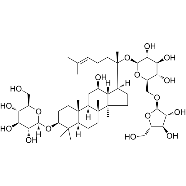
Notoginsenoside Fe (Notoginseng triterpenes; Ginsenoside Mb) is a saponin with anti-obesity and anti-neuroblastoma activities. Notoginsenoside Fe can be isolated from leaves of Panax notoginseng. Notoginsenoside Fe specifically activates paraventricular nucleus neurons in the hypothalamus, effectively reducing body weight, improving fasting blood glucose and protecting liver function by decreasing food intake, increasing resting metabolic rate and enhancing energy expenditure. Notoginsenoside Fe also inhibits the c-Src signaling pathway, blocks the proliferation and viability of human neuroblastoma cells, while improving mitochondrial dysfunction and alleviating apoptosis. Notoginsenoside Fe can be used in studies related to diet-induced obesity and neuroblastoma.
Nur für Forschungszwecke. Wir verkaufen nicht an Patienten.
- Reinheit : 99.99%
- CAS. Nr.: 88105-29-7
- Formel: C47H80O17
- Molecular Weight:917.13
-
Speicherung:Powder -20°C, 3 years , 4°C, 2 years ; In solvent -80°C, 6 months , -20°C, 1 month

To place orders, for customer services and technical support, please contact: MedChemExpress USA
Tel: 609-228-6898 E-mail: [email protected] [email protected]


-
 Biologische Aktivität
-
Chemical Information
-
Lösungsmittel & Löslichkeit
- Protokoll
- Reinheit & Dokumentation
- Verweise
-
 Help & FAQs




Biologische Aktivität
Beschreibung
In Vitro
Notoginsenoside Fe (50 μM; 24 h) significantly reduces cell viability of human neuroblastoma SH-SY5Y cells, with a weaker inhibitory effect than notoginsenoside Ft1 (HY-N0910)[3].
MedChemExpress (MCE) has not independently confirmed the accuracy of these methods. They are for reference only. Further protocols information, click here.
-
Cell Line:human neuroblastoma SH-SY5Y cells
-
Concentration:50 μM
-
Incubation Time:24 h
-
Result:Significantly inhibited the viability of SH-SY5Y cells, though its inhibitory effect was weaker than that of notoginsenoside Ft1.
In Vivo
Notoginsenoside Fe (5 mM in 2 μl per mouse; i.c.v.; single dose) reduces food intake in diet-induced obese male C57BL/6 mice and specifically activates paraventricular nucleus neurons[1].
Notoginsenoside Fe (10 mg/kg; i.p.; daily; 6 days) has no effect on body weight, food intake, or hypothalamic neuron activation in healthy lean male C57BL/6 mice[1].
MedChemExpress (MCE) has not independently confirmed the accuracy of these methods. They are for reference only.
-
Animal Model:C57BL/6 mice with Obesity (male, 6 weeks old, diet-induced obesity model)[1]
-
Dosage:10 mg/kg
-
Administration:i.p.; daily; 14 days
-
Result:Reduced body weight by 12% compared to untreated diet-induced obese mice.
Significantly decreased daily food intake.
Lowered fasting blood glucose.
Improved glucose tolerance, with reduced blood glucose levels at 60, 90, and 120 minutes following glucose injection.
Ameliorated hepatic steatosis, reduced hepatic triglyceride levels, and lowered serum alanine transaminase (ALT) levels compared to untreated obese mice.
Reduced adipose tissue adipocyte size and fat mass percentage, while increasing lean mass percentage relative to body weight.
Reduced mRNA expression levels of lipogenesis-related genes FAS, CD36, SREBP-1c, and MCP-1 in white adipose tissue.
Significantly increased oxygen consumption (VO2) and carbon dioxide production (VCO2) in both light and dark cycles, with no change in respiratory exchange ratio (RER).
-
Animal Model:C57BL/6 mice wiht Obesity (male, 6 weeks old, diet-induced obesity model, stereotaxic guide cannula implantation)[1]
-
Dosage:5 mM in 2 μl per mouse
-
Administration:i.c.v.; single dose
-
Result:Significantly reduced food intake within 6 hours post-injection compared to saline-treated mice.
Induced a marked increase in C-Fos-positive neuron counts in the hypothalamic paraventricular nucleus (PVH), with no change in C-Fos expression in the arcuate nucleus (ARC).


Chemical Information
-
CAS. Nr. 88105-29-7
-
Appearance Solid
-
Molecular Weight 917.13
-
Formel C47H80O17
-
Color Light yellow to yellow
-
SMILES
C[C@]12[C@@](C)([C@]3([H])C[C@@H](O)[C@]1([H])[C@@]([H])([C@](C)(CC/C=C(C)/C)O[C@@H]4O[C@H](CO[C@@H]5O[C@@H](CO)[C@H](O)[C@H]5O)[C@@H](O)[C@H](O)[C@H]4O)CC2)CC[C@@]([C@]3(C)CC6)([H])C(C)(C)[C@H]6O[C@@]7([H])[C@H](O)[C@@H](O)[C@H](O)[C@@H](CO)O7
-
Synonyms
Notoginseng triterpenes; Ginsenoside Mb
-
Structure Classification
-
Versand
Room temperature in continental US; may vary elsewhere.
-
Speicherung
Powder -20°C 3 years 4°C 2 years In solvent -80°C 6 months -20°C 1 month


Lösungsmittel & Löslichkeit
In Vitro:
H2O : 50 mg/mL (54.52 mM; Need ultrasonic)
DMSO : < 1 mg/mL (insoluble or slightly soluble)
Please refer to the solubility information to select the appropriate solvent. Once prepared, please aliquot and store the solution to prevent product inactivation from repeated freeze-thaw cycles.
Storage method and period of stock solution: -80°C, 6 months; -20°C, 1 month. When stored at -80°C, please use it within 6 months. When stored at -20°C, please use it within 1 month.
* Note: If you choose water as the stock solution, please dilute it to the working solution, then filter and sterilize it with a 0.22 μm filter before use.
Please refer to the solubility information to select the appropriate solvent. Once prepared, please aliquot and store the solution to prevent product inactivation from repeated freeze-thaw cycles.
Storage method and period of stock solution: -80°C, 6 months; -20°C, 1 month. When stored at -80°C, please use it within 6 months. When stored at -20°C, please use it within 1 month.
* Note: If you choose water as the stock solution, please dilute it to the working solution, then filter and sterilize it with a 0.22 μm filter before use.
Konzentration (Stammlösung) × Volumen (Stammlösung) = Konzentration (Ziellösung) × Volumen (Ziellösung)

Protokoll
-
Apoptosis
Apoptosis, also called programmed cell death, is generally characterized by distinct morphological characteristics.
-
TUNEL staining for apoptotic DNA fragmentation
TUNEL staining detects DNA strand breaks by using terminal deoxynucleotidyl transferase to add labeled nucleotides to exposed 3′-OH DNA termini, generating either microscopic staining in fixed cells or tissue sections, or fluorescence/cytometric signal in cell suspensions. TUNEL positivity reflects DNA fragmentation but should not be interpreted alone as definitive apoptosis, because TUNEL can also label necrotic, autolytic, mechanically damaged, or DNA-repair-associated DNA breaks.
-
Annexin V plus membrane-impermeant dye apoptosis staining
Annexin V-based apoptosis assays rely on the detection of phosphatidylserine (PS) externalization from the inner leaflet of the plasma membrane to the outer leaflet, an early biochemical hallmark of apoptosis. Fluorescently labeled Annexin V binds PS in a calcium-dependent manner, enabling identification of early apoptotic cells by flow cytometry or fluorescence microscopy. When combined with a membrane-impermeant DNA-binding dye (e. g. , propidium iodide), this approach allows discrimination between viable (Annexin V−/dye−), early apoptotic (Annexin V+/dye−), and late apoptotic or necrotic (Annexin V+/dye+) cell populations by assessing membrane integrity and PS exposure.
-
Mitochondrial membrane-potential and mitochondrial mass staining
Mitochondrial membrane potential staining measures the electrochemical polarization across the mitochondrial inner membrane in live cells using lipophilic cationic fluorescent probes; early rhodamine-based work showed that selective mitochondrial dye accumulation is lost when the mitochondrial transmembrane potential is dissipated. JC-1 reports mitochondrial polarization by shifting from green monomer fluorescence to red J-aggregate fluorescence as dye concentration increases within energized mitochondria; therefore, the red/green fluorescence ratio is used as a relative readout of mitochondrial membrane potential. TMRE or TMRM staining provides a single-channel relative readout because these cationic rhodamine esters accumulate in polarized mitochondria, and lower fluorescence indicates reduced mitochondrial polarization when acquisition and dye-loading conditions are controlled. Mitochondrial mass staining is commonly performed with MitoTracker Green FM or related MitoTracker dyes as
-
Apoptosis Solutions
Apoptosis is a regulated, generally non-lytic cell-death pathway that removes unwanted, damaged, infected, or abnormal cells through coordinated morphological changes, caspase activation, DNA fragmentation, and membrane remodeling. The intrinsic apoptosis pathway is controlled mainly by mitochondrial outer membrane permeabilization, BCL-2 family proteins, cytochrome c release, apoptosome formation, caspase-9 activation, and downstream executioner caspase-3/7 activation. The extrinsic apoptosis pathway is initiated by death receptors such as Fas, TNFR, and TRAIL receptors, which recruit adaptor proteins and activate caspase-8 before engaging executioner caspases or mitochondrial amplification through BID cleavage. Apoptosis is linked to many phenotypes, including cancer cell killing, tissue homeostasis, immune regulation, neurodegeneration, infection response, and treatment-induced cytotoxicity; unresolved questions include how apoptosis interacts with necroptosis, pyroptosis, ferroptos
-
Research Protocol for Metabolic Diseases
AMP-activated protein kinase, AMPK, is a conserved cellular energy sensor that responds to reduced cellular energy status and coordinates metabolism by increasing ATP-generating catabolic pathways while suppressing ATP-consuming anabolic processes. In metabolic disease research, the AMPK pathway is experimentally relevant because it regulates hepatic lipid synthesis, fatty acid oxidation, glucose production, skeletal-muscle glucose disposal, mTORC1-linked biosynthesis, autophagy, mitochondrial homeostasis, and whole-body energy balance. The central pathway logic is that energy stress, metformin, exercise-like stimulation, or direct AMPK activators increase AMPKα Thr172 phosphorylation and downstream substrate phosphorylation, including ACC and RAPTOR. Phosphorylation of ACC suppresses lipogenesis and supports fatty acid oxidation, whereas phosphorylation of RAPTOR suppresses mTORC1 signaling and links cellular energy status to growth and protein synthesis control. The pathway is linked
-
Cell Viability Determination by MTT Colorimetric Assay
The following protocol uses the MTT colorimetric assay as a classic literature-established method for assessing cell viability/metabolic activity in cultured mammalian cells. MTT[3-(4,5-dimethylthiazol-2-yl)-2,5-diphenyltetrazolium bromide] is reduced by metabolically active cells to a colored formazan product; the amount of formazan is quantified spectrophotometrically and provides an indirect measure of metabolically active viable cells. Importantly, MTT reduction reflects cellular oxidoreductase/metabolic activity rather than an absolute direct count of living cells, so changes in cellular metabolism can alter the signal independently of cell number.


Reinheit & Dokumentation
-
Data Sheet (283 KB)
-
SDS (252 KB)
- English - EN (252 KB)
- Français - FR (252 KB)
- Deutsch - DE (252 KB)
- Norwegian - NO (252 KB)
- Español - ES (252 KB)
- Swedish - SV (252 KB)
- Italian - IT (252 KB)
- Korean - KR (252 KB)
- Portuguese - PT (252 KB)
-
Handling Instructions (2659 KB)
Verweise
[1]. Li H, et al. Notoginsenoside Fe suppresses diet induced obesity and activates paraventricular hypothalamic neurons. RSC Adv. 2019;9(3):1290-1298. Published 2019 Jan 11. [Content Brief]
[2]. Guo X, et al. Protective effects of Notoginsenoside R2 on reducing lipid accumulation and mitochondrial dysfunction in diabetic nephropathy through regulation of c-Src. Chin Med. 2025;20(1):10. Published 2025 Jan 15. [Content Brief]
[3]. Gao B, et al. p38 MAPK and ERK1/2 pathways are involved in the pro-apoptotic effect of notoginsenoside Ft1 on human neuroblastoma SH-SY5Y cells. Life Sci. 2014;108(2):63-70. [Content Brief]


Complete Stock Solution Preparation Table
Please refer to the solubility information to select the appropriate solvent. Once prepared, please aliquot and store the solution to prevent product inactivation from repeated freeze-thaw cycles.
Storage method and period of stock solution: -80°C, 6 months; -20°C, 1 month. When stored at -80°C, please use it within 6 months. When stored at -20°C, please use it within 1 month.
| Optional Solvent | Concentration Solvent Mass | 1 mg | 5 mg | 10 mg | 25 mg |
|---|---|---|---|---|---|
| H2O | 1 mM | 1.0904 mL | 5.4518 mL | 10.9036 mL | 27.2589 mL |
| 5 mM | 0.2181 mL | 1.0904 mL | 2.1807 mL | 5.4518 mL | |
| 10 mM | 0.1090 mL | 0.5452 mL | 1.0904 mL | 2.7259 mL | |
| 15 mM | 0.0727 mL | 0.3635 mL | 0.7269 mL | 1.8173 mL | |
| 20 mM | 0.0545 mL | 0.2726 mL | 0.5452 mL | 1.3629 mL | |
| 25 mM | 0.0436 mL | 0.2181 mL | 0.4361 mL | 1.0904 mL | |
| 30 mM | 0.0363 mL | 0.1817 mL | 0.3635 mL | 0.9086 mL | |
| 40 mM | 0.0273 mL | 0.1363 mL | 0.2726 mL | 0.6815 mL | |
| 50 mM | 0.0218 mL | 0.1090 mL | 0.2181 mL | 0.5452 mL |
* Note: If you choose water as the stock solution, please dilute it to the working solution, then filter and sterilize it with a 0.22 μm filter before use.


Notoginsenoside Fe Related Classifications
HY-N0046 Related Classifications
 Powered by Bioz