



R-268712

Based on 1 Customer Validation
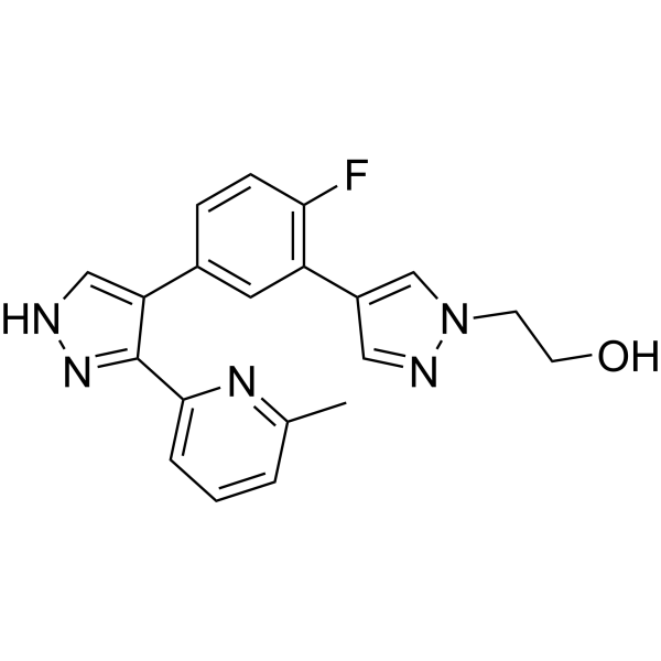
R-268712 is an orally active and selective ALK-5 inhibitor, with an IC50 of 2.5 nM. R-268712 inhibits the phosphorylation of Smad3 in a dose-dependent manner with an IC50 of 10.4 nM. R-268712 suppresses glomerulonephritis as well as glomerulosclerosis by inhibiting TGF-β signaling, which can be used in studies of renal fibrosis and cancer.
Nur für Forschungszwecke. Wir verkaufen nicht an Patienten.
- Reinheit : 99.92%
- CAS. Nr.: 879487-87-3
- Formel: C20H18FN5O
- Molecular Weight:363.39
-
Speicherung:Powder -20°C, 3 years , 4°C, 2 years ; In solvent -80°C, 2 years , -20°C, 1 year

To place orders, for customer services and technical support, please contact: MedChemExpress USA
Tel: 609-228-6898 E-mail: [email protected] [email protected]


-
 Biologische Aktivität
-
Chemical Information
-
Lösungsmittel & Löslichkeit
- Protokoll
- Reinheit & Dokumentation
- Verweise
-
 Help & FAQs


-
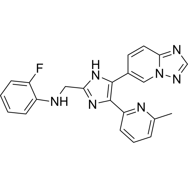
HY-19928
VactosertibALK5 Inhibitor -
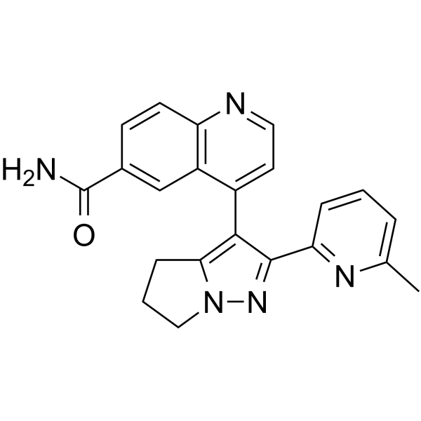
HY-13226
GalunisertibTGFβRI Inhibitor -
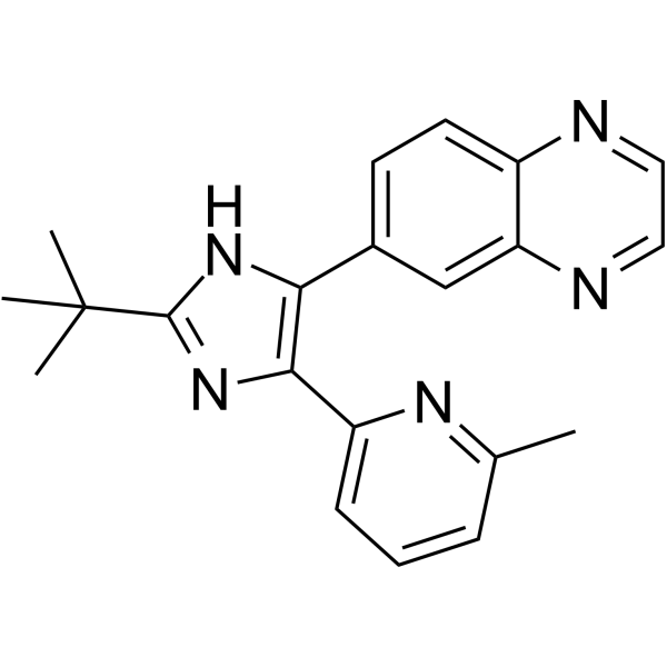
HY-12043
SB 525334ALK5 Inhibitor -
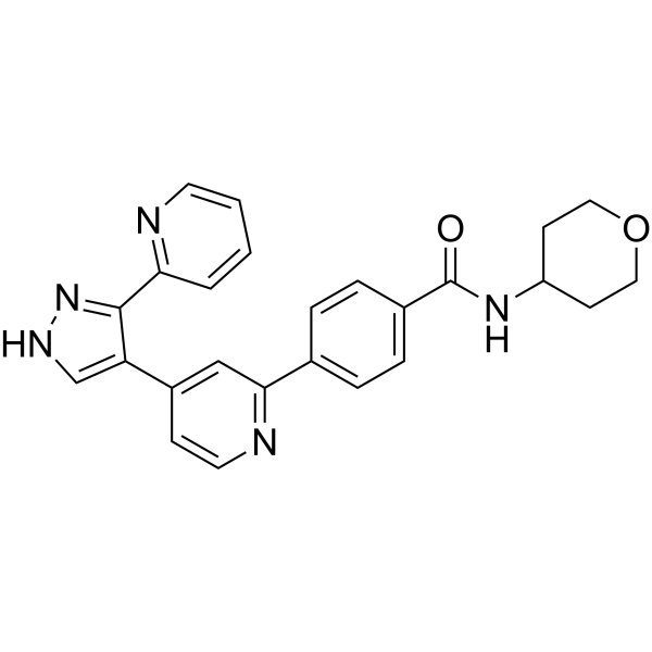
HY-10326
GW788388ALK5 Inhibitor -
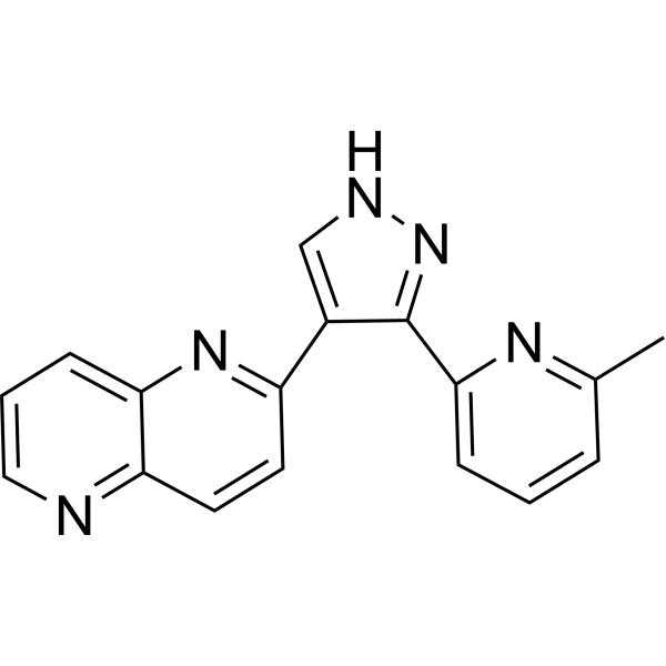
HY-13012
RepSoxTGFβR-1/ALK5 Inhibitor -

HY-10431
SB-431542TGF-β Receptor Kinase Inhibitor -
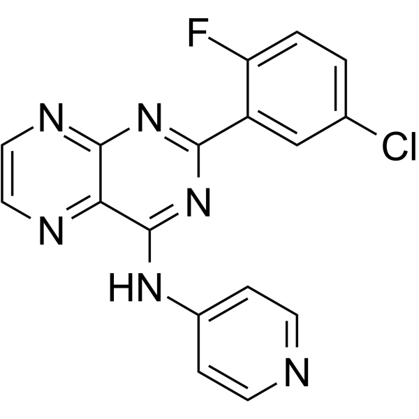
HY-13227
SD-208TGF-β Receptor Inhibitor -
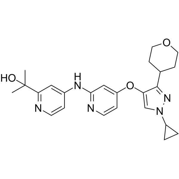
HY-103021
LY3200882ALK5 Inhibitor -
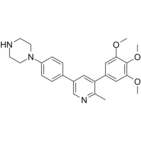
HY-16712
LDN-214117ALK2 Inhibitor -
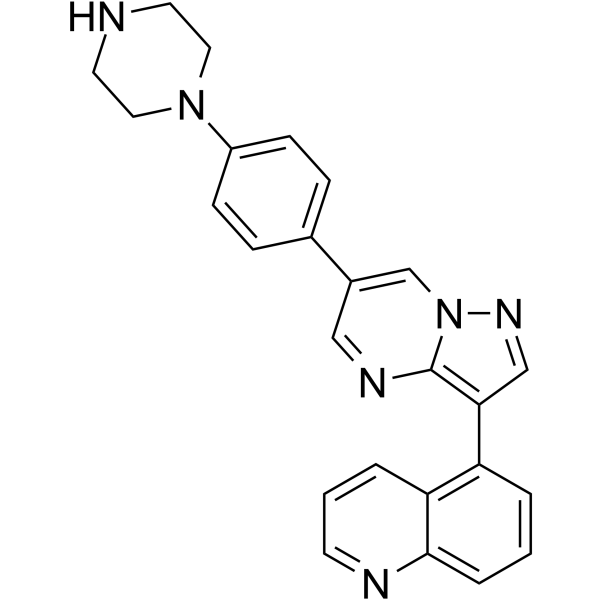
HY-15897
LDN-212854ALK2 Inhibitor -
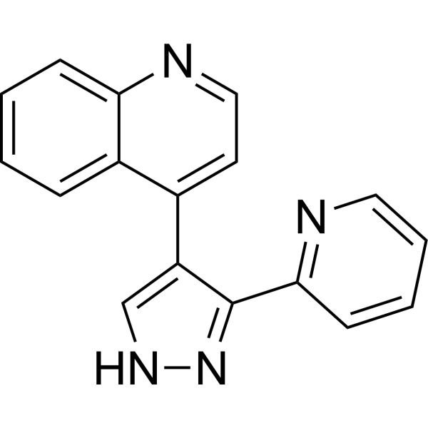
HY-13462
LY-364947TGFβR-I Inhibitor -
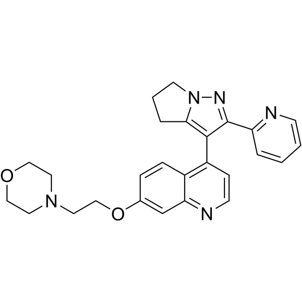
HY-12075
LY2109761TGF-βR I/II Inhibitor


Biologische Aktivität
Beschreibung
IC50 & Target
|
TGFBR1 2.5 nM (IC50) |
Smad3 10.4 nM (IC50) |
In Vitro
R-268712 (3, 10, 30, 100, 300 nM; 1 h) inhibits the phosphorylation of Smad3 in a dose-dependent manner with an IC50 of 10.4 nM in HFL-1 cells[1]. R-268712 (3, 10, 30, 100, 300 nM; 72 h) inhibits myofibroblast transdifferentiation (MTD) from fibroblasts in a dose-dependent manner without inhibition of cell growth in HFL-1cells[1].
MedChemExpress (MCE) has not independently confirmed the accuracy of these methods. They are for reference only.
-
Cell Line:HFL-1 cells
-
Concentration:3, 10, 30, 100, 300 nM
-
Incubation Time:1 or 72 h
-
Result:Inhibited the phosphorylation of Smad3 in a dose-dependent manner with an IC50 of 10.4 nM when incubation 1 h.
Suppressed myofibroblast transdifferentiation (MTD) from fibroblasts in a dose-dependent manner without inhibition of cell growth when incubation after 72 h.
In Vivo
R-268712 (1, 3, 10 mg/kg; p.o.; single daily for 3 days) inhibits renal luciferase activity in a dose-dependent manner in UUO model[2].
R-268712 (0.3, 1 mg/kg; p.o.; single daily for 33 days) shows renoprotective effects (improves and maintains renal function as well as inhibits glomerular sclerosis) on Thy1 nephritis model when at dosage of 1 mg/kg[2].
MedChemExpress (MCE) has not independently confirmed the accuracy of these methods. They are for reference only.
-
Animal Model:Male WKY/Hos rats[2].
-
Dosage:0.3, 1, 3, and 10 mg/kg
-
Administration:Oral administration; single.
-
Result:
1.19 Pharmacokinetic Parameters of R-268712 in male WKY/Hos rats (n=4)[2].
PO (0.3 mg/kg) PO (1 mg/kg) PO (3 mg/kg) PO (10 mg/kg) AUC0-24 (µg•h/mL) 0.075 0.28 1.6 8.2
-
Animal Model:Male Col1a1-Luc Tg rats (10 to14-week-old; UUO model; n=5-6)[2].
-
Dosage:1, 3, 10 mg/kg
-
Administration:Oral administration; single daily for 3 days.
-
Result:Suppressed activity of renal luciferase in a dose-dependent manner.
-
Animal Model:Male WKY/Hos rats (4-week-old; Thy1 nephritis model; n=7)[2].
-
Dosage:0.3, 1 mg/kg
-
Administration:Oral administration; single daily for 33 days.
-
Result:Significantly reduced proteinuria at day 21( the repression continued until day 28), and serum creatinine level (dosage at 1 mg/kg).
Apparently suppressed glomerular sclerosis by 28% and reduced the increase of the hydroxyproline content when at 1 mg/kg.
Suppressed the activation of mesangial parenchymal cell and the injury of podocyte on the basis of TGF-β signaling inhibition at 1 mg/kg.


Chemical Information
-
CAS. Nr. 879487-87-3
-
Appearance Solid
-
Molecular Weight 363.39
-
Formel C20H18FN5O
-
Color White to light yellow
-
SMILES
FC1=C(C2=CN(CCO)N=C2)C=C(C3=CNN=C3C4=CC=CC(C)=N4)C=C1
-
Versand
Room temperature in continental US; may vary elsewhere.
-
Speicherung
Powder -20°C 3 years 4°C 2 years In solvent -80°C 2 years -20°C 1 year


Lösungsmittel & Löslichkeit
In Vitro:
DMSO : 125 mg/mL (343.98 mM; Need ultrasonic; Hygroscopic DMSO has a significant impact on the solubility of product, please use newly opened DMSO)
Please refer to the solubility information to select the appropriate solvent. Once prepared, please aliquot and store the solution to prevent product inactivation from repeated freeze-thaw cycles.
Storage method and period of stock solution: -80°C, 2 years; -20°C, 1 year. When stored at -80°C, please use it within 2 years. When stored at -20°C, please use it within 1 year.
Please refer to the solubility information to select the appropriate solvent. Once prepared, please aliquot and store the solution to prevent product inactivation from repeated freeze-thaw cycles.
Storage method and period of stock solution: -80°C, 2 years; -20°C, 1 year. When stored at -80°C, please use it within 2 years. When stored at -20°C, please use it within 1 year.
Konzentration (Stammlösung) × Volumen (Stammlösung) = Konzentration (Ziellösung) × Volumen (Ziellösung)
In Vivo:
Select the appropriate dissolution method based on your experimental animal and administration route.
- For the following dissolution methods, please ensure to first prepare a clear stock solution using an In Vitro approach and then sequentially add co-solvents:
- To ensure reliable experimental results, the clarified stock solution can be appropriately stored based on storage conditions. As for the working solution for In Vivo experiments, it is recommended to prepare freshly and use it on the same day.
- The percentages shown for the solvents indicate their volumetric ratio in the final prepared solution. If precipitation or phase separation occurs during preparation, heat and/or sonication can be used to aid dissolution.
Add each solvent one by one: 10% DMSO 40% PEG300 5% Tween-80 45% Saline
Solubility: ≥ 2.08 mg/mL (5.72 mM); Clear solution
This protocol yields a clear solution of ≥ 2.08 mg/mL (saturation unknown).
Taking 1 mL working solution as an example, add 100 μL DMSO stock solution (20.8 mg/mL) to 400 μL PEG300, and mix evenly; then add 50 μL Tween-80 and mix evenly; then add 450 μL Saline to adjust the volume to 1 mL.
Preparation of Saline: Dissolve 0.9 g sodium chloride in ddH₂O and dilute to 100 mL to obtain a clear Saline solution.
Add each solvent one by one: 10% DMSO 90% (20% SBE-β-CD in Saline)
Solubility: ≥ 2.08 mg/mL (5.72 mM); Clear solution
This protocol yields a clear solution of ≥ 2.08 mg/mL (saturation unknown).
Taking 1 mL working solution as an example, add 100 μL DMSO stock solution (20.8 mg/mL) to 900 μL 20% SBE-β-CD in Saline, and mix evenly.
Preparation of 20% SBE-β-CD in Saline (4°C, storage for one week): 2 g SBE-β-CD powder is dissolved in 10 mL Saline, completely dissolve until clear.
In Vivo Dissolution Calculator
Please enter the basic information of animal experiments:
-
-
-
-
Recommended: Prepare an additional quantity of animals to account for potential losses during experiments.
Please enter your animal formula composition:
-
%DMSO +
Recommended: Keep the proportion of DMSO in working solution below 2% if your animal is weak.
-
%+
-
+%Tween-80 + +
-
%Saline +
The co-solvents required include: DMSO, . All of co-solvents are available by MedChemExpress (MCE). , Tween 80. All of co-solvents are available by MedChemExpress (MCE).
Working solution concentration: 0.22 mg/mL
Method for preparing stock solution: mg drug dissolved in μL DMSO. Stock solution concentration: mg/mL.
1. Take μL DMSO stock solution;
2. Add μL .
μL , mix evenly;
3. Then add μL Tween 80, mix evenly;
4. Then add μL
Please ensure that the stock solution in the first step is dissolved to a clear state, and add co-solvents in sequence. You can use ultrasonic heating (ultrasonic cleaner, recommended frequency 20-40 kHz), vortexing, etc. to assist dissolution.


Reinheit & Dokumentation
-
Data Sheet (280 KB)
-
SDS (393 KB)
- English - EN (393 KB)
- Français - FR (393 KB)
- Deutsch - DE (393 KB)
- Norwegian - NO (393 KB)
- Español - ES (393 KB)
- Swedish - SV (393 KB)
- Italian - IT (393 KB)
- Korean - KR (393 KB)
- Portuguese - PT (393 KB)
-
Handling Instructions (2659 KB)
Verweise
[1]. Terashima H, et al. Attenuation of pulmonary fibrosis in type I collagen-targeted reporter mice with ALK-5 inhibitors. Pulm Pharmacol Ther. 2019 Feb;54:31-38. [Content Brief]
[2]. Terashima H, et al. R-268712, an orally active transforming growth factor-β type I receptor inhibitor, prevents glomerular sclerosis in a Thy1 nephritis model. Eur J Pharmacol. 2014 Jul 5;734:60-6. [Content Brief]
[3]. Wang H, et al. Development of small molecule inhibitors targeting TGF-β ligand and receptor: Structures, mechanism, preclinical studies and clinical usage. Eur J Med Chem. 2020 Apr 1;191:112154. [Content Brief]


Complete Stock Solution Preparation Table
Please refer to the solubility information to select the appropriate solvent. Once prepared, please aliquot and store the solution to prevent product inactivation from repeated freeze-thaw cycles.
Storage method and period of stock solution: -80°C, 2 years; -20°C, 1 year. When stored at -80°C, please use it within 2 years. When stored at -20°C, please use it within 1 year.
| Optional Solvent | Concentration Solvent Mass | 1 mg | 5 mg | 10 mg | 25 mg |
|---|---|---|---|---|---|
| DMSO | 1 mM | 2.7519 mL | 13.7593 mL | 27.5186 mL | 68.7966 mL |
| 5 mM | 0.5504 mL | 2.7519 mL | 5.5037 mL | 13.7593 mL | |
| 10 mM | 0.2752 mL | 1.3759 mL | 2.7519 mL | 6.8797 mL | |
| 15 mM | 0.1835 mL | 0.9173 mL | 1.8346 mL | 4.5864 mL | |
| 20 mM | 0.1376 mL | 0.6880 mL | 1.3759 mL | 3.4398 mL | |
| 25 mM | 0.1101 mL | 0.5504 mL | 1.1007 mL | 2.7519 mL | |
| 30 mM | 0.0917 mL | 0.4586 mL | 0.9173 mL | 2.2932 mL | |
| 40 mM | 0.0688 mL | 0.3440 mL | 0.6880 mL | 1.7199 mL | |
| 50 mM | 0.0550 mL | 0.2752 mL | 0.5504 mL | 1.3759 mL | |
| 60 mM | 0.0459 mL | 0.2293 mL | 0.4586 mL | 1.1466 mL | |
| 80 mM | 0.0344 mL | 0.1720 mL | 0.3440 mL | 0.8600 mL | |
| 100 mM | 0.0275 mL | 0.1376 mL | 0.2752 mL | 0.6880 mL |


R-268712 Related Classifications
HY-12953 Related Classifications
 Powered by Bioz