



BC1618
Based on 1 publication(s) in Google Scholar
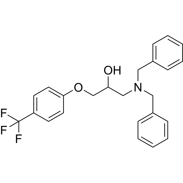
BC1618, an orally active Fbxo48 inhibitory compound, stimulates Ampk-dependent signaling (via preventing activated pAmpkα from Fbxo48-mediated degradation). BC1618 promotes mitochondrial fission, facilitates autophagy and improves hepatic insulin sensitivity.
For research use only. We do not sell to patients.
- Purity : 99.74%
- CAS No.: 2222094-18-8
- Formula: C24H24F3NO2
- Molecular Weight:415.45
-
Storage:Powder -20°C, 3 years ; In solvent -80°C, 6 months , -20°C, 1 month

To place orders, for customer services and technical support, please contact: MedChemExpress USA
Tel: 609-228-6898 E-mail: [email protected] [email protected]


-
 Biological Activity
-
Chemical Information
-
Solvent & Solubility
- Protocols
- Purity & Documentation
- References
-
 Help & FAQs


-
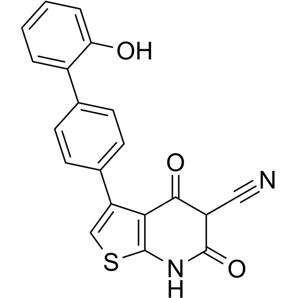
HY-50662
A-769662AMPK Activator -
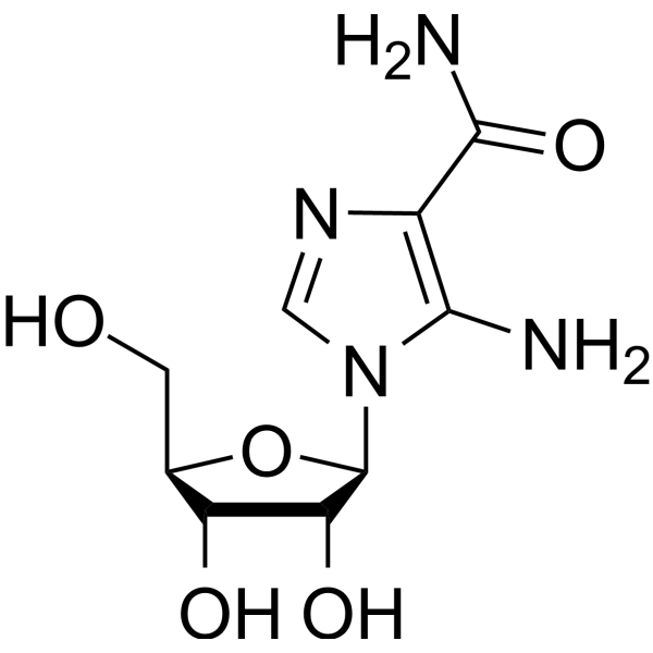
HY-13417
AICARAMPK Activator -
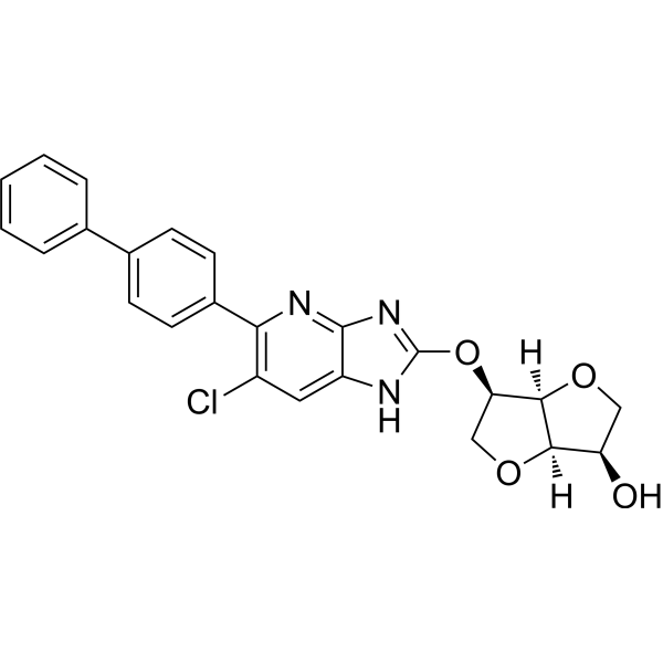
HY-111363
MK8722AMPK Activator -
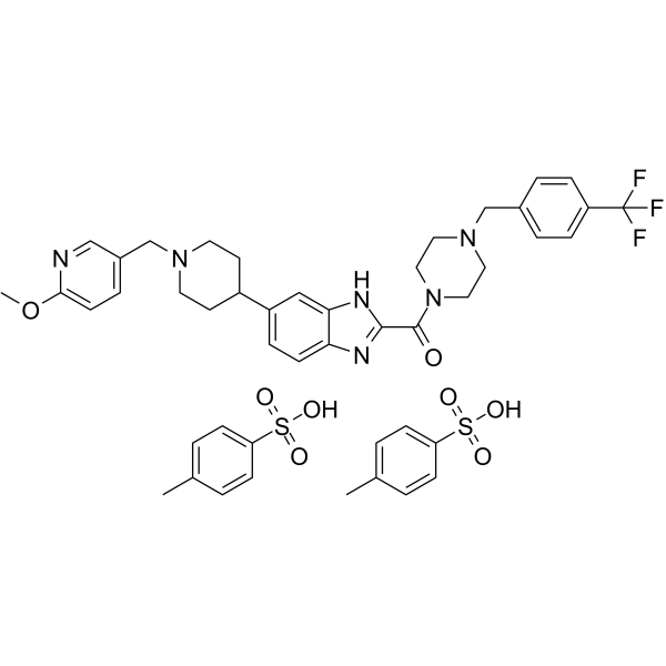
HY-136447
ASP4132AMPK Activator -

HY-Y0320
Dimethyl sulfoxide, meets analytical specification of Ch.P.Aprotic Solvent -
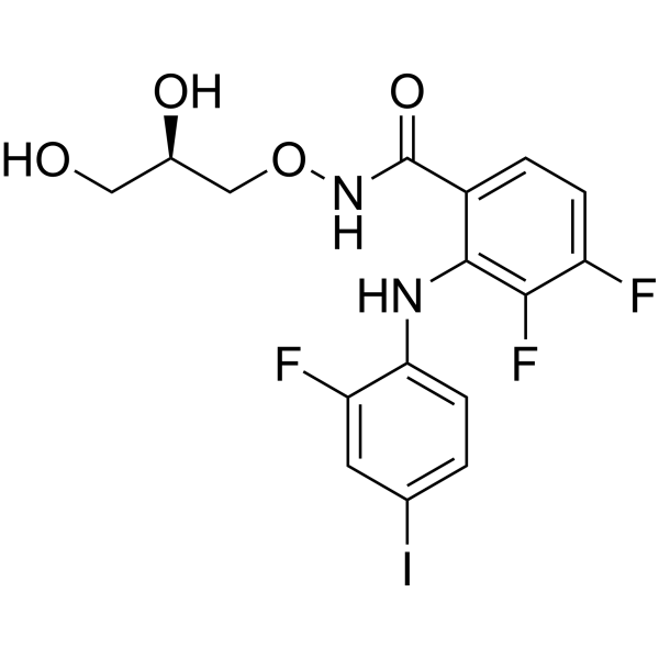
HY-10254
MirdametinibMEK Inhibitor -
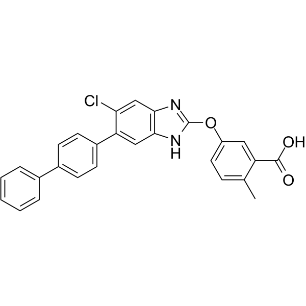
HY-107988
MK-3903AMPK Activator -
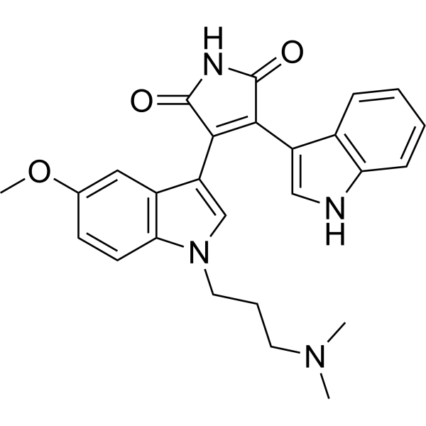
HY-13689
Go 6983PKC Inhibitor -
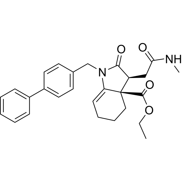
HY-100004
Fumarate hydratase-IN-1Fumarate Hydratase Inhibitor -
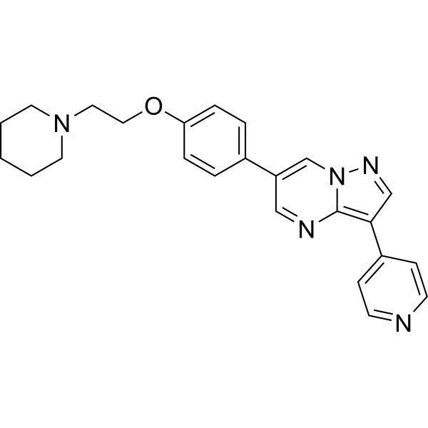
HY-13418A
DorsomorphinAMPK Inhibitor -
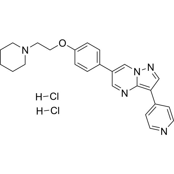
HY-13418
Dorsomorphin dihydrochlorideAMPK Inhibitor -
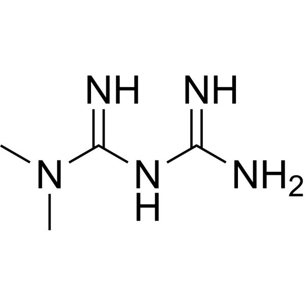
HY-B0627
MetforminAntihyperglycemic Agent
Publications Citing Use of MedChemExpress (MCE) BC1618
MoreAll AMPK Isoforms
More

Biological Activity
Description
In Vitro
BC1618 enhances pAmpkα protein stability during CHX treatment[1].
BC1618 displays more than 1,000-fold enhanced activity to stimulate pAmpkα in cells than metformin[1].
BC1618 (0.1-2 μM, 16 h) induced dose- and time-dependent increases in pAmpkα and pACC protein levels are also confirmed in human primary-like hepatocytes[1].
BC1618 (1 μM) effectively disrupts the interaction between Fbxo48 and pAmpkα, and has no effect on Fbxo48, Ampkα1 or Ampkα2 messenger RNAs[1].
BC1618 increases the abundance of a series of autophagic marker proteins during glucose depletion. BC1618 induces phosphorylation of the mTORC1 associated protein Raptor, reducing pS6 levels, all consistent with the known mTOR inhibitory effects exerted by activated Ampk[1].
MedChemExpress (MCE) has not independently confirmed the accuracy of these methods. They are for reference only.
-
Cell Line:BEAS-2B cells.
-
Concentration:0-2 μM.
-
Incubation Time:16 h.
-
Result:Induced pAmpkα and pACC protein levels dose-dependently.
In Vivo
BC1618, appears to be ~1,000-fold more potent than metformin and is extremely well tolerated in mice[1].
BC1618 displays excellent oral bioavailability with a peak of 2,000 ng/mL within 0.5h and 500 ng/mL in plasma at 4h after an oral load of 20mg/kg[1].
MedChemExpress (MCE) has not independently confirmed the accuracy of these methods. They are for reference only.
-
Animal Model:C57BL/6 mice[1].
-
Dosage:2 or 10 mg/kg (challenged with LPS (3 mg/kg) for an additional 18 h).
-
Administration:IP, once.
-
Result:Reduced lung inflammation in endotoxin treated mice.
-
Animal Model:C57BL/6 mice[1].
-
Dosage:15 and 30 mg/kg/d.
-
Administration:Drinking water for 3 months.
-
Result:Exhibited no obvious toxicity.


Chemical Information
-
CAS No. 2222094-18-8
-
Appearance Solid
-
Molecular Weight 415.45
-
Formula C24H24F3NO2
-
Color White to off-white
-
SMILES
OC(COC1=CC=C(C(F)(F)F)C=C1)CN(CC2=CC=CC=C2)CC3=CC=CC=C3
-
Shipping
Room temperature in continental US; may vary elsewhere.
-
Storage
Powder -20°C 3 years In solvent -80°C 6 months -20°C 1 month


Publications (1)
-
Journal Impact Factor
-
Most Recent
-
Res Sq
Mycobacterium tuberculosis resides in lysosome-poor monocyte-derived lung cells during chronic infection. [Abstract]2023 Jun 15:rs.3.rs-3049913. PMID: 37398178


Solvent & Solubility
In Vitro:
DMSO : 100 mg/mL (240.70 mM; Need ultrasonic; Hygroscopic DMSO has a significant impact on the solubility of product, please use newly opened DMSO)
H2O : < 0.1 mg/mL (insoluble)
Please refer to the solubility information to select the appropriate solvent. Once prepared, please aliquot and store the solution to prevent product inactivation from repeated freeze-thaw cycles.
Storage method and period of stock solution: -80°C, 6 months; -20°C, 1 month. When stored at -80°C, please use it within 6 months. When stored at -20°C, please use it within 1 month.
Please refer to the solubility information to select the appropriate solvent. Once prepared, please aliquot and store the solution to prevent product inactivation from repeated freeze-thaw cycles.
Storage method and period of stock solution: -80°C, 6 months; -20°C, 1 month. When stored at -80°C, please use it within 6 months. When stored at -20°C, please use it within 1 month.
Concentration (start) × Volume (start) = Concentration (final) × Volume (final)
In Vivo:
Select the appropriate dissolution method based on your experimental animal and administration route.
- For the following dissolution methods, please ensure to first prepare a clear stock solution using an In Vitro approach and then sequentially add co-solvents:
- To ensure reliable experimental results, the clarified stock solution can be appropriately stored based on storage conditions. As for the working solution for In Vivo experiments, it is recommended to prepare freshly and use it on the same day.
- The percentages shown for the solvents indicate their volumetric ratio in the final prepared solution. If precipitation or phase separation occurs during preparation, heat and/or sonication can be used to aid dissolution.
Add each solvent one by one: 10% DMSO 90% (20% SBE-β-CD in Saline)
Solubility: 2.5 mg/mL (6.02 mM); Suspended solution; Need ultrasonic
This protocol yields a suspended solution of 2.5 mg/mL. Suspended solution can be used for oral and intraperitoneal injection.
Taking 1 mL working solution as an example, add 100 μL DMSO stock solution (25.0 mg/mL) to 900 μL 20% SBE-β-CD in Saline, and mix evenly.
Preparation of 20% SBE-β-CD in Saline (4°C, storage for one week): 2 g SBE-β-CD powder is dissolved in 10 mL Saline, completely dissolve until clear.
For the following dissolution methods, please prepare the working solution directly:
It is recommended to prepare fresh solutions and use them promptly within a short period of time.
The percentages shown for the solvents indicate their volumetric ratio in the final prepared solution. If precipitation or phase separation occurs during preparation, heat and/or sonication can be used to aid dissolution.
Add each solvent one by one: 50% PEG300 50% Saline
Solubility: 5 mg/mL (12.04 mM); Suspended solution; Need ultrasonic
In Vivo Dissolution Calculator
Please enter the basic information of animal experiments:
-
-
-
-
Recommended: Prepare an additional quantity of animals to account for potential losses during experiments.
Please enter your animal formula composition:
-
%DMSO +
Recommended: Keep the proportion of DMSO in working solution below 2% if your animal is weak.
-
%+
-
+%Tween-80 + +
-
%Saline +
The co-solvents required include: DMSO, . All of co-solvents are available by MedChemExpress (MCE). , Tween 80. All of co-solvents are available by MedChemExpress (MCE).
Working solution concentration: 0.22 mg/mL
Method for preparing stock solution: mg drug dissolved in μL DMSO. Stock solution concentration: mg/mL.
1. Take μL DMSO stock solution;
2. Add μL .
μL , mix evenly;
3. Then add μL Tween 80, mix evenly;
4. Then add μL
Please ensure that the stock solution in the first step is dissolved to a clear state, and add co-solvents in sequence. You can use ultrasonic heating (ultrasonic cleaner, recommended frequency 20-40 kHz), vortexing, etc. to assist dissolution.


Purity & Documentation
-
Data Sheet (275 KB)
-
SDS (393 KB)
- English - EN (393 KB)
- Français - FR (393 KB)
- Deutsch - DE (393 KB)
- Norwegian - NO (393 KB)
- Español - ES (393 KB)
- Swedish - SV (393 KB)
- Italian - IT (393 KB)
- Korean - KR (393 KB)
- Portuguese - PT (393 KB)
-
Handling Instructions (2659 KB)
References


Complete Stock Solution Preparation Table
Please refer to the solubility information to select the appropriate solvent. Once prepared, please aliquot and store the solution to prevent product inactivation from repeated freeze-thaw cycles.
Storage method and period of stock solution: -80°C, 6 months; -20°C, 1 month. When stored at -80°C, please use it within 6 months. When stored at -20°C, please use it within 1 month.
| Optional Solvent | Concentration Solvent Mass | 1 mg | 5 mg | 10 mg | 25 mg |
|---|---|---|---|---|---|
| DMSO | 1 mM | 2.4070 mL | 12.0351 mL | 24.0703 mL | 60.1757 mL |
| 5 mM | 0.4814 mL | 2.4070 mL | 4.8141 mL | 12.0351 mL | |
| 10 mM | 0.2407 mL | 1.2035 mL | 2.4070 mL | 6.0176 mL | |
| 15 mM | 0.1605 mL | 0.8023 mL | 1.6047 mL | 4.0117 mL | |
| 20 mM | 0.1204 mL | 0.6018 mL | 1.2035 mL | 3.0088 mL | |
| 25 mM | 0.0963 mL | 0.4814 mL | 0.9628 mL | 2.4070 mL | |
| 30 mM | 0.0802 mL | 0.4012 mL | 0.8023 mL | 2.0059 mL | |
| 40 mM | 0.0602 mL | 0.3009 mL | 0.6018 mL | 1.5044 mL | |
| 50 mM | 0.0481 mL | 0.2407 mL | 0.4814 mL | 1.2035 mL | |
| 60 mM | 0.0401 mL | 0.2006 mL | 0.4012 mL | 1.0029 mL | |
| 80 mM | 0.0301 mL | 0.1504 mL | 0.3009 mL | 0.7522 mL | |
| 100 mM | 0.0241 mL | 0.1204 mL | 0.2407 mL | 0.6018 mL |


BC1618 Related Classifications
HY-134656 Related Classifications
 Powered by Bioz