



Dynorphin A TFA
Based on 2 publication(s) in Google Scholar
Dynorphin A TFA is an endogenous opioid peptide involved in inhibitory neurotransmission in the central nervous system (CNS). Dynorphin A TFA is a highy potent kappa opioid receptor (KOR) agonist, and is also an agonist for other opioid receptors, such as mu (MOR) and delta (DOR). Dynorphin A TFA can induce neuronal death, and can be used in the research of neurological disease.
For research use only. We do not sell to patients.
- Formula: C101H156F3N31O25
- Molecular Weight:2261.50
-
Storage:
Please store the product under the recommended conditions in the Certificate of Analysis.

To place orders, for customer services and technical support, please contact: MedChemExpress USA
Tel: 609-228-6898 E-mail: [email protected] [email protected]


-
 Biological Activity
-
Chemical Information
- Protocols
- Purity & Documentation
- References
-
 Help & FAQs


Publications Citing Use of MedChemExpress (MCE) Dynorphin A TFA
MoreAll Opioid Receptor Isoforms
MoreAll Caspase Isoforms
MoreAll Endogenous Metabolite Isoforms
More

Biological Activity
Description
IC50 & Target
[3]|
Human Endogenous Metabolite |
Caspase-3 |
κ Opioid Receptor/KOR |
In Vitro
Dynorphin A TFA (10 μM, 4 h/72 h) increases caspase-3 activity and the level of cytochrome c released from mitochondria in mouse striatal neurons, and induces neuronal death[3].
dynorphin A TFA (33 μM, 4 h) elevates [Ca2+]i and causes a significant loss of neurons[4].
dynorphin A TFA (1 μM) inhibits the release of vasopressin (VP) from the isolated neural lobe[5].
MedChemExpress (MCE) has not independently confirmed the accuracy of these methods. They are for reference only. Further protocols information, click here.
-
Cell Line:Mouse striatal neurons
-
Concentration:10 μM
-
Incubation Time:0, 24, 48, 72 h
-
Result:Induced neuronal death (identified by the fragmentation and destruction of the cell body and neurites).
In Vivo
Dynorphin A TFA (intracerebroventricular injection, 500 pmol/5 μL per day for 4 d) alleviates stress-induced behavioral impairments in ddY mice accompanied by regulation of the 5-HTergic system in the brain[6].
MedChemExpress (MCE) has not independently confirmed the accuracy of these methods. They are for reference only.
-
Animal Model:24 h water-deprived male rats[5]
-
Dosage:1 μg of 2 μL
-
Administration:Intracerebroventricular injection
-
Result:Inhibited vasopressin (VP) release 30 min upon injection.
-
Animal Model:Male ddY mice[6]
-
Dosage:15, 150, 1500 pmol/5 μL per day for 4 days
-
Administration:Intracerebroventricular injection
-
Result:Attenuated the repeated stress-induced escape failures from the shock.


Chemical Information
-
Molecular Weight 2261.50
-
Formula C101H156F3N31O25
-
Sequence
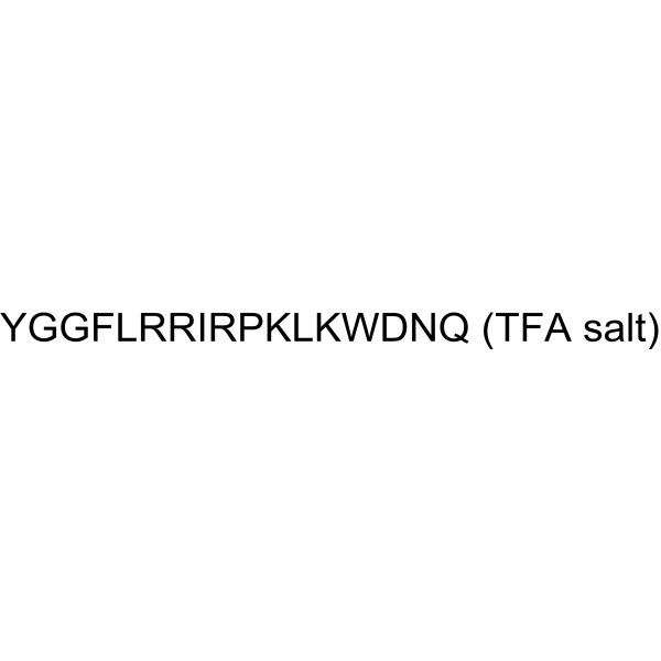
Tyr-Gly-Gly-Phe-Leu-Arg-Arg-Ile-Arg-Pro-Lys-Leu-Lys-Trp-Asp-Asn-Gln
-
Sequence Shortening
YGGFLRRIRPKLKWDNQ
-
Shipping
Room temperature in continental US; may vary elsewhere.
-
Storage
Please store the product under the recommended conditions in the Certificate of Analysis.


Publications (2)
-
Journal Impact Factor
-
Most Recent
-
Neuron
Astrocyte-mediated central amygdala microcircuit gates comorbid anxiety symptoms in chronic pain. [Abstract]2025 Oct 2:S0896-6273(25)00670-1. PMID: 41043420 -

Protocols
-
Research Protocol for Neurological Diseases
PINK1/Parkin-mediated mitophagy pathway is a mitochondrial quality-control signaling axis in which mitochondrial depolarization stabilizes PINK1 on damaged mitochondria, activates Parkin recruitment and E3 ubiquitin ligase activity, promotes ubiquitination of outer mitochondrial membrane proteins, recruits selective autophagy adaptors, and drives lysosomal degradation of damaged mitochondria. In neurological disease research, this pathway is experimentally important because neurons, especially dopaminergic neurons, are highly dependent on mitochondrial integrity, and defective mitochondrial turnover can lead to mitochondrial dysfunction, oxidative stress, impaired neuronal survival, α-synuclein accumulation, and neuroinflammatory damage-associated signals. The genetic disease link is strongest in Parkinson’s disease because mutations in PRKN/parkin cause autosomal recessive juvenile parkinsonism, mutations in PINK1 cause hereditary early-onset Parkinson’s disease, and Drosophila studie


Purity & Documentation
References
[1]. Zhang, et al.Dynorphin A as a Potential Endogenous Ligand for Four Members of the Opioid Receptor Gene Family. J Pharmacol Exp Ther. 1998 Jul;286(1):136-41. [Content Brief]
[2]. Aruna Sharma, et al. Monoclonal antibodies as novel neurotherapeutic agents in CNS injury and repair. Int Rev Neurobiol. 2012;102:23-45. [Content Brief]
[3]. I. N. SINGH, et al. Dynorphin A (1–17) induces apoptosis in striatal neurons in vitro through AMPA/kainate receptor-mediated cytochrome c release and caspase-3 activation. Neuroscience. 2003;122(4):1013-23. [Content Brief]
[4]. B J Van de Heijning, et al. Dynorphin-A and vasopressin release in the rat: a structure-activity study. Neuropeptides. 1994 Jun;26(6):371-8. [Content Brief]
[5]. Takayoshi Mamiya, et al. Dynorphin a (1-13) alleviated stress-induced behavioral impairments in mice. Biol Pharm Bull. 2014;37(8):1269-73. [Content Brief]
[6]. K F Hauser, et al. Dynorphin A (1-13) neurotoxicity in vitro: opioid and non-opioid mechanisms in mouse spinal cord neurons. Exp Neurol. 1999 Dec;160(2):361-75. [Content Brief]

Calculators
Concentration (start) × Volume (start) = Concentration (final) × Volume (final)


Dynorphin A TFA Related Classifications
HY-P1333A Related Classifications
 Powered by Bioz