



Aurantiamide

Based on 1 Customer Validation
Aurantiamide is a non-covalent, orally active, blood-brain-permeable GRPR selective antagonist with anti-inflammatory and neuroprotective effects. Aurantiamide reduces inflammation and oxidative stress in renal tissue by inhibiting GRPR-mediated renal necrosis pathways (such as RIPK3/MLKL signaling) and NF-κB inflammatory pathways, exerting anti-acute kidney injury and endothelial function activities. Aurantiamide also inhibits the M1 polarization of microglia and inhibits NLRP3 activation, thereby improving AD mouse models. Aurantiamide has in vivo inhibitory efficacy in acute kidney injury models such as ischemia/reperfusion, sepsis, and hypertension models.
For research use only. We do not sell to patients.
- Purity : 99.56%
- CAS No.: 58115-31-4
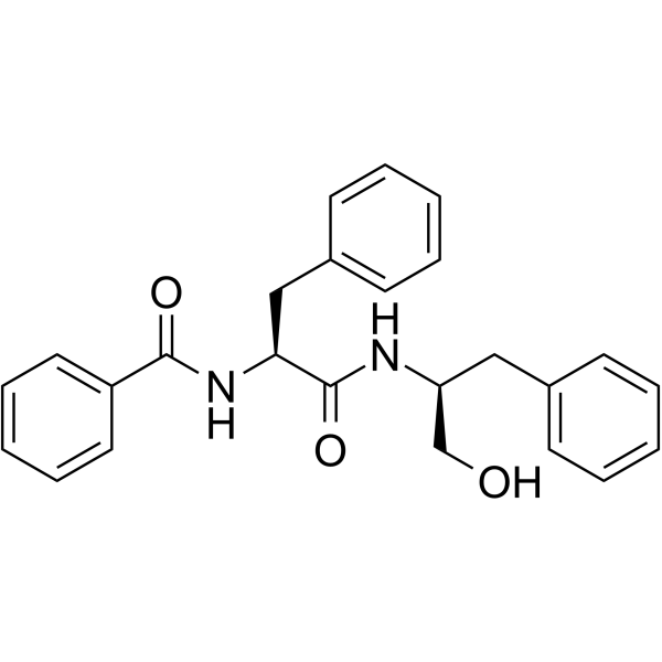
- Formula: C25H26N2O3
- Molecular Weight:402.49
-
Storage:
-20°C, protect from light
* In solvent : -80°C, 6 months; -20°C, 1 month (protect from light)

To place orders, for customer services and technical support, please contact: MedChemExpress USA
Tel: 609-228-6898 E-mail: [email protected] [email protected]


-
 Biological Activity
-
Chemical Information
-
Solvent & Solubility
- Protocols
- Purity & Documentation
- References
-
 Help & FAQs




Biological Activity
Description
IC50 & Target
|
NF-κB |
RIPK3 |
Cellular Effect
|
Cell Line

|
Type | Value | Description | References |
|---|---|---|---|---|
| N9 | IC50 |
14.91 μM
Compound: 23
|
Antineuroinflammatory activity in mouse N9 cells assessed as inhibition of LPS-induced nitric oxide production after 24 hrs by Griess assay
Antineuroinflammatory activity in mouse N9 cells assessed as inhibition of LPS-induced nitric oxide production after 24 hrs by Griess assay
|
[PMID: 28073678] |
In Vitro
Aurantiamide (25, 50, 100 μM; 12 h) significantly reduces the protein and mRNA expression of the renal injury marker KIM1 in the human renal proximal tubular epithelial cell (HK-2) hypoxia/reoxygenation and LPS (1 mg/mL) stimulation model, inhibits the phosphorylation of proteins related to the RIPK3/MLKL necrosis pathway and NF-κB inflammatory pathway, and alleviates cell damage and inflammatory response[1].
Aurantiamide (10, 20 μM; 24 h) inhibits LPS and IFN-γ-induced BV2 activation and M1 polarization in mouse BV2 cells, inhibited the activation of NLRP3, and thus exerted anti-CNS inflammatory activity[2].
Aurantiamide promotes M2 polarization of microglia and improves cognitive ability of Alzheimer's disease mice[3].
Aurantiamide (3 μM; 18 h) significantly upregulates the mRNA and protein expression of endothelial nitric oxide synthase (eNOS) in human umbilical vein endothelial cells (HUVECs), promotes the production of nitric oxide (NO), and has no significant toxic effect on cell viability[4].
MedChemExpress (MCE) has not independently confirmed the accuracy of these methods. They are for reference only. Further protocols information, click here.
-
Cell Line:Human proximal tubule epithelial cells (HK-2) [LPS-induced model]
-
Concentration:25 μM, 50 μM, 100 μM
-
Incubation Time:24 h
-
Result:WB (Western blot) : Reduced protein expression of renal injury marker KIM1, phosphorylated RIPK3 (p-RIPK3), phosphorylated MLKL (p-MLKL), and phosphorylated P65 (p-P65) in a dose-dependent manner, compared to H/R or LPS-stimulated controls.
qPCR : Downregulated mRNA levels of KIM1, MCP-1, and IL-6, with significant inhibition at 50 μM and 100 μM.
IF (Immunofluorescence) : Decreased immunofluorescent intensity of p-MLKL and KIM1 in cytoplasm, indicating reduced necroptosis and tubular injury.
-
Cell Line:Human umbilical vein endothelial cells (HUVEC)
-
Concentration:3 μM
-
Incubation Time:18 h
-
Result:No significant cytotoxicity observed, with cell viability maintaining over 90% compared to control.
WB (Western blot) : Upregulated eNOS protein expression by 1.8-fold compared to control, with enhanced NO production detected by nitrite/nitrate assay.
\qPCR : Increased eNOS mRNA expression by 2.3-fold, confirming transcriptional activation of eNOS.
In Vivo
Aurantiamide (0.5 mg/kg; intraperitoneal injection; once a day, 5 days a week; 4 weeks) significantly reduces mean arterial blood pressure, improves endothelium-dependent vasodilation, upregulates aortic endothelial nitric oxide synthase (eNOS) protein expression and promotes nitric oxide (NO) production in the two-kidney-one-clip (2K-1C) renovascular hypertension model in Sprague-Dawley rats[4].
The metabolic characteristics of Aurantiamide (0.1 mg/kg; oral gavage; single dose) and Aurantiamide acetate (HY-N2905) (0.2 mg/kg; oral gavage; single dose) in rats shows that they have the characteristics of rapid diffusion, wide distribution, and can pass through the blood-brain barrier, with a peak time of 0.5 h. In addition, the decline rate of aurantiamide acetate is faster than that of aurantiamide[5].
MedChemExpress (MCE) has not independently confirmed the accuracy of these methods. They are for reference only.
-
Animal Model:Male C57BL/6 mice (6-8 weeks old, 20-22 g) + cisplatin-induced, I/R, or CLP-induced acute kidney injury model[1]
-
Dosage:2.5, 5, 10 mg/kg (dissolved in 0.5% carboxymethylcellulose + 0.1% Tween 80)
-
Administration:Oral gavage, three times before the surgery, with a 24 h interval between each administration
-
Result:Renal function : Reduced serum creatinine and BUN levels by 30-45% compared to model controls, with the 10 mg/kg dose showing the most pronounced effect.
Histopathology : PAS staining revealed decreased tubular dilation, glycogen deposition, and interstitial fibrosis; immunofluorescence showed reduced KIM1 (renal injury marker) and F4/80+ macrophage infiltration in renal tissues.
Protein expression : Western blot demonstrated dose-dependent inhibition of p-RIPK3, p-MLKL, and p-P65 (NF-κB) in renal lysates, with corresponding reduction in pro-inflammatory cytokines (IL-6, TNF-α) by qPCR.
-
Animal Model:Male Sprague-Dawley rats (8 weeks old, 230-250 g) + two-kidney one-clip (2K-1C) renovascular hypertension model[4]
-
Dosage:0.5 mg/kg (dissolved in DMSO, final concentration 0.1%)
-
Administration:Intraperitoneal injection, once daily for 5 days/week, total 4 weeks
-
Result:Blood pressure : Reduced mean arterial pressure (MAP) by 20-25% compared to hypertensive controls, with significant improvement in endothelium-dependent relaxation to acetylcholine (ACh) and reduced constriction to phenylephrine (Phe).
Vascular function : Organ bath assays showed enhanced ACh-induced vasodilation and attenuated Phe-induced vasoconstriction in aortic rings, correlated with increased eNOS protein expression (1.5-fold by Western blot) and NO production (measured as nitrite/nitrate levels).
Red blood cell deformability : Ektacytometry revealed increased erythrocyte deformability (Elmax) in treated rats, indicating improved blood fluidity and microvascular flow.


Chemical Information
-
CAS No. 58115-31-4
-
Appearance Solid
-
Molecular Weight 402.49
-
Formula C25H26N2O3
-
Color White to off-white
-
SMILES
O=C(N[C@H](CO)CC1=CC=CC=C1)[C@@H](NC(C2=CC=CC=C2)=O)CC3=CC=CC=C3
-
Structure Classification
-
Shipping
Room temperature in continental US; may vary elsewhere.
-
Storage
-20°C, protect from light
* In solvent : -80°C, 6 months; -20°C, 1 month (protect from light)


Solvent & Solubility
In Vitro:
DMSO : 50 mg/mL (124.23 mM; Need ultrasonic; Hygroscopic DMSO has a significant impact on the solubility of product, please use newly opened DMSO)
Please refer to the solubility information to select the appropriate solvent. Once prepared, please aliquot and store the solution to prevent product inactivation from repeated freeze-thaw cycles.
Storage method and period of stock solution: -80°C, 6 months; -20°C, 1 month (protect from light). When stored at -80°C, please use it within 6 months. When stored at -20°C, please use it within 1 month.
Please refer to the solubility information to select the appropriate solvent. Once prepared, please aliquot and store the solution to prevent product inactivation from repeated freeze-thaw cycles.
Storage method and period of stock solution: -80°C, 6 months; -20°C, 1 month (protect from light). When stored at -80°C, please use it within 6 months. When stored at -20°C, please use it within 1 month.
Concentration (start) × Volume (start) = Concentration (final) × Volume (final)
In Vivo:
Select the appropriate dissolution method based on your experimental animal and administration route.
- For the following dissolution methods, please ensure to first prepare a clear stock solution using an In Vitro approach and then sequentially add co-solvents:
- To ensure reliable experimental results, the clarified stock solution can be appropriately stored based on storage conditions. As for the working solution for In Vivo experiments, it is recommended to prepare freshly and use it on the same day.
- The percentages shown for the solvents indicate their volumetric ratio in the final prepared solution. If precipitation or phase separation occurs during preparation, heat and/or sonication can be used to aid dissolution.
Add each solvent one by one: 10% DMSO 40% PEG300 5% Tween-80 45% Saline
Solubility: ≥ 1.25 mg/mL (3.11 mM); Clear solution
This protocol yields a clear solution of ≥ 1.25 mg/mL (saturation unknown).
Taking 1 mL working solution as an example, add 100 μL DMSO stock solution (12.5 mg/mL) to 400 μL PEG300, and mix evenly; then add 50 μL Tween-80 and mix evenly; then add 450 μL Saline to adjust the volume to 1 mL.
Preparation of Saline: Dissolve 0.9 g sodium chloride in ddH₂O and dilute to 100 mL to obtain a clear Saline solution.
In Vivo Dissolution Calculator
Please enter the basic information of animal experiments:
-
-
-
-
Recommended: Prepare an additional quantity of animals to account for potential losses during experiments.
Please enter your animal formula composition:
-
%DMSO +
Recommended: Keep the proportion of DMSO in working solution below 2% if your animal is weak.
-
%+
-
+%Tween-80 + +
-
%Saline +
The co-solvents required include: DMSO, . All of co-solvents are available by MedChemExpress (MCE). , Tween 80. All of co-solvents are available by MedChemExpress (MCE).
Working solution concentration: 0.22 mg/mL
Method for preparing stock solution: mg drug dissolved in μL DMSO. Stock solution concentration: mg/mL. * In solvent : -80°C, 6 months; -20°C, 1 month (protect from light)
1. Take μL DMSO stock solution;
2. Add μL .
μL , mix evenly;
3. Then add μL Tween 80, mix evenly;
4. Then add μL
Please ensure that the stock solution in the first step is dissolved to a clear state, and add co-solvents in sequence. You can use ultrasonic heating (ultrasonic cleaner, recommended frequency 20-40 kHz), vortexing, etc. to assist dissolution.

Protocols
-
Nephrotoxicity Study
This protocol assesses nephrotoxicity by combining functional kidney injury readouts, urinary/tissue injury biomarkers, and renal histopathology. Serum creatinine and BUN reflect impaired kidney function, while KIM-1, NGAL, clusterin, osteopontin, IL-18, cystatin C, nephrin, Oat5, urinary protein, glucose, and alkaline phosphatase have been used to detect tubular injury in cisplatin-, gentamicin-, and acetaminophen-induced nephrotoxicity models.
-
LPS-Induced Endotoxemia/Systemic Inflammation
Lipopolysaccharide (LPS)-induced endotoxemia is a widely used in vivo model of acute systemic inflammation in which LPS, a Gram-negative bacterial endotoxin, activates innate immune signaling primarily through TLR4, leading to rapid and transient induction of pro-inflammatory cytokines such as TNF-α, IL-6, and IL-1β in circulation and tissues. This cytokine surge is commonly used as a measurable readout of systemic inflammatory activation and immune dysregulation, and is typically assessed within hours after intraperitoneal LPS administration in mouse models of endotoxemia. The model captures key features of systemic inflammatory response syndrome, including cytokine release, immune cell activation, and downstream tissue responses, and has been used to evaluate anti-inflammatory interventions such as cytokine modulation, lipid mediators, and immune cell-targeting therapies.
-
ROS/oxidative-stress fluorescent staining
ROS/oxidative-stress fluorescent staining uses cell-permeant fluorogenic probes that become fluorescent after oxidation inside cells or tissues; commonly used examples include DCFH-DA/DCFDA for broad cellular oxidant detection, DHE for superoxide-related signal detection, MitoSOX for mitochondrial superoxide-related signal detection, and CellROX probes for oxidative-stress-associated fluorescence readouts. The assay detects probe oxidation rather than a single ROS species unless the probe and analysis method have been chemically validated for that species. DCFH-DA enters cells, is deacetylated by intracellular esterases to DCFH, and produces fluorescent DCF after oxidation, so the readout is used as an operational measure of total cellular oxidative stress rather than a species-specific ROS measurement. DHE and MitoSOX can report superoxide-related oxidation, but red fluorescence alone can include non-specific ethidium-like oxidation products; HPLC or optimized spectral approaches are
-
Research Protocol for Cardiovascular Diseases
Cardiovascular disease can be modeled as maladaptive cardiac remodeling, where ischemic injury or pressure overload activates inflammatory signaling, fibroblast activation, extracellular-matrix deposition, cardiomyocyte hypertrophy, vascular remodeling, and progressive ventricular dysfunction. The TGF-β/SMAD axis is a central profibrotic pathway after myocardial injury and pressure overload, while innate immune and cytokine pathways regulate leukocyte recruitment, scar formation, and adverse remodeling. Key unresolved questions include which inflammatory signals are reparative versus harmful, when fibrosis is protective versus maladaptive, and whether pathway inhibition improves function without weakening necessary infarct healing or compensatory remodeling.
-
Research Protocol for Inflammation-related Diseases
The NLRP3 inflammasome is a cytosolic innate immune signaling platform that integrates priming signals and danger-signal activation to promote caspase-1 activation, maturation of IL-1β and IL-18, and gasdermin D-mediated pyroptotic cell death. The core experimental logic is to determine whether inflammatory disease phenotypes are driven by increased NLRP3 expression, ASC-containing inflammasome assembly, caspase-1 cleavage, GSDMD cleavage, and extracellular release of IL-1β/IL-18 rather than by nonspecific cell injury alone. The pathway is strongly linked to inflammation-related disease phenotypes because monosodium urate crystals activate NALP3/NLRP3 inflammasome signaling in gout-like crystal inflammation, cholesterol crystals activate NLRP3 inflammasomes in atherogenesis models, and DSS-induced intestinal inflammation has been reported to involve NLRP3 inflammasome activity. However, experimental colitis studies also show context-dependent protective effects of NLRP3 inflammasome co


Purity & Documentation
-
Data Sheet (283 KB)
-
SDS (393 KB)
- English - EN (393 KB)
- Français - FR (393 KB)
- Deutsch - DE (393 KB)
- Norwegian - NO (393 KB)
- Español - ES (393 KB)
- Swedish - SV (393 KB)
- Italian - IT (393 KB)
- Korean - KR (393 KB)
- Portuguese - PT (393 KB)
-
Handling Instructions (2659 KB)
References
[1]. He RB, et al. Aurantiamide mitigates acute kidney injury by suppressing renal necroptosis and inflammation via GRPR-dependent mechanism. [Content Brief]
[2]. Shen H, et al. Aurantiamide suppresses the activation of NLRP3 inflammasome to improve the cognitive function and central inflammation in mice with Alzheimer's disease. CNS Neurosci Ther. 2023 Apr;29(4):1075-1085. [Content Brief]
[3]. Shen H, et al. Aurantiamide promotes M2 polarization of microglial cells to improve the cognitive ability of mice with Alzheimer's disease. Phytother Res. 2023 Jan;37(1):101-110. [Content Brief]
[4]. Aslan M, et al. Effects of aurantiamide on a rat model of renovascular arterial hypertension. Pflugers Arch. 2023 Oct;475(10):1177-1192. [Content Brief]
[5]. Lijiang Chen, et al. Pharmacokinetics and Biodistribution of Aurantiamide and Aurantiamide Acetate in Rats After Oral Administration of Portulaca Oleracea L. Extracts. J Agric Food Chem. 2016 May 4;64(17):3445-55. [Content Brief]


Complete Stock Solution Preparation Table
Please refer to the solubility information to select the appropriate solvent. Once prepared, please aliquot and store the solution to prevent product inactivation from repeated freeze-thaw cycles.
Storage method and period of stock solution: -80°C, 6 months; -20°C, 1 month (protect from light). When stored at -80°C, please use it within 6 months. When stored at -20°C, please use it within 1 month.
| Optional Solvent | Concentration Solvent Mass | 1 mg | 5 mg | 10 mg | 25 mg |
|---|---|---|---|---|---|
| DMSO | 1 mM | 2.4845 mL | 12.4227 mL | 24.8453 mL | 62.1133 mL |
| 5 mM | 0.4969 mL | 2.4845 mL | 4.9691 mL | 12.4227 mL | |
| 10 mM | 0.2485 mL | 1.2423 mL | 2.4845 mL | 6.2113 mL | |
| 15 mM | 0.1656 mL | 0.8282 mL | 1.6564 mL | 4.1409 mL | |
| 20 mM | 0.1242 mL | 0.6211 mL | 1.2423 mL | 3.1057 mL | |
| 25 mM | 0.0994 mL | 0.4969 mL | 0.9938 mL | 2.4845 mL | |
| 30 mM | 0.0828 mL | 0.4141 mL | 0.8282 mL | 2.0704 mL | |
| 40 mM | 0.0621 mL | 0.3106 mL | 0.6211 mL | 1.5528 mL | |
| 50 mM | 0.0497 mL | 0.2485 mL | 0.4969 mL | 1.2423 mL | |
| 60 mM | 0.0414 mL | 0.2070 mL | 0.4141 mL | 1.0352 mL | |
| 80 mM | 0.0311 mL | 0.1553 mL | 0.3106 mL | 0.7764 mL | |
| 100 mM | 0.0248 mL | 0.1242 mL | 0.2485 mL | 0.6211 mL |


Aurantiamide Related Classifications
HY-N2909 Related Classifications
 Powered by Bioz