



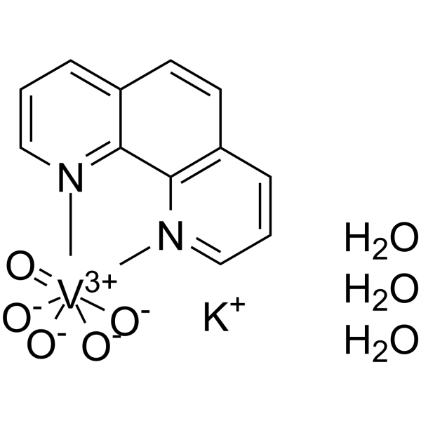
BpV(phen) trihydrate
Based on 3 publication(s) in Google Scholar
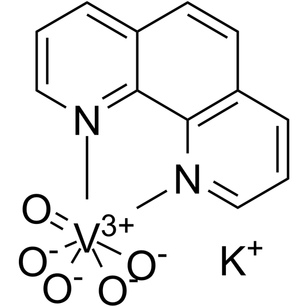
BpV(phen) trihydrate, a insulin-mimetic agent, is a potent protein tyrosine phosphatase (PTP) and PTEN inhibitor with IC50s of 38 nM, 343 nM and 920 nM for PTEN, PTP-β and PTP-1B, respectively. BpV(phen) trihydrate inhibits proliferation of the protozoan parasite Leishmania in vitro. bpV(phen) trihydrate strongly induces the secretion of a large number of chemokines and pro-inflammatory cytokines, and it activates a Th1-type pathway (IL-12, IFNγ). BpV(phen) trihydrate can also induce cell apoptosis, and has anti-angiogenic and anti-tumor activity.
For research use only. We do not sell to patients.
- Purity : 98.0%
- CAS No.: 171202-16-7
- Formula: C12H14KN2O8V
- Molecular Weight:404.29
-
Storage:
4°C, sealed storage, away from moisture
* In solvent : -80°C, 6 months; -20°C, 1 month (sealed storage, away from moisture)

To place orders, for customer services and technical support, please contact: MedChemExpress USA
Tel: 609-228-6898 E-mail: [email protected] [email protected]


-
 Biological Activity
-
Chemical Information
-
Solvent & Solubility
- Protocols
- Purity & Documentation
- References
-
 Help & FAQs


-
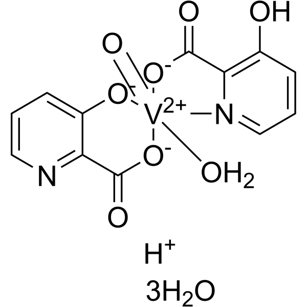
HY-13074
VO-Ohpic trihydrate -
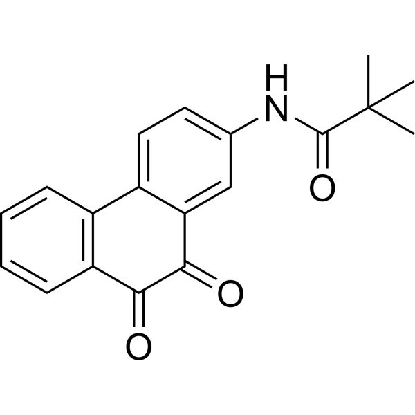
HY-15842
SF1670PTEN Inhibitor -
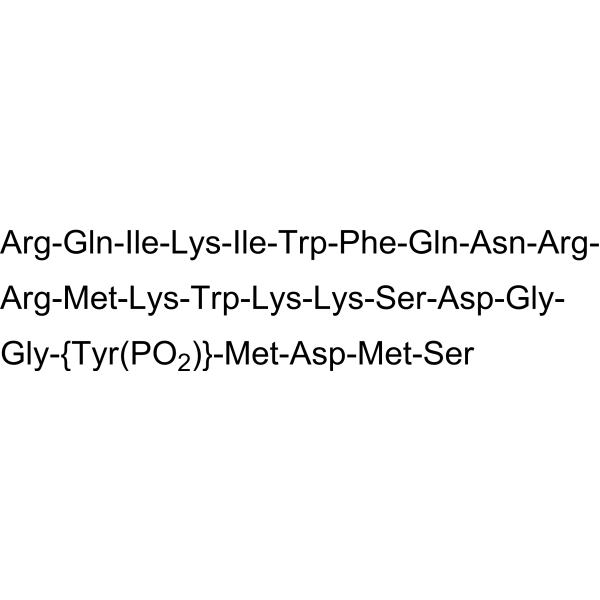
HY-P0175
740 Y-PPI3K Activator -

HY-18764
BpV(pic) potassium hydratePTEN Inhibitor -
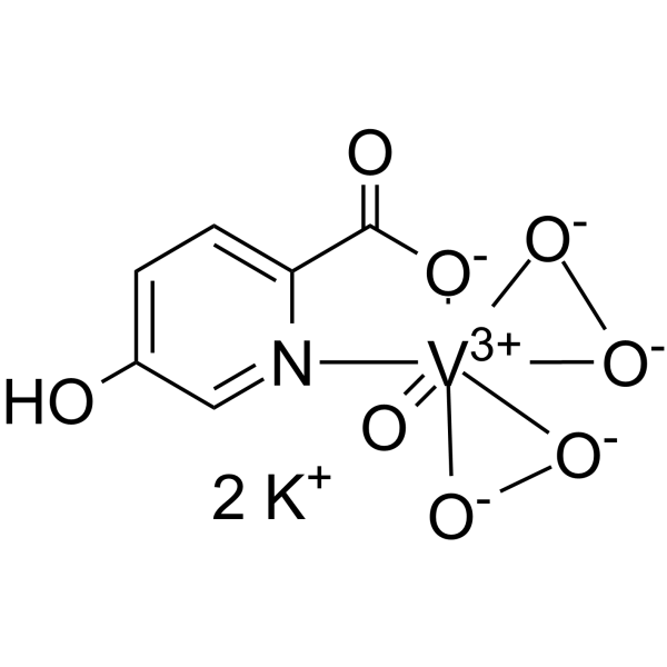
HY-128693
BpV(HOpic)PTEN Inhibitor -
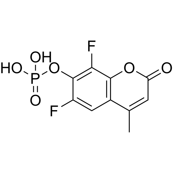
HY-120166
DiFMUPFluorogenic Substrate -
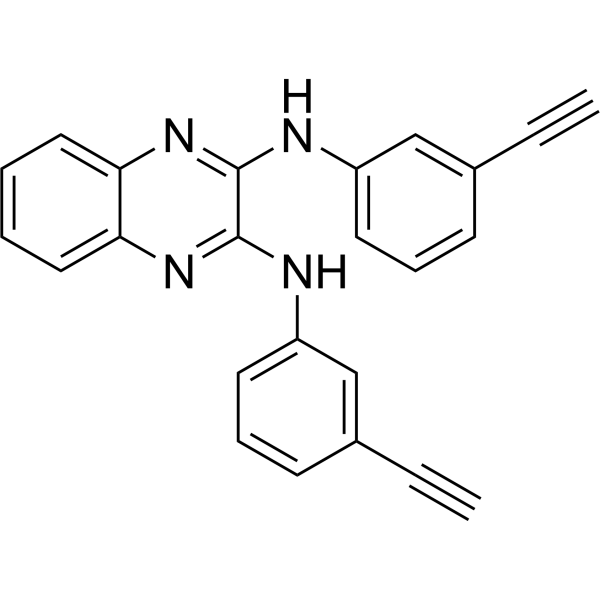
HY-135699
TD52CIP2A Inhibitor -
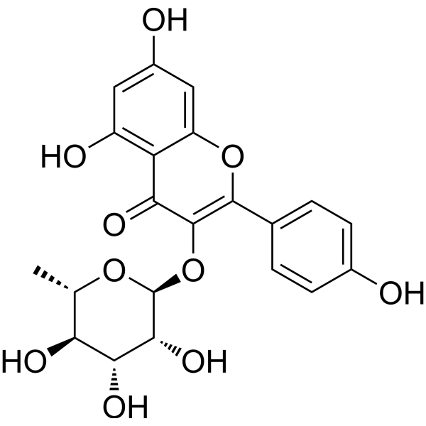
HY-N1441
AfzelinFlavonol Glycoside -
HY-N2116
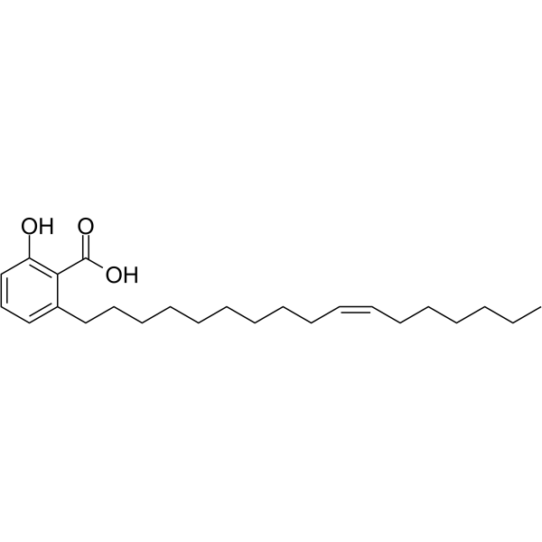
Ginkgolic acid C17:1Antitumor Agent -
HY-136065
bpV(phen)PTP/PTEN Inhibitor -
HY-160093
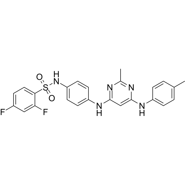
SQLE-IN-1SQLE Inhibitor -
HY-B0849
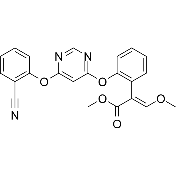
AzoxystrobinFungicide
Publications Citing Use of MedChemExpress (MCE) BpV(phen) trihydrate
MoreAll Parasite Isoforms
More

Biological Activity
Description
IC50 & Target
|
Leishmania |
In Vitro
BpV(phen) (5 μM; 24.5 hours; H9c2 cells) treatment causes a further decrease of cell viability in H/R-injured H9c2 cells[1].
BpV(phen) (5 μM; 24.5 hours; H9c2 cells) treatment increases the apoptosis of H/R-injured H9c2 cells[1].
BpV(phen) (5 μM; 24.5 hours; H9c2 cells) treatment significantly promotes the accumulation of cytoplasmic Cytochrome C in H/R-injured H9c2 cells[1].
After stimulation of BpV(phen), PTEN-induced putative kinase protein 1 (PINK1)/Parkin-mediated mitophagy is inhibited[1].
BpV(phen) is an insulin-mimetic agent following insulin-receptor tyrosine kinase hyperphosphorylation and activation[4].
MedChemExpress (MCE) has not independently confirmed the accuracy of these methods. They are for reference only. Further protocols information, click here.
-
Cell Line:Hypoxia/reoxygenation (H/R)-injured H9c2 cells
-
Concentration:5 μM
-
Incubation Time:24.5 hours (hypoxia for 24 h; reoxygenation for 30 minutes)
-
Result:Caused a further decrease of cell viability.
-
Cell Line:Hypoxia/reoxygenation (H/R)-injured H9c2 cells
-
Concentration:5 μM
-
Incubation Time:24.5 hours (hypoxia for 24 h; reoxygenation for 30 minutes)
-
Result:Increased the apoptosis of H/R-injured H9c2 cells.
-
Cell Line:Hypoxia/reoxygenation (H/R)-injured H9c2 cells
-
Concentration:5 μM
-
Incubation Time:24.5 hours (hypoxia for 24 h; reoxygenation for 30 minutes)
-
Result:Showed an increased release of Cytochrome C.
In Vivo
MedChemExpress (MCE) has not independently confirmed the accuracy of these methods. They are for reference only.
-
Animal Model:Male BALB/c nude (nu/nu) athymic mice (6-7 weeks old) injected with PC-3 cells[2]
-
Dosage:5 mg/kg
-
Administration:Intraperitoneal injection; daily; for 38 days
-
Result:Caused a significant reduction in average tumor volume.


Chemical Information
-
CAS No. 171202-16-7
-
Appearance Solid
-
Molecular Weight 404.29
-
Formula C12H14KN2O8V
-
Color Yellow to orange
-
SMILES
O=[V+3]12([N]3=CC=C4)([O-][O-]1)([O-][O-]2)[N]5=C6C3=C4C=CC6=CC=C5.O.O.O.[K+]
-
Synonyms
Bisperoxovanadium(phen) trihydrate
-
Shipping
Room temperature in continental US; may vary elsewhere.
-
Storage
4°C, sealed storage, away from moisture
* In solvent : -80°C, 6 months; -20°C, 1 month (sealed storage, away from moisture)


Publications (3)
-
Journal Impact Factor
-
Most Recent
-
Nat Commun
The phosphatase PTEN links platelets with immune regulatory functions of mouse T follicular helper cells. [Abstract]2022 May 19;13(1):2762. PMID: 35589797 -
Biochem Biophys Res Commun
2020 Sep 3;529(4):1045-1052. PMID: 32819563 -
Am J Mens Health
The Guangsi Yulin Decoction Inhibits Oxidative Stress and Inflammation-Induced Spermatogenesis Dysfunction via the PTEN/ PI3K/ AKT/ FoxO1 Pathway. [Abstract]2026 Mar-Apr;20(2):15579883261427164. PMID: 41807111


Solvent & Solubility
In Vitro:
H2O : 100 mg/mL (247.35 mM; Need ultrasonic)
Please refer to the solubility information to select the appropriate solvent. Once prepared, please aliquot and store the solution to prevent product inactivation from repeated freeze-thaw cycles.
Storage method and period of stock solution: -80°C, 6 months; -20°C, 1 month (sealed storage, away from moisture). When stored at -80°C, please use it within 6 months. When stored at -20°C, please use it within 1 month.
* Note: If you choose water as the stock solution, please dilute it to the working solution, then filter and sterilize it with a 0.22 μm filter before use.
Please refer to the solubility information to select the appropriate solvent. Once prepared, please aliquot and store the solution to prevent product inactivation from repeated freeze-thaw cycles.
Storage method and period of stock solution: -80°C, 6 months; -20°C, 1 month (sealed storage, away from moisture). When stored at -80°C, please use it within 6 months. When stored at -20°C, please use it within 1 month.
* Note: If you choose water as the stock solution, please dilute it to the working solution, then filter and sterilize it with a 0.22 μm filter before use.
Concentration (start) × Volume (start) = Concentration (final) × Volume (final)
In Vivo:
For the following dissolution methods, please prepare the working solution directly:
It is recommended to prepare fresh solutions and use them promptly within a short period of time.
The percentages shown for the solvents indicate their volumetric ratio in the final prepared solution. If precipitation or phase separation occurs during preparation, heat and/or sonication can be used to aid dissolution.
Add each solvent one by one: PBS
Solubility: 25 mg/mL (61.84 mM); Clear solution; Need ultrasonic and warming

Protocols
-
Apoptosis
Apoptosis, also called programmed cell death, is generally characterized by distinct morphological characteristics.
-
TUNEL staining for apoptotic DNA fragmentation
TUNEL staining detects DNA strand breaks by using terminal deoxynucleotidyl transferase to add labeled nucleotides to exposed 3′-OH DNA termini, generating either microscopic staining in fixed cells or tissue sections, or fluorescence/cytometric signal in cell suspensions. TUNEL positivity reflects DNA fragmentation but should not be interpreted alone as definitive apoptosis, because TUNEL can also label necrotic, autolytic, mechanically damaged, or DNA-repair-associated DNA breaks.
-
LPS-Induced Endotoxemia/Systemic Inflammation
Lipopolysaccharide (LPS)-induced endotoxemia is a widely used in vivo model of acute systemic inflammation in which LPS, a Gram-negative bacterial endotoxin, activates innate immune signaling primarily through TLR4, leading to rapid and transient induction of pro-inflammatory cytokines such as TNF-α, IL-6, and IL-1β in circulation and tissues. This cytokine surge is commonly used as a measurable readout of systemic inflammatory activation and immune dysregulation, and is typically assessed within hours after intraperitoneal LPS administration in mouse models of endotoxemia. The model captures key features of systemic inflammatory response syndrome, including cytokine release, immune cell activation, and downstream tissue responses, and has been used to evaluate anti-inflammatory interventions such as cytokine modulation, lipid mediators, and immune cell-targeting therapies.
-
Naïve CD4+ T-cell subset differentiation/polarization
Naïve CD4+ T-cell subset differentiation/polarization is an in vitro assay in which purified naïve CD4+ T cells are activated through TCR and CD28 costimulation and cultured with defined cytokines and neutralizing antibodies to generate Th0, Th1, Th2, Th17, or induced Treg-like populations. Differentiation is detected by subset-associated cytokines and transcription factors: IFN-γ/T-bet for Th1, IL-4/GATA3 for Th2, IL-17A/RORγt for Th17, and Foxp3 for induced Treg cells. The assay readout is usually generated by intracellular cytokine staining after restimulation, transcription-factor staining by flow cytometry, ELISA of secreted cytokines, or gene-expression analysis. The result reflects cytokine-directed lineage commitment or polarization rather than antigen-specific immune protection by itself.
-
Annexin V plus membrane-impermeant dye apoptosis staining
Annexin V-based apoptosis assays rely on the detection of phosphatidylserine (PS) externalization from the inner leaflet of the plasma membrane to the outer leaflet, an early biochemical hallmark of apoptosis. Fluorescently labeled Annexin V binds PS in a calcium-dependent manner, enabling identification of early apoptotic cells by flow cytometry or fluorescence microscopy. When combined with a membrane-impermeant DNA-binding dye (e. g. , propidium iodide), this approach allows discrimination between viable (Annexin V−/dye−), early apoptotic (Annexin V+/dye−), and late apoptotic or necrotic (Annexin V+/dye+) cell populations by assessing membrane integrity and PS exposure.
-
Research Protocol for Endocrine Diseases
Endocrine diseases often arise from disrupted hormone production, hormone signaling, or target-tissue responsiveness; for diabetes-focused endocrine disease models, insulin signaling regulates glucose uptake, hepatic glucose output, lipid metabolism, and β-cell compensation. Type 2 diabetes develops through interacting defects in insulin resistance, β-cell dysfunction, adipose inflammation, hepatic glucose overproduction, altered incretin signaling, and ectopic lipid metabolism. A major unresolved question is whether endocrine dysfunction is driven primarily by target-tissue insulin resistance, intrinsic β-cell failure, immune/inflammatory stress, or combined multi-organ failure that differs by disease stage.
-
Apoptosis Solutions
Apoptosis is a regulated, generally non-lytic cell-death pathway that removes unwanted, damaged, infected, or abnormal cells through coordinated morphological changes, caspase activation, DNA fragmentation, and membrane remodeling. The intrinsic apoptosis pathway is controlled mainly by mitochondrial outer membrane permeabilization, BCL-2 family proteins, cytochrome c release, apoptosome formation, caspase-9 activation, and downstream executioner caspase-3/7 activation. The extrinsic apoptosis pathway is initiated by death receptors such as Fas, TNFR, and TRAIL receptors, which recruit adaptor proteins and activate caspase-8 before engaging executioner caspases or mitochondrial amplification through BID cleavage. Apoptosis is linked to many phenotypes, including cancer cell killing, tissue homeostasis, immune regulation, neurodegeneration, infection response, and treatment-induced cytotoxicity; unresolved questions include how apoptosis interacts with necroptosis, pyroptosis, ferroptos
-
Research Protocol for Inflammation-related Diseases
The NLRP3 inflammasome is a cytosolic innate immune signaling platform that integrates priming signals and danger-signal activation to promote caspase-1 activation, maturation of IL-1β and IL-18, and gasdermin D-mediated pyroptotic cell death. The core experimental logic is to determine whether inflammatory disease phenotypes are driven by increased NLRP3 expression, ASC-containing inflammasome assembly, caspase-1 cleavage, GSDMD cleavage, and extracellular release of IL-1β/IL-18 rather than by nonspecific cell injury alone. The pathway is strongly linked to inflammation-related disease phenotypes because monosodium urate crystals activate NALP3/NLRP3 inflammasome signaling in gout-like crystal inflammation, cholesterol crystals activate NLRP3 inflammasomes in atherogenesis models, and DSS-induced intestinal inflammation has been reported to involve NLRP3 inflammasome activity. However, experimental colitis studies also show context-dependent protective effects of NLRP3 inflammasome co
-
Cell Viability Determination by MTT Colorimetric Assay
The following protocol uses the MTT colorimetric assay as a classic literature-established method for assessing cell viability/metabolic activity in cultured mammalian cells. MTT[3-(4,5-dimethylthiazol-2-yl)-2,5-diphenyltetrazolium bromide] is reduced by metabolically active cells to a colored formazan product; the amount of formazan is quantified spectrophotometrically and provides an indirect measure of metabolically active viable cells. Importantly, MTT reduction reflects cellular oxidoreductase/metabolic activity rather than an absolute direct count of living cells, so changes in cellular metabolism can alter the signal independently of cell number.


Purity & Documentation
-
Data Sheet (298 KB)
-
SDS (252 KB)
- English - EN (252 KB)
- Français - FR (252 KB)
- Deutsch - DE (252 KB)
- Norwegian - NO (252 KB)
- Español - ES (252 KB)
- Swedish - SV (252 KB)
- Italian - IT (252 KB)
- Korean - KR (252 KB)
- Portuguese - PT (252 KB)
-
Handling Instructions (2659 KB)
References
[1]. Tang W, et al. PTEN-mediated mitophagy and APE1 overexpression protects against cardiac hypoxia/reoxygenation injury. In Vitro Cell Dev Biol Anim. 2019 Oct;55(9):741-748. [Content Brief]
[2]. Caron D, et al. Protein tyrosine phosphatase inhibition induces anti-tumor activity: evidence of Cdk2/p27 kip1 and Cdk2/SHP-1 complex formation in human ovarian cancer cells. Cancer Lett. 2008 Apr 18;262(2):265-75. [Content Brief]
[3]. Schmid AC, et al. Bisperoxovanadium compounds are potent PTEN inhibitors. FEBS Lett. 2004 May 21;566(1-3):35-8. [Content Brief]
[4]. Band CJ, et al. Early signaling events triggered by peroxovanadium [bpV(phen)] are insulin receptor kinase (IRK)-dependent: specificity of inhibition of IRK-associated protein tyrosine phosphatase(s) by bpV(phen). Mol Endocrinol. 1997 Dec;11(13):1899-910. [Content Brief]
[5]. Chen Q, et al. Potassium Bisperoxo(1,10-phenanthroline)oxovanadate (bpV(phen)) Induces Apoptosis and Pyroptosis and Disrupts the P62-HDAC6 Protein Interaction to Suppress the Acetylated Microtubule-dependent Degradation of Autophagosomes. J Biol Chem. 2015 Oct 23;290(43):26051-8. [Content Brief]


Complete Stock Solution Preparation Table
Please refer to the solubility information to select the appropriate solvent. Once prepared, please aliquot and store the solution to prevent product inactivation from repeated freeze-thaw cycles.
Storage method and period of stock solution: -80°C, 6 months; -20°C, 1 month (sealed storage, away from moisture). When stored at -80°C, please use it within 6 months. When stored at -20°C, please use it within 1 month.
| Optional Solvent | Concentration Solvent Mass | 1 mg | 5 mg | 10 mg | 25 mg |
|---|---|---|---|---|---|
| H2O | 1 mM | 2.4735 mL | 12.3674 mL | 24.7347 mL | 61.8368 mL |
| 5 mM | 0.4947 mL | 2.4735 mL | 4.9469 mL | 12.3674 mL | |
| 10 mM | 0.2473 mL | 1.2367 mL | 2.4735 mL | 6.1837 mL | |
| 15 mM | 0.1649 mL | 0.8245 mL | 1.6490 mL | 4.1225 mL | |
| 20 mM | 0.1237 mL | 0.6184 mL | 1.2367 mL | 3.0918 mL | |
| 25 mM | 0.0989 mL | 0.4947 mL | 0.9894 mL | 2.4735 mL | |
| 30 mM | 0.0824 mL | 0.4122 mL | 0.8245 mL | 2.0612 mL | |
| 40 mM | 0.0618 mL | 0.3092 mL | 0.6184 mL | 1.5459 mL | |
| 50 mM | 0.0495 mL | 0.2473 mL | 0.4947 mL | 1.2367 mL | |
| 60 mM | 0.0412 mL | 0.2061 mL | 0.4122 mL | 1.0306 mL | |
| 80 mM | 0.0309 mL | 0.1546 mL | 0.3092 mL | 0.7730 mL | |
| 100 mM | 0.0247 mL | 0.1237 mL | 0.2473 mL | 0.6184 mL |
* Note: If you choose water as the stock solution, please dilute it to the working solution, then filter and sterilize it with a 0.22 μm filter before use.


BpV(phen) trihydrate Related Classifications
HY-122818 Related Classifications
 Powered by Bioz
Keywords
- BpV(phen)
- 171202-16-7
- Bisperoxovanadium(phen)
- PTEN
- Phosphatase
- Parasite
- Apoptosis
- PTP
- Cytochrome C
- mitochondria
- hypoxia
- reoxygenation
- PINK1
- Peroxovanadiums
- anti-tumor
- anti-proliferative Hypoxia/reoxygenation (H/R)-injured H9c2 cells[1] 5 μM 24.5 hours (hypoxia for 24 h
- reoxygenation for 30 minutes) Increased the apoptosis of H/R-injured H9c2 cells. Hypoxia/reoxygenation (H/R)-injured H9c2 cells[1] 5 μM 24.5 hours (hypoxia for 24 h
- reoxygenation for 30 minutes) Showed an increased release of Cytochrome C.
- Inhibitor
- inhibitor
- inhibit