



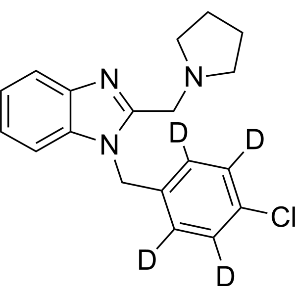
Clemizole-d4

Based on 1 Customer Validation
Clemizole-d4 is the deuterated-labeled Clemizole (HY-30234). Clemizole is an orally active, blood-brain barrier permeable TRPC5 inhibitor, with an IC50 value of 1.05-1.34 μM against mouse TRPC5. Clemizole blocks TRPC1:TRPC5, TRPC3, TRPC4, TRPC6, TRPC7, hERG, hKCNQ1/hKCNE1 and hKv1.5 channels, and activates TRPA1; it modulates 5HT-2B and HTR2A receptors; it inhibits HCV RNA replication, CrtN enzymatic activity, oxidative stress, neuroinflammation, cell apoptosis and bacterial virulence; it maintains blood-brain barrier (BBB) integrity; it enhances DNA repair capacity; it improves cell viability; and it alters cardiac electrophysiological properties. Clemizole can be used in the research of Dravet syndrome, hepatitis C virus infection, Staphylococcus aureus skin infection, Cisplatin (HY-17394)-induced nephrotoxicity, STXBP1-related diseases, traumatic brain injury and xeroderma pigmentosum type C.
For research use only. We do not sell to patients.
- Purity : 99.82%
- CAS No.: 1251554-64-9
- Formula: C19H16D4ClN3
- Molecular Weight:329.86
-
Storage:Powder -20°C, 3 years , 4°C, 2 years ; In solvent -80°C, 6 months , -20°C, 1 month

To place orders, for customer services and technical support, please contact: MedChemExpress USA
Tel: 609-228-6898 E-mail: [email protected] [email protected]


-
 Biological Activity
-
Chemical Information
-
Solvent & Solubility
- Protocols
- Purity & Documentation
- References
-
 Help & FAQs




Biological Activity
Description
IC50 & Target
|
TRPC5 1.05-1.34 μM (IC50) |
Application
1. This compound can be used as a tracer
2. This compound can be used as an internal standard for quantitative analysis by NMR, GC-MS, or LC-MS.


Chemical Information
-
CAS No. 1251554-64-9
-
Unlabeled CAS 442-52-4
-
Appearance Solid
-
Molecular Weight 329.86
-
Formula C19H16D4ClN3
-
Color White to off-white
-
SMILES
ClC1=C([2H])C([2H])=C(C([2H])=C1[2H])CN2C(CN3CCCC3)=NC4=CC=CC=C24
-
Shipping
Room temperature in continental US; may vary elsewhere.
-
Storage
Powder -20°C 3 years 4°C 2 years In solvent -80°C 6 months -20°C 1 month


Solvent & Solubility
In Vitro:
DMSO : 100 mg/mL (303.16 mM; ultrasonic and warming and heat to 80°C; Hygroscopic DMSO has a significant impact on the solubility of product, please use newly opened DMSO)
Please refer to the solubility information to select the appropriate solvent. Once prepared, please aliquot and store the solution to prevent product inactivation from repeated freeze-thaw cycles.
Storage method and period of stock solution: -80°C, 6 months; -20°C, 1 month. When stored at -80°C, please use it within 6 months. When stored at -20°C, please use it within 1 month.
Please refer to the solubility information to select the appropriate solvent. Once prepared, please aliquot and store the solution to prevent product inactivation from repeated freeze-thaw cycles.
Storage method and period of stock solution: -80°C, 6 months; -20°C, 1 month. When stored at -80°C, please use it within 6 months. When stored at -20°C, please use it within 1 month.
Concentration (start) × Volume (start) = Concentration (final) × Volume (final)

Protocols
-
Research Protocol for Infectious Diseases
Infectious-disease experiments test how pathogens interact with host barriers, innate immune receptors, inflammatory signaling, pathogen replication, and tissue injury; pattern-recognition receptors such as TLRs, RIG-I-like receptors, NOD-like receptors, and inflammasomes detect microbial molecules and activate NF-κB, interferon, and cytokine responses. The central hypothesis is that infection severity reflects the balance between pathogen burden and host response: protective inflammation restricts pathogen growth, whereas excessive or mislocalized inflammation contributes to tissue damage and disease phenotype. Unresolved questions include which host pathways are protective versus pathogenic, why some infection models fail to translate to human disease, and which combined readouts best predict clinically relevant infection outcomes.
-
Nephrotoxicity Study
This protocol assesses nephrotoxicity by combining functional kidney injury readouts, urinary/tissue injury biomarkers, and renal histopathology. Serum creatinine and BUN reflect impaired kidney function, while KIM-1, NGAL, clusterin, osteopontin, IL-18, cystatin C, nephrin, Oat5, urinary protein, glucose, and alkaline phosphatase have been used to detect tubular injury in cisplatin-, gentamicin-, and acetaminophen-induced nephrotoxicity models.
-
Mammalian live/dead viability and cytotoxicity staining
Live/dead viability and cytotoxicity staining assays are based on the simultaneous detection of intracellular esterase activity in metabolically active (viable) cells and membrane integrity loss in non-viable cells. In commonly used dual-staining approaches, membrane-permeant fluorogenic substrates are converted by intracellular esterases into fluorescent products in live cells, while impermeant DNA-binding dyes selectively enter cells with compromised plasma membranes and label nucleic acids in dead or dying cells, enabling discrimination between viable and non-viable populations by fluorescence microscopy or flow cytometry.
-
Annexin V plus membrane-impermeant dye apoptosis staining
Annexin V-based apoptosis assays rely on the detection of phosphatidylserine (PS) externalization from the inner leaflet of the plasma membrane to the outer leaflet, an early biochemical hallmark of apoptosis. Fluorescently labeled Annexin V binds PS in a calcium-dependent manner, enabling identification of early apoptotic cells by flow cytometry or fluorescence microscopy. When combined with a membrane-impermeant DNA-binding dye (e. g. , propidium iodide), this approach allows discrimination between viable (Annexin V−/dye−), early apoptotic (Annexin V+/dye−), and late apoptotic or necrotic (Annexin V+/dye+) cell populations by assessing membrane integrity and PS exposure.
-
ROS/oxidative-stress fluorescent staining
ROS/oxidative-stress fluorescent staining uses cell-permeant fluorogenic probes that become fluorescent after oxidation inside cells or tissues; commonly used examples include DCFH-DA/DCFDA for broad cellular oxidant detection, DHE for superoxide-related signal detection, MitoSOX for mitochondrial superoxide-related signal detection, and CellROX probes for oxidative-stress-associated fluorescence readouts. The assay detects probe oxidation rather than a single ROS species unless the probe and analysis method have been chemically validated for that species. DCFH-DA enters cells, is deacetylated by intracellular esterases to DCFH, and produces fluorescent DCF after oxidation, so the readout is used as an operational measure of total cellular oxidative stress rather than a species-specific ROS measurement. DHE and MitoSOX can report superoxide-related oxidation, but red fluorescence alone can include non-specific ethidium-like oxidation products; HPLC or optimized spectral approaches are
-
CCK-8/WST-8 Cell Proliferation Assay
The CCK-8/WST-8 assay is based on the reduction of the water-soluble tetrazolium salt WST-8 to a water-soluble formazan product by cellular dehydrogenases in metabolically active cells, where the generated formazan amount is proportional to the number of living cells and is quantified by measuring absorbance in the visible range, providing a colorimetric readout for cell viability and proliferation assessment. This class of tetrazolium-based assays improves upon earlier MTT-based systems by producing a water-soluble formazan, eliminating the need for organic solubilization steps and enabling direct spectrophotometric measurement in culture medium.
-
Research Protocol for Neurological Diseases
PINK1/Parkin-mediated mitophagy pathway is a mitochondrial quality-control signaling axis in which mitochondrial depolarization stabilizes PINK1 on damaged mitochondria, activates Parkin recruitment and E3 ubiquitin ligase activity, promotes ubiquitination of outer mitochondrial membrane proteins, recruits selective autophagy adaptors, and drives lysosomal degradation of damaged mitochondria. In neurological disease research, this pathway is experimentally important because neurons, especially dopaminergic neurons, are highly dependent on mitochondrial integrity, and defective mitochondrial turnover can lead to mitochondrial dysfunction, oxidative stress, impaired neuronal survival, α-synuclein accumulation, and neuroinflammatory damage-associated signals. The genetic disease link is strongest in Parkinson’s disease because mutations in PRKN/parkin cause autosomal recessive juvenile parkinsonism, mutations in PINK1 cause hereditary early-onset Parkinson’s disease, and Drosophila studie
-
Cell Viability Determination by MTT Colorimetric Assay
The following protocol uses the MTT colorimetric assay as a classic literature-established method for assessing cell viability/metabolic activity in cultured mammalian cells. MTT[3-(4,5-dimethylthiazol-2-yl)-2,5-diphenyltetrazolium bromide] is reduced by metabolically active cells to a colored formazan product; the amount of formazan is quantified spectrophotometrically and provides an indirect measure of metabolically active viable cells. Importantly, MTT reduction reflects cellular oxidoreductase/metabolic activity rather than an absolute direct count of living cells, so changes in cellular metabolism can alter the signal independently of cell number.
-
Apoptosis
Apoptosis, also called programmed cell death, is generally characterized by distinct morphological characteristics.
-
TUNEL staining for apoptotic DNA fragmentation
TUNEL staining detects DNA strand breaks by using terminal deoxynucleotidyl transferase to add labeled nucleotides to exposed 3′-OH DNA termini, generating either microscopic staining in fixed cells or tissue sections, or fluorescence/cytometric signal in cell suspensions. TUNEL positivity reflects DNA fragmentation but should not be interpreted alone as definitive apoptosis, because TUNEL can also label necrotic, autolytic, mechanically damaged, or DNA-repair-associated DNA breaks.
-
Cell Cytotoxicity Assay
Cytotoxicity assays are usually based on the assessment of cell membrane damage, which can also be indirectly detected by measuring cell viability. Detection methods include MTT assay, CKK-8 assay, LDH assay and ATP assay, etc.
-
Apoptosis Solutions
Apoptosis is a regulated, generally non-lytic cell-death pathway that removes unwanted, damaged, infected, or abnormal cells through coordinated morphological changes, caspase activation, DNA fragmentation, and membrane remodeling. The intrinsic apoptosis pathway is controlled mainly by mitochondrial outer membrane permeabilization, BCL-2 family proteins, cytochrome c release, apoptosome formation, caspase-9 activation, and downstream executioner caspase-3/7 activation. The extrinsic apoptosis pathway is initiated by death receptors such as Fas, TNFR, and TRAIL receptors, which recruit adaptor proteins and activate caspase-8 before engaging executioner caspases or mitochondrial amplification through BID cleavage. Apoptosis is linked to many phenotypes, including cancer cell killing, tissue homeostasis, immune regulation, neurodegeneration, infection response, and treatment-induced cytotoxicity; unresolved questions include how apoptosis interacts with necroptosis, pyroptosis, ferroptos
-
MTT Cell Proliferation Assay
The MTT assay is a colorimetric endpoint assay for estimating viable cell number, cell growth, cytotoxicity, or cell activation in cultured mammalian cells. Living cells reduce the yellow tetrazolium salt MTT into purple/blue formazan, while dead cells do not generate the same signal; the resulting color can be quantified with a multiwell spectrophotometer. MTT reduction is commonly interpreted as a readout of metabolic activity that often correlates with viable cell number, but it should not be treated as a direct cell-counting method unless the assay is optimized for the cell type and experimental condition. Studies show that MTT reduction can involve mitochondrial and non-mitochondrial reducing systems, and formazan may accumulate in intracellular lipid droplets rather than simply marking mitochondria.


Purity & Documentation
-
Data Sheet (282 KB)
-
SDS (251 KB)
- English - EN (251 KB)
- Français - FR (251 KB)
- Deutsch - DE (251 KB)
- Norwegian - NO (251 KB)
- Español - ES (251 KB)
- Swedish - SV (251 KB)
- Italian - IT (251 KB)
- Korean - KR (251 KB)
- Portuguese - PT (251 KB)
-
Handling Instructions (2659 KB)
References
[1]. Richter JM, et al. Clemizole hydrochloride is a novel and potent inhibitor of transient receptor potential channel TRPC5. Molecular pharmacology. 2014 Nov;86(5):514-21. [Content Brief]
[2]. Baraban SC, et al. Drug screening in Scn1a zebrafish mutant identifies clemizole as a potential Dravet syndrome treatment. Nature communications. 2013;4:2410. [Content Brief]
[3]. Jie LJ, et al. Clemizole hydrochloride blocks cardiac potassium currents stably expressed in HEK 293 cells. British journal of pharmacology. 2017 Feb;174(3):254-266. [Content Brief]
[4]. Einav S, et al. Discovery of a hepatitis C target and its pharmacological inhibitors by microfluidic affinity analysis. Nature biotechnology. 2008 Sep;26(9):1019-27. [Content Brief]
[5]. Yu H, et al. Clemizole inhibits CrtN-driven staphyloxanthin biosynthesis in Staphylococcus aureus to enhance host immune clearance. Communications biology. 2026 Feb 25;9(1):484. [Content Brief]
[6]. Kumaş-Kulualp M, et al. Clemizole hydrochloride, a potent TRPC5 calcium channel inhibitor, prevents cisplatin-induced nephrotoxicity in Spraque-Dawley rats. Journal of biochemical and molecular toxicology. 2023 Jul;37(7):e23372. [Content Brief]
[7]. Moog M, et al. Clemizole and trazodone are effective antiseizure treatments in a zebrafish model of STXBP1 disorder. Epilepsia open. 2022 Sep;7(3):504-511. [Content Brief]
[8]. Chauhan C, et al. Clemizole Mitigates Traumatic Brain Injury by Inhibiting Oxidative Stress, Neuroinflammation, and Apoptosis. ACS chemical neuroscience. 2026 May 06;17(9):1787-1801. [Content Brief]
[9]. Nishimura T, et al. Using chimeric mice with humanized livers to predict human drug metabolism and a drug-drug interaction. The Journal of pharmacology and experimental therapeutics. 2013 Feb;344(2):388-96. [Content Brief]
[10]. Griffin A, et al. Clemizole and modulators of serotonin signalling suppress seizures in Dravet syndrome. Brain : a journal of neurology. 2017 Mar 01;140(3):669-683. [Content Brief]
[11]. Kobaisi F, et al. Isoconazole and Clemizole Hydrochloride Partially Reverse the Xeroderma Pigmentosum C Phenotype. International journal of molecular sciences. 2021 Jul 29;22(15):8156. [Content Brief]


Complete Stock Solution Preparation Table
Please refer to the solubility information to select the appropriate solvent. Once prepared, please aliquot and store the solution to prevent product inactivation from repeated freeze-thaw cycles.
Storage method and period of stock solution: -80°C, 6 months; -20°C, 1 month. When stored at -80°C, please use it within 6 months. When stored at -20°C, please use it within 1 month.
| Optional Solvent | Concentration Solvent Mass | 1 mg | 5 mg | 10 mg | 25 mg |
|---|---|---|---|---|---|
| DMSO | 1 mM | 3.0316 mL | 15.1579 mL | 30.3159 mL | 75.7897 mL |
| 5 mM | 0.6063 mL | 3.0316 mL | 6.0632 mL | 15.1579 mL | |
| 10 mM | 0.3032 mL | 1.5158 mL | 3.0316 mL | 7.5790 mL | |
| 15 mM | 0.2021 mL | 1.0105 mL | 2.0211 mL | 5.0526 mL | |
| 20 mM | 0.1516 mL | 0.7579 mL | 1.5158 mL | 3.7895 mL | |
| 25 mM | 0.1213 mL | 0.6063 mL | 1.2126 mL | 3.0316 mL | |
| 30 mM | 0.1011 mL | 0.5053 mL | 1.0105 mL | 2.5263 mL | |
| 40 mM | 0.0758 mL | 0.3789 mL | 0.7579 mL | 1.8947 mL | |
| 50 mM | 0.0606 mL | 0.3032 mL | 0.6063 mL | 1.5158 mL | |
| 60 mM | 0.0505 mL | 0.2526 mL | 0.5053 mL | 1.2632 mL | |
| 80 mM | 0.0379 mL | 0.1895 mL | 0.3789 mL | 0.9474 mL | |
| 100 mM | 0.0303 mL | 0.1516 mL | 0.3032 mL | 0.7579 mL |


Clemizole-d4 Related Classifications
HY-30234S Related Classifications
 Powered by Bioz