



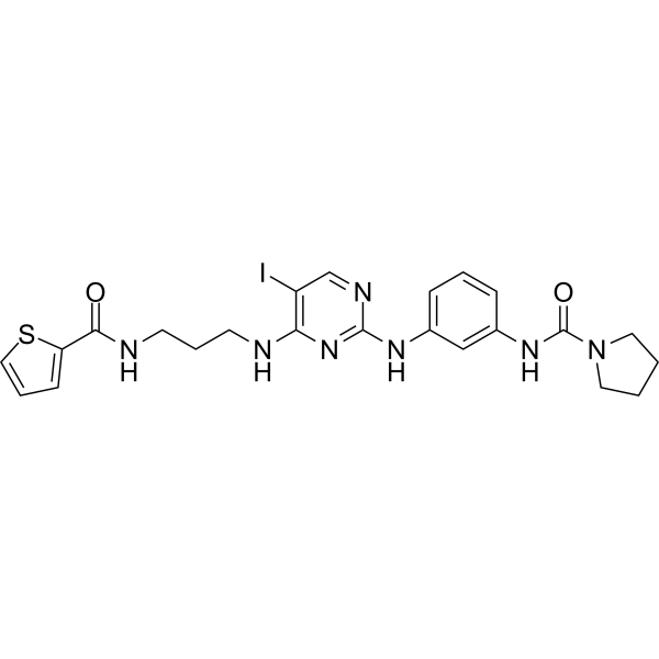
BX795
Based on 42 publication(s) in Google Scholar
BX795 is a potent and selective inhibitor of PDK1, with an IC50 of 6 nM. BX795 is also a potent and relatively specific inhibitor of TBK1 and IKKε, with an IC50 of 6 and 41 nM, respectively. BX795 blocks phosphorylation of S6K1, Akt, PKCδ, and GSK3β, and has lower selectivity over PKA, PKC, c-Kit, GSK3β etc. BX795 modulates autophagy.
Nos produits utilisent uniquement pour la recherche. Nous ne vendons pas aux patients.
- Pureté : 99.84%
- CAS No.: 702675-74-9
- Formule: C23H26IN7O2S
- Masse moléculaire:591.47
-
Stockage:Powder -20°C, 3 years , 4°C, 2 years ; In solvent -80°C, 6 months , -20°C, 1 month

To place orders, for customer services and technical support, please contact: MedChemExpress USA
Tel: 609-228-6898 E-mail: [email protected] [email protected]


-
 Activité biologique
-
Chemical Information
-
Solvant et solubilité
- Protocole
- Pureté et documentation
- Références
-
 Help & FAQs


-
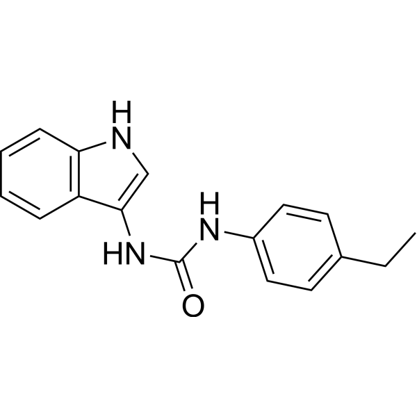
HY-112693
H-151STING Antagonist -
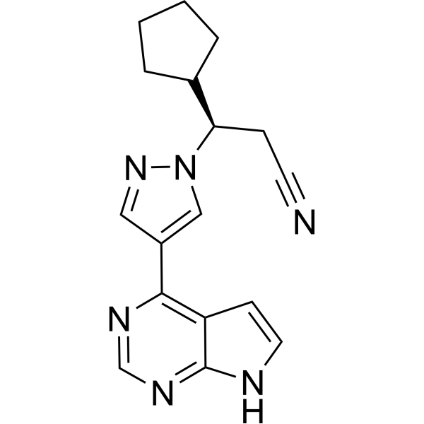
HY-50856
RuxolitinibJAK1/2 Inhibitor -
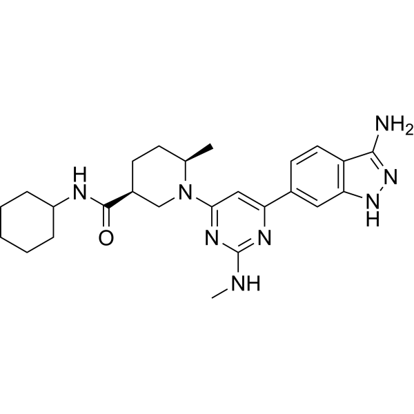
HY-14981
GSK2334470PDK1 Inhibitor -
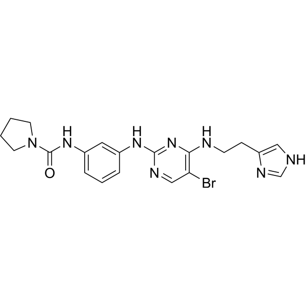
HY-11005
BX-912Apoptosis Inducer -
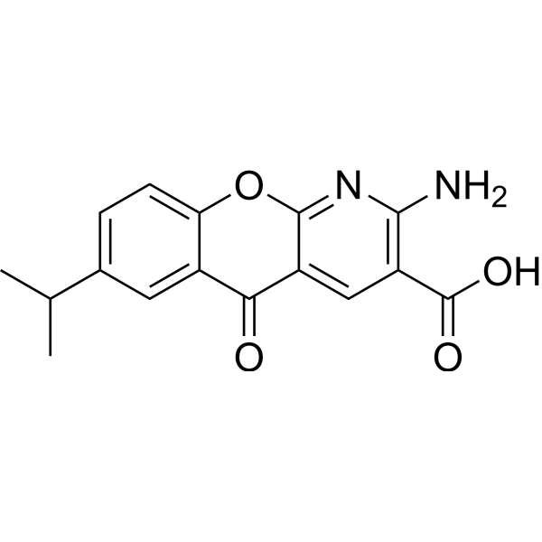
HY-B0713
AmlexanoxIKK Inhibitor -
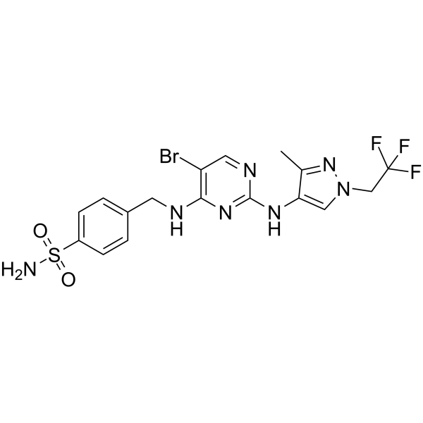
HY-111941
GSK8612TBK1 Inhibitor -
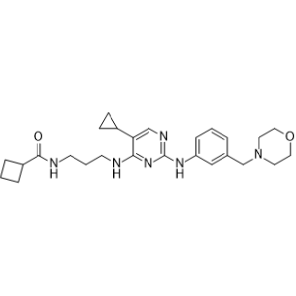
HY-13018
MRT67307IKKε/TBK-1 Inhibitor -

HY-10219
RapamycinmTOR Inhibitor -
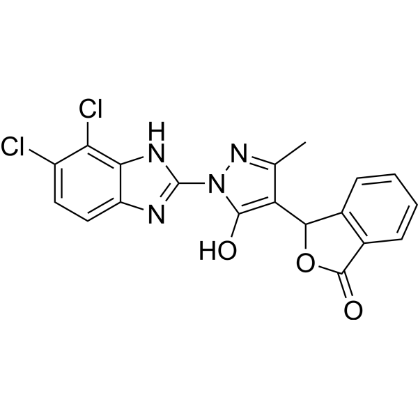
HY-114180
RU.521cGAS Inhibitor -
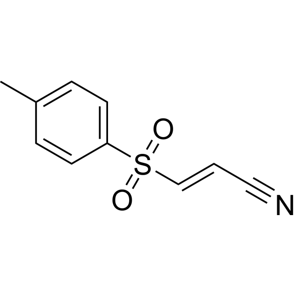
HY-13453
BAY 11-7082IκBα/NF-κB Inhibitor -
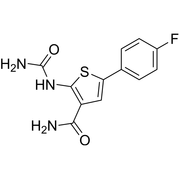
HY-10074
TPCA-1IKK-2 Inhibitor -
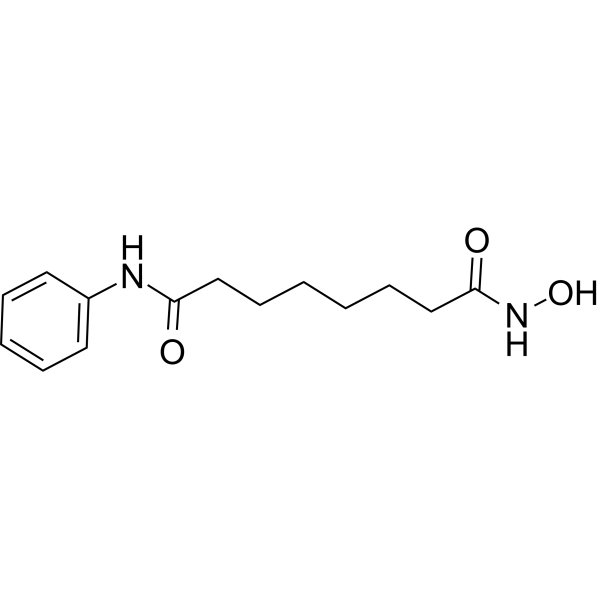
HY-10221
VorinostatHDAC Inhibitor
Publications Citing Use of MedChemExpress (MCE) BX795
More- Nature. 2022 Oct;610(7933):761-767. [Abstract]
- Immunity. 2025 Dec 5:S1074-7613(25)00506-0. [Abstract]
- Autophagy. 2025 Jan;21(1):178-190. [Abstract]
- Autophagy. 2017 Jan 2;13(1):133-148. [Abstract]
- Nat Commun. 2024 Jan 2;15(1):130. [Abstract]
- Mol Cell. 2026 Feb 19;86(4):722-739.e8. [Abstract]
- Mol Cell. 2025 Mar 27:S1097-2765(25)00201-1. [Abstract]
- Adv Sci (Weinh). 2026 Mar;13(13):e15546. [Abstract]
- Cell Mol Biol Lett. 2023 Oct 24;28(1):85. [Abstract]
- Pharmacol Res. 2023 Jan:187:106577. [Abstract]
- J Orthop Translat. 2024 Jun 28:47:207-222. [Abstract]
- Cell Chem Biol. 2022 Jul 21;29(7):1113-1125.e6. [Abstract]
- Food Res Int. 2026 Jul 18;242:120075.
- EMBO J. 2025 Sep 1. [Abstract]
- J Anim Sci Biotechnol. 2022 Jul 4;13(1):75. [Abstract]
- Cell Rep. 2022 Sep 13;40(11):111342. [Abstract]
- mLife. 2025 Jun 18;4(3):249-258. [Abstract]
- Mol Cell Proteomics. 2018 Dec;17(12):2434-2447. [Abstract]
- J Cell Biol. 2021 Feb 1;220(2):e201911025. [Abstract]
- Cancer Immunol Immunother. 2025 Jan 3;74(2):73. [Abstract]
- Transl Oncol. 2022 Jan;15(1):101272. [Abstract]
- Oncol Rep. 2022 Jan;47(1):11. [Abstract]
- iScience. 2023 Jun 10;26(7):107090. [Abstract]
- Poultry Sci. 2023 May;102(5):102597. [Abstract]
- Microbiol Spectr. 2023 Jun 15;11(3):e0070123. [Abstract]
- J Biotechnol. 2025 Mar:399:9-18. [Abstract]
- J Cell Sci. 2018 Nov 30;131(23):jcs226241. [Abstract]
- Vet Res. 2024 Jun 28;55(1):83. [Abstract]
- J Proteome Res. 2021 Aug 6;20(8):4113-4130. [Abstract]
- Viruses. 2021 Jun 28;13(7):1255. [Abstract]
- Appl Biochem Biotechnol. 2024 Nov;196(11):7908-7927. [Abstract]
- Tissue Cell. 2026 Apr 22:102:103550. [Abstract]
- Fitoterapia. 2019 Apr:134:14-22. [Abstract]
- PLoS One. 2025 Jun 16;20(6):e0325700. [Abstract]
- Res Sq. 2026 May 25.
- bioRxiv. 2026 Apr 23.
- bioRxiv. 2026 Mar 12.
- bioRxiv. 2025 Dec 25.
- Res Sq. 2025 Oct 6.
- University of Greifswald. 2025.
- Oxid Med Cell Longev. 2021 Jan 25:2021:8836058. [Abstract]
- Patent. US20200268864A1.
 Customer Validation & Images

-
WB
-
WB
-
WB
-
WB
-
WB


Activité biologique
Description
|
PDK1 6 nM (IC50) |
TBK1 6 nM (IC50) |
IKKε 41 nM (IC50) |
Cellular Effect
|
Cell Line

|
Type | Value | Description | References |
|---|---|---|---|---|
| HeLa | CC50 |
46 μM
Compound: 73; BX795
|
Cytotoxicity against human HeLa cells
Cytotoxicity against human HeLa cells
|
[PMID: 33539089] |
In Vitro
BX795 effectively blocks PDK1 activity in PC-3 cells, as shown by their ability to block phosphorylation of S6K1, Akt, PKCδ, and GSK3β. BX795 potently inhibits tumor cell growth on plastic with IC50 of 1.6, 1.4, and 1.9 μM for MDA-468, HCT-116, and MiaPaca cells, respectively.In soft agar, BX795 displays higher growth inhibition with IC50 of 0.72, and 0.25 μM for MDA-468, and PC-3 cells, respectively[1]. In addition, BX795, as an inhibitor of the TBK1/IKKε, blocks TBK1- and IKKε-mediated activation of IRF3 and production of IFN-β[2]. In platelet physiological responses, BX795 produces inhibitory effect on 2-MeSADP-induced or collagen-induced aggregation, ATP secretion, and thromboxane generation[3].
MedChemExpress (MCE) has not independently confirmed the accuracy of these methods. They are for reference only.


Chemical Information
-
CAS No. 702675-74-9
-
Appearance Solid
-
Masse moléculaire 591.47
-
Formule C23H26IN7O2S
-
Color White to light brown
-
SMILES
O=C(NCCCNC1=C(C=NC(NC2=CC=CC(NC(N3CCCC3)=O)=C2)=N1)I)C4=CC=CS4
-
Livraison
Room temperature in continental US; may vary elsewhere.
-
Stockage
Powder -20°C 3 years 4°C 2 years In solvent -80°C 6 months -20°C 1 month


Publications (42)
-
Journal Impact Factor
-
Most Recent
-
Nature
2022 Oct;610(7933):761-767. PMID: 36261523
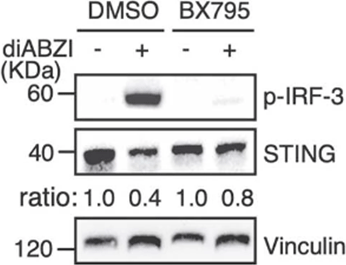
BX795 purchased from MedChemExpress. Usage Cited in: Nature. 2022 Oct;610(7933):761-767. [Abstract]
HeLa cells pretreated with DMSO or 2 µM BX795 for 24 h are stimulated with 2.5 µM diABZI or not and analysed by Western blot.
-
Immunity
A chemical agonist and the Golgi-resident lipid PI4P activate STING by inducing transmembrane helix rearrangement. [Abstract]2025 Dec 5:S1074-7613(25)00506-0. PMID: 41352342 -
Autophagy
TRIM21-mediated ubiquitination of SQSTM1/p62 abolishes its Ser403 phosphorylation and enhances palmitic acid cytotoxicity. [Abstract]2025 Jan;21(1):178-190. PMID: 39172027 -
Autophagy
HSF1 stress response pathway regulates autophagy receptor SQSTM1/p62-associated proteostasis. [Abstract]2017 Jan 2;13(1):133-148. PMID: 27846364
BX795 purchased from MedChemExpress. Usage Cited in: Autophagy. 2017 Jan 2;13(1):133-148. [Abstract]
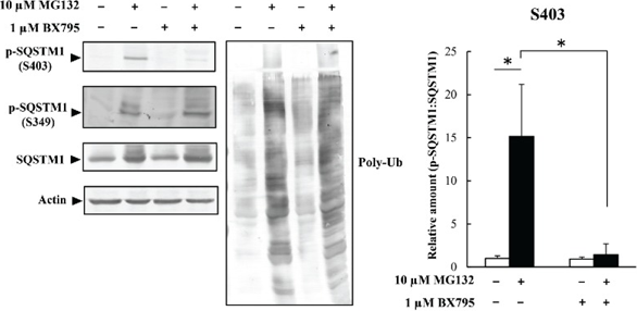
HeLa cells are treated with a BX795 TBK1 inhibitor (1 μM) and MG132 (10 μM) for 12 h. Cell lysates are analyzed by immunoblot analysis. Band intensities are measured, and phosphorylated-SQSTM1 values are normalized to total SQSTM1. The combined MG132/BX795 treatment results in a 90% reduction in S403 phosphorylation but has no effect on S349 phosphorylation.
-
Nat Commun
2024 Jan 2;15(1):130. PMID: 38167258 -
Mol Cell
Asparagine sensing by TBK1 controls its phase separation to drive antiviral innate immune responses. [Abstract]2026 Feb 19;86(4):722-739.e8. PMID: 41653919 -
Mol Cell
STING mediates lysosomal quality control and recovery through its proton channel function and TFEB activation in lysosomal storage disorders. [Abstract]2025 Mar 27:S1097-2765(25)00201-1. PMID: 40185098 -
Adv Sci (Weinh)
EGR Proteins Mediate Interferon-Independent Anti-HSV-1 Responses Through Viral and Host Targets. [Abstract]2026 Mar;13(13):e15546. PMID: 41486724 -
Cell Mol Biol Lett
Inhibition of SQSTM1 S403 phosphorylation facilitates the aggresome formation of ubiquitinated proteins during proteasome dysfunction. [Abstract]2023 Oct 24;28(1):85. PMID: 37872526 -
Pharmacol Res
Fluvoxamine alleviates bleomycin-induced lung fibrosis via regulating the cGAS-STING pathway. [Abstract]2023 Jan:187:106577. PMID: 36435270 -
J Orthop Translat
TBK1 pharmacological inhibition mitigates osteoarthritis through attenuating inflammation and cellular senescence in chondrocytes. [Abstract]2024 Jun 28:47:207-222. PMID: 39040492 -
Cell Chem Biol
Identifying enhancers of innate immune signaling as broad-spectrum antivirals active against emerging viruses. [Abstract]2022 Jul 21;29(7):1113-1125.e6. PMID: 35728599 -
-
EMBO J
2025 Sep 1. PMID: 40890487 -
J Anim Sci Biotechnol
Genome-wide identification of functional enhancers and their potential roles in pig breeding. [Abstract]2022 Jul 4;13(1):75. PMID: 35781353 -
Cell Rep
In vivo G-CSF treatment activates the GR-SOCS1 axis to suppress IFN-γ secretion by natural killer cells. [Abstract]2022 Sep 13;40(11):111342. PMID: 36103837 -
mLife
Potential antiviral effects of the marine probiotic Paraliobacillus zengyii on the respiratory syncytial virus. [Abstract]2025 Jun 18;4(3):249-258. PMID: 40600055 -
Mol Cell Proteomics
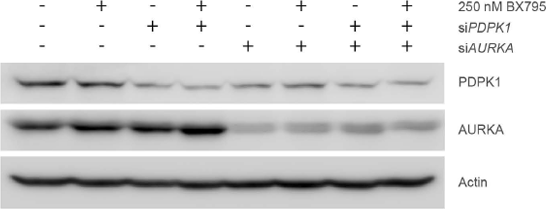
Functional Proteomics and Deep Network Interrogation Reveal a Complex Mechanism of Action of Midostaurin in Lung Cancer Cells. [Abstract]2018 Dec;17(12):2434-2447. PMID: 30217950
BX795 purchased from MedChemExpress. Usage Cited in: Mol Cell Proteomics. 2018 Dec;17(12):2434-2447. [Abstract]
Relative A427 cell counts upon 96 hrs siRNA-mediated knockdown of PDPK1 and/or AURKA and/or 72 hrs treatment with 250 nM BX795.
-
J Cell Biol
2021 Feb 1;220(2):e201911025. PMID: 33404607 -
Cancer Immunol Immunother
Therapeutic potential of anti-ErbB3 chimeric antigen receptor natural killer cells against breast cancer. [Abstract]2025 Jan 3;74(2):73. PMID: 39751931 -
Transl Oncol
2022 Jan;15(1):101272. PMID: 34823094 -
Oncol Rep
PIK3CA mutation affects the proliferation of colorectal cancer cells through the PI3K-MEK/PDK1-GPT2 pathway. [Abstract]2022 Jan;47(1):11. PMID: 34751411 -
iScience
Suppression of TREX1 deficiency-induced cellular senescence and interferonopathies by inhibition of DNA damage response. [Abstract]2023 Jun 10;26(7):107090. PMID: 37416470 -
Poultry Sci
2023 May;102(5):102597. PMID: 36931072 -
Microbiol Spectr
Replication of Porcine Astrovirus Type 1-Infected PK-15 Cells In Vitro Affected by RIG-I and MDA5 Signaling Pathways. [Abstract]2023 Jun 15;11(3):e0070123. PMID: 37140381 -
J Biotechnol
2025 Mar:399:9-18. PMID: 39824361 -
J Cell Sci
Alpha-synuclein fibrils recruit TBK1 and OPTN to lysosomal damage sites and induce autophagy in microglial cells. [Abstract]2018 Nov 30;131(23):jcs226241. PMID: 30404831 -
Vet Res
Duck STING mediates antiviral autophagy directing the interferon signaling pathway to inhibit duck plague virus infection. [Abstract]2024 Jun 28;55(1):83. PMID: 38943190 -
J Proteome Res
Quantitative Proteomics Reveals a Novel Role of the E3 Ubiquitin-Protein Ligase FANCL in the Activation of the Innate Immune Response through Regulation of TBK1 Phosphorylation during Peste des Petits Ruminants Virus Infection. [Abstract]2021 Aug 6;20(8):4113-4130. PMID: 34289691 -
Viruses
Screening and Identification of Lujo Virus Inhibitors Using a Recombinant Reporter Virus Platform. [Abstract]2021 Jun 28;13(7):1255. PMID: 34203149 -
Appl Biochem Biotechnol
Angiopoietin 1 Relieves Osteolysis by Promoting Macrophage Mitophagy Through the TBK1-SQSTM1 Pathway to Inhibit AIM2 Inflammasome-Mediated Pyroptosis. [Abstract]2024 Nov;196(11):7908-7927. PMID: 38662322 -
Tissue Cell
Bi-magnolignan suppresses triple-negative breast cancer by triggering TBK1-dependent mitophagy. [Abstract]2026 Apr 22:102:103550. PMID: 42035609 -
Fitoterapia
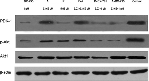
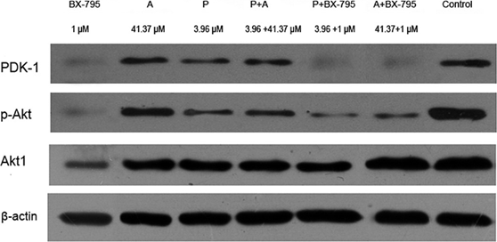
The antagonism between apigenin and protoapigenone to the PDK-1 target in Macrothelypteris torresiana. [Abstract]2019 Apr:134:14-22. PMID: 30731149
BX795 purchased from MedChemExpress. Usage Cited in: Fitoterapia. 2019 Apr:134:14-22. [Abstract]
BT474 cells are exposed to protoapigenone, apigenin and BX-795 using different administration combination and analyzed by Western blot, β-actin was used as internal control.
BX795 purchased from MedChemExpress. Usage Cited in: Fitoterapia. 2019 Apr:134:14-22. [Abstract]
MDA-MB-231 cells are exposed to protoapigenone, apigenin and BX-795 using different administration combination and analyzed by Western blot, β-actin was used as internal control.
-
PLoS One
Drug repositioning for pan-cancers of the digestive system: Identification of amonafide and BX795 as potential therapeutics via integrative Omics analysis. [Abstract]2025 Jun 16;20(6):e0325700. PMID: 40522954 -
-
-
-
-
-
-
Oxid Med Cell Longev
Apelin/APJ-Manipulated CaMKK/AMPK/GSK3 β Signaling Works as an Endogenous Counterinjury Mechanism in Promoting the Vitality of Random-Pattern Skin Flaps. [Abstract]2021 Jan 25:2021:8836058. PMID: 33574981 -


Solvant et solubilité
In Vitro:
DMSO : 33.33 mg/mL (56.35 mM; Need ultrasonic; Hygroscopic DMSO has a significant impact on the solubility of product, please use newly opened DMSO)
Please refer to the solubility information to select the appropriate solvent. Once prepared, please aliquot and store the solution to prevent product inactivation from repeated freeze-thaw cycles.
Storage method and period of stock solution: -80°C, 6 months; -20°C, 1 month. When stored at -80°C, please use it within 6 months. When stored at -20°C, please use it within 1 month.
Please refer to the solubility information to select the appropriate solvent. Once prepared, please aliquot and store the solution to prevent product inactivation from repeated freeze-thaw cycles.
Storage method and period of stock solution: -80°C, 6 months; -20°C, 1 month. When stored at -80°C, please use it within 6 months. When stored at -20°C, please use it within 1 month.
Concentration (start) × Volume (start) = Concentration (final) × Volume (final)
In Vivo:
Select the appropriate dissolution method based on your experimental animal and administration route.
- For the following dissolution methods, please ensure to first prepare a clear stock solution using an In Vitro approach and then sequentially add co-solvents:
- To ensure reliable experimental results, the clarified stock solution can be appropriately stored based on storage conditions. As for the working solution for In Vivo experiments, it is recommended to prepare freshly and use it on the same day.
- The percentages shown for the solvents indicate their volumetric ratio in the final prepared solution. If precipitation or phase separation occurs during preparation, heat and/or sonication can be used to aid dissolution.
Add each solvent one by one: 10% DMSO 40% PEG300 5% Tween-80 45% Saline
Solubility: ≥ 2.5 mg/mL (4.23 mM); Clear solution
This protocol yields a clear solution of ≥ 2.5 mg/mL (saturation unknown).
Taking 1 mL working solution as an example, add 100 μL DMSO stock solution (25.0 mg/mL) to 400 μL PEG300, and mix evenly; then add 50 μL Tween-80 and mix evenly; then add 450 μL Saline to adjust the volume to 1 mL.
Preparation of Saline: Dissolve 0.9 g sodium chloride in ddH₂O and dilute to 100 mL to obtain a clear Saline solution.
In Vivo Dissolution Calculator
Please enter the basic information of animal experiments:
-
-
-
-
Recommended: Prepare an additional quantity of animals to account for potential losses during experiments.
Please enter your animal formula composition:
-
%DMSO +
Recommended: Keep the proportion of DMSO in working solution below 2% if your animal is weak.
-
%+
-
+%Tween-80 + +
-
%Saline +
The co-solvents required include: DMSO, . All of co-solvents are available by MedChemExpress (MCE). , Tween 80. All of co-solvents are available by MedChemExpress (MCE).
Working solution concentration: 0.22 mg/mL
Method for preparing stock solution: mg drug dissolved in μL DMSO. Stock solution concentration: mg/mL.
1. Take μL DMSO stock solution;
2. Add μL .
μL , mix evenly;
3. Then add μL Tween 80, mix evenly;
4. Then add μL
Please ensure that the stock solution in the first step is dissolved to a clear state, and add co-solvents in sequence. You can use ultrasonic heating (ultrasonic cleaner, recommended frequency 20-40 kHz), vortexing, etc. to assist dissolution.


Pureté et documentation
-
Fiche technique (277 KB)
-
SDS (396 KB)
- English - EN (396 KB)
- Français - FR (396 KB)
- Deutsch - DE (396 KB)
- Norwegian - NO (396 KB)
- Español - ES (396 KB)
- Swedish - SV (396 KB)
- Italian - IT (396 KB)
- Korean - KR (396 KB)
- Portuguese - PT (396 KB)
-
Instruction de manipulation (2659 KB)
Références
[1]. Feldman RI, et al. Novel small molecule inhibitors of 3-phosphoinositide-dependent kinase-1. J Biol Chem. 2005 May 20;280(20):19867-74. [Content Brief]
[2]. Clark K, et al. Use of the pharmacological inhibitor BX795 to study the regulation and physiological roles of TBK1 and IkappaB kinase epsilon: a distinct upstream kinase mediates Ser-172 phosphorylation and activation. J Biol Chem. 2009 May 22;284(21):141 [Content Brief]
[3]. Dangelmaier C, et al. PDK1 selectively phosphorylates Thr(308) on Akt and contributes to human platelet functional responses. Thromb Haemost. 2014 Mar 3;111(3):508-17. [Content Brief]


Complete Stock Solution Preparation Table
Please refer to the solubility information to select the appropriate solvent. Once prepared, please aliquot and store the solution to prevent product inactivation from repeated freeze-thaw cycles.
Storage method and period of stock solution: -80°C, 6 months; -20°C, 1 month. When stored at -80°C, please use it within 6 months. When stored at -20°C, please use it within 1 month.
| Optional Solvent | Concentration Solvent Mass | 1 mg | 5 mg | 10 mg | 25 mg |
|---|---|---|---|---|---|
| DMSO | 1 mM | 1.6907 mL | 8.4535 mL | 16.9070 mL | 42.2676 mL |
| 5 mM | 0.3381 mL | 1.6907 mL | 3.3814 mL | 8.4535 mL | |
| 10 mM | 0.1691 mL | 0.8454 mL | 1.6907 mL | 4.2268 mL | |
| 15 mM | 0.1127 mL | 0.5636 mL | 1.1271 mL | 2.8178 mL | |
| 20 mM | 0.0845 mL | 0.4227 mL | 0.8454 mL | 2.1134 mL | |
| 25 mM | 0.0676 mL | 0.3381 mL | 0.6763 mL | 1.6907 mL | |
| 30 mM | 0.0564 mL | 0.2818 mL | 0.5636 mL | 1.4089 mL | |
| 40 mM | 0.0423 mL | 0.2113 mL | 0.4227 mL | 1.0567 mL | |
| 50 mM | 0.0338 mL | 0.1691 mL | 0.3381 mL | 0.8454 mL |


BX795 Related Classifications
HY-10514 Related Classifications
 Powered by Bioz