



CCG-222740
Based on 1 publication(s) in Google Scholar
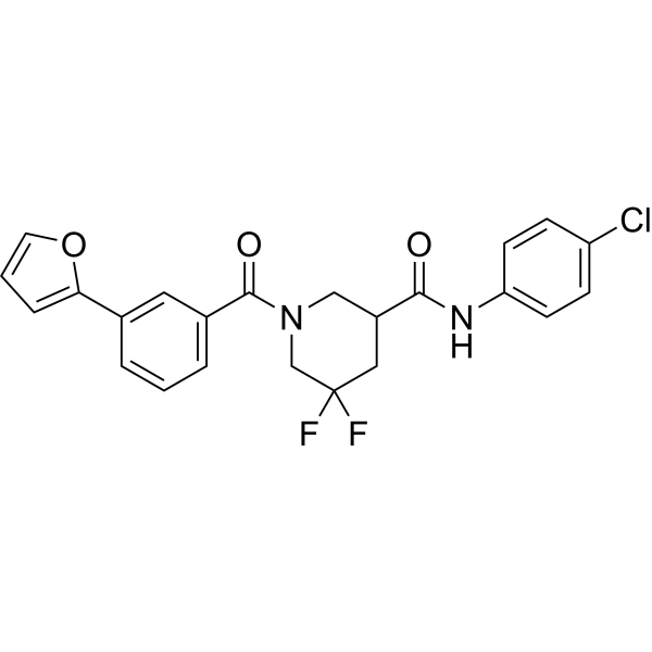
CCG-222740 is an orally active and selective Rho/myocardin-related transcription factor (MRTF) pathway inhibitor. CCG-222740 is also a potent inhibitor of alpha-smooth muscle actin protein expression. CCG-222740 effectively reduces fibrosis in skin and blocks melanoma metastasis.
Nur für Forschungszwecke. Wir verkaufen nicht an Patienten.
- Reinheit : 99.53%
- CAS. Nr.: 1922098-69-8
- Formel: C23H19ClF2N2O3
- Molecular Weight:444.86
-
Speicherung:Powder -20°C, 3 years , 4°C, 2 years ; In solvent -80°C, 2 years , -20°C, 1 year

To place orders, for customer services and technical support, please contact: MedChemExpress USA
Tel: 609-228-6898 E-mail: [email protected] [email protected]


-
 Biologische Aktivität
-
Chemical Information
-
Lösungsmittel & Löslichkeit
- Protokoll
- Reinheit & Dokumentation
- Verweise
-
 Help & FAQs


-
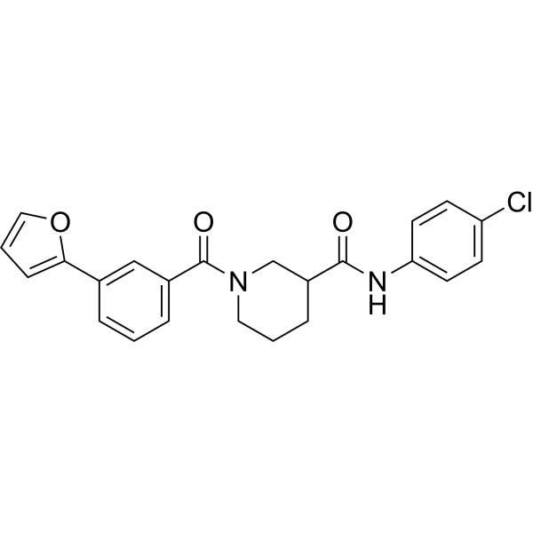
HY-108361
CCG-203971Rho/MRTF/SRF Inhibitor -
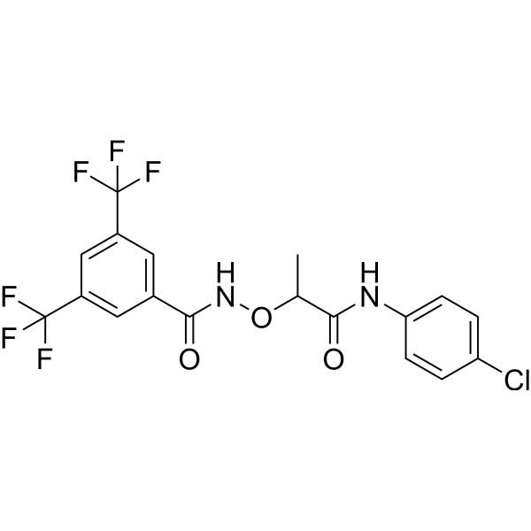
HY-13991
CCG-1423Rho/MRTF/SRF Pathway Inhibitor -
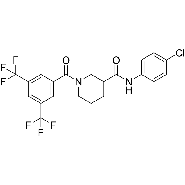
HY-120855
CCG-100602MRTF-A/SRF Inhibitor -
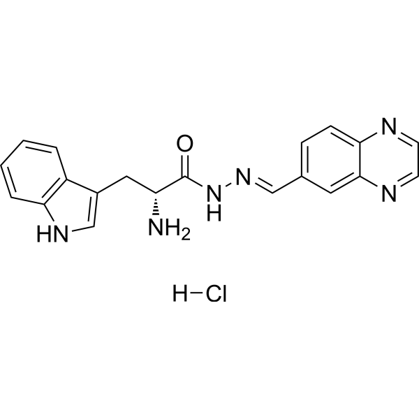
HY-12646
Rhosin hydrochlorideRho GTPases Inhibitor -

HY-10071
Y-27632ROCK Inhibitor -
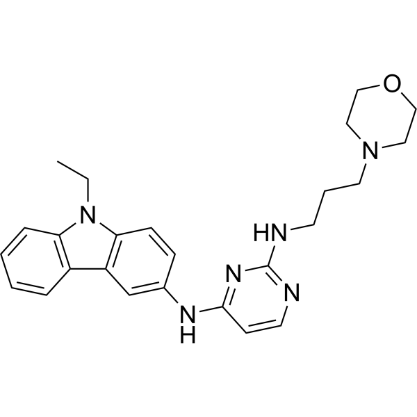
HY-12810
EHop-016Rac1/Rac3 Inhibitor -
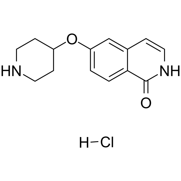
HY-15687
SAR407899 hydrochlorideROCK Inhibitor -
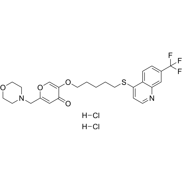
HY-16659
EHT 1864Rac Family Inhibitor -
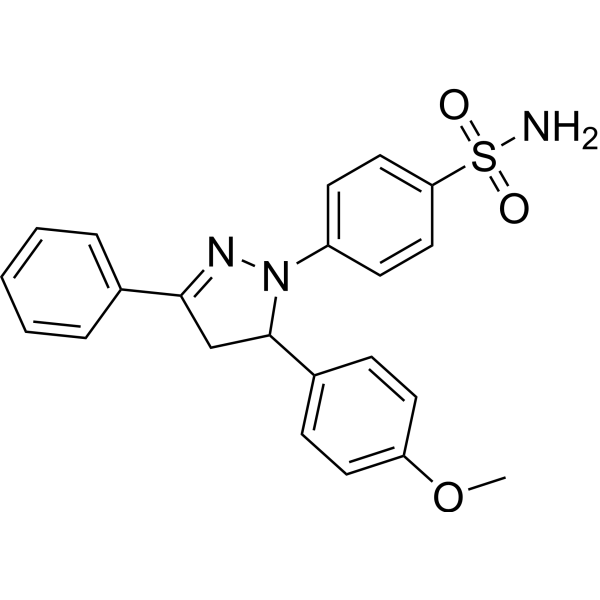
HY-12755
ML141Cdc42 GTPase Inhibitor -

HY-10583
Y-27632 dihydrochlorideROCK Inhibitor -
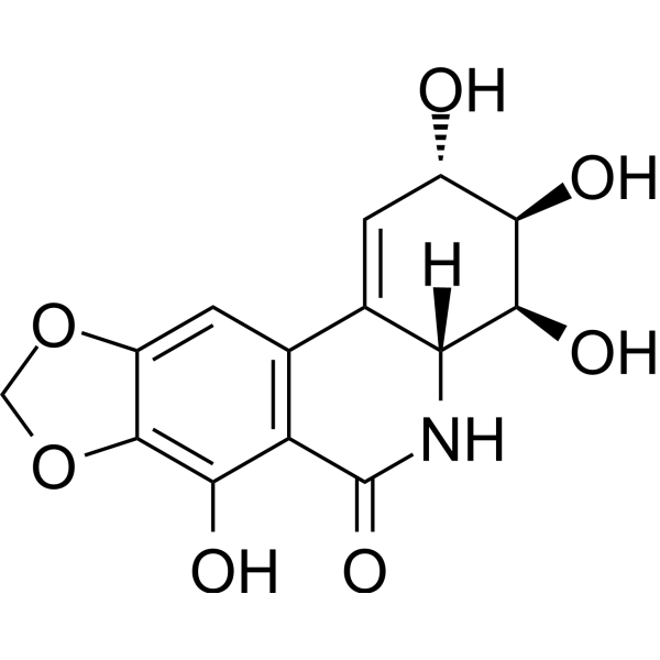
HY-16563
NarciclasineROCK Activator -
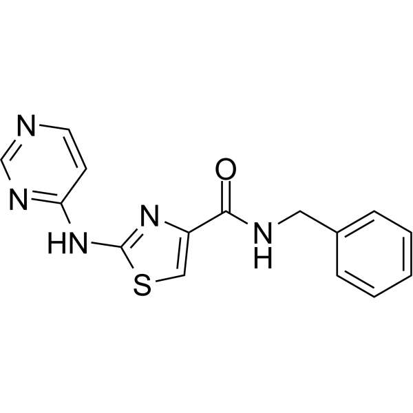
HY-13257
ThiazovivinROCK Inhibitor
Publications Citing Use of MedChemExpress (MCE) CCG-222740
More

Biologische Aktivität
Beschreibung
IC50 & Target
Rho/MRTF pathway[1]
In Vitro
CCG-222740 (10, 20 μM; for 72 hours) increases the protein levels of p27 and decreases cyclin D1. CCG-222740 decreases cell viability of CAFs, with an IC50 of ~10 μM[1].
CCG-222740 (10, 25 μM) is potent at decreasing αSMA protein expression in human conjunctival fibroblasts[2].
CCG-222740 has an IC50 of 5 μM in a fibroblast-mediated collagen contraction assay, and it is less cytotoxic[2].
MedChemExpress (MCE) has not independently confirmed the accuracy of these methods. They are for reference only.
-
Cell Line:Cancer associated fibroblasts (CAFs)
-
Concentration:10, 20 μM
-
Incubation Time:72 hours
-
Result:Increased the protein levels of p27 and decreased cyclin D1.
In Vivo
MedChemExpress (MCE) has not independently confirmed the accuracy of these methods. They are for reference only.
-
Animal Model:KC mice (LSL-KrasG12D/+; Pdx-1-Cre) of age at 9 weeks[1]
-
Dosage:100 mg/kg
-
Administration:Oral gavage; daily; for 7 days
-
Result:Reduced α-SMA levels in the pancreas of caerulein-stimulated KC mice.
- Nuclear Protein Extraction (High-Salt/Hypotonic Fractionation)
- Fibrosis/Collagen Morphometry
- RNA extraction experimental
- Western Blot
- Collagen: Sirius Red Staining
- Connective Tissue: Masson's Trichrome/Collagen Trichrome Staining
- Orthotopic Cell-Line Xenograft
- Protocol For Protein Expression And Purification


Chemical Information
-
CAS. Nr. 1922098-69-8
-
Appearance Solid
-
Molecular Weight 444.86
-
Formel C23H19ClF2N2O3
-
Color Light yellow to yellow
-
SMILES
O=C(C1CN(C(C2=CC=CC(C3=CC=CO3)=C2)=O)CC(F)(F)C1)NC4=CC=C(Cl)C=C4
-
Versand
Room temperature in continental US; may vary elsewhere.
-
Speicherung
Powder -20°C 3 years 4°C 2 years In solvent -80°C 2 years -20°C 1 year


Publications (1)
-
Journal Impact Factor
-
Most Recent
-
Science
2025 Dec 11;390(6778):eadl1988. PMID: 40997217


Lösungsmittel & Löslichkeit
In Vitro:
DMSO : 125 mg/mL (280.99 mM; Need ultrasonic; Hygroscopic DMSO has a significant impact on the solubility of product, please use newly opened DMSO)
Please refer to the solubility information to select the appropriate solvent. Once prepared, please aliquot and store the solution to prevent product inactivation from repeated freeze-thaw cycles.
Storage method and period of stock solution: -80°C, 2 years; -20°C, 1 year. When stored at -80°C, please use it within 2 years. When stored at -20°C, please use it within 1 year.
Please refer to the solubility information to select the appropriate solvent. Once prepared, please aliquot and store the solution to prevent product inactivation from repeated freeze-thaw cycles.
Storage method and period of stock solution: -80°C, 2 years; -20°C, 1 year. When stored at -80°C, please use it within 2 years. When stored at -20°C, please use it within 1 year.
Konzentration (Stammlösung) × Volumen (Stammlösung) = Konzentration (Ziellösung) × Volumen (Ziellösung)
In Vivo:
Select the appropriate dissolution method based on your experimental animal and administration route.
- For the following dissolution methods, please ensure to first prepare a clear stock solution using an In Vitro approach and then sequentially add co-solvents:
- To ensure reliable experimental results, the clarified stock solution can be appropriately stored based on storage conditions. As for the working solution for In Vivo experiments, it is recommended to prepare freshly and use it on the same day.
- The percentages shown for the solvents indicate their volumetric ratio in the final prepared solution. If precipitation or phase separation occurs during preparation, heat and/or sonication can be used to aid dissolution.
Add each solvent one by one: 10% DMSO 40% PEG300 5% Tween-80 45% Saline
Solubility: ≥ 2.25 mg/mL (5.06 mM); Clear solution
This protocol yields a clear solution of ≥ 2.25 mg/mL (saturation unknown).
Taking 1 mL working solution as an example, add 100 μL DMSO stock solution (22.5 mg/mL) to 400 μL PEG300, and mix evenly; then add 50 μL Tween-80 and mix evenly; then add 450 μL Saline to adjust the volume to 1 mL.
Preparation of Saline: Dissolve 0.9 g sodium chloride in ddH₂O and dilute to 100 mL to obtain a clear Saline solution.
Add each solvent one by one: 10% DMSO 90% (20% SBE-β-CD in Saline)
Solubility: 2.25 mg/mL (5.06 mM); Suspended solution; Need ultrasonic
This protocol yields a suspended solution of 2.25 mg/mL. Suspended solution can be used for oral and intraperitoneal injection.
Taking 1 mL working solution as an example, add 100 μL DMSO stock solution (22.5 mg/mL) to 900 μL 20% SBE-β-CD in Saline, and mix evenly.
Preparation of 20% SBE-β-CD in Saline (4°C, storage for one week): 2 g SBE-β-CD powder is dissolved in 10 mL Saline, completely dissolve until clear.
In Vivo Dissolution Calculator
Please enter the basic information of animal experiments:
-
-
-
-
Recommended: Prepare an additional quantity of animals to account for potential losses during experiments.
Please enter your animal formula composition:
-
%DMSO +
Recommended: Keep the proportion of DMSO in working solution below 2% if your animal is weak.
-
%+
-
+%Tween-80 + +
-
%Saline +
The co-solvents required include: DMSO, . All of co-solvents are available by MedChemExpress (MCE). , Tween 80. All of co-solvents are available by MedChemExpress (MCE).
Working solution concentration: 0.22 mg/mL
Method for preparing stock solution: mg drug dissolved in μL DMSO. Stock solution concentration: mg/mL.
1. Take μL DMSO stock solution;
2. Add μL .
μL , mix evenly;
3. Then add μL Tween 80, mix evenly;
4. Then add μL
Please ensure that the stock solution in the first step is dissolved to a clear state, and add co-solvents in sequence. You can use ultrasonic heating (ultrasonic cleaner, recommended frequency 20-40 kHz), vortexing, etc. to assist dissolution.


Reinheit & Dokumentation
-
Data Sheet (274 KB)
-
SDS (254 KB)
- English - EN (254 KB)
- Français - FR (254 KB)
- Deutsch - DE (254 KB)
- Norwegian - NO (254 KB)
- Español - ES (254 KB)
- Swedish - SV (254 KB)
- Italian - IT (254 KB)
- Korean - KR (254 KB)
- Portuguese - PT (254 KB)
-
Handling Instructions (2659 KB)
Verweise
[1]. Leal AS, et al. The Rho/MRTF pathway inhibitor CCG-222740 reduces stellate cell activation and modulates immune cell populations in KrasG12D; Pdx1-Cre (KC) mice. Sci Rep. 2019 May 8;9(1):7072. [Content Brief]
[2]. Yu-Wai-Man C, et al. Local delivery of novel MRTF/SRF inhibitors prevents scar tissue formation in a preclinical model of fibrosis. Sci Rep. 2017 Mar 31;7(1):518. [Content Brief]


Complete Stock Solution Preparation Table
Please refer to the solubility information to select the appropriate solvent. Once prepared, please aliquot and store the solution to prevent product inactivation from repeated freeze-thaw cycles.
Storage method and period of stock solution: -80°C, 2 years; -20°C, 1 year. When stored at -80°C, please use it within 2 years. When stored at -20°C, please use it within 1 year.
| Optional Solvent | Concentration Solvent Mass | 1 mg | 5 mg | 10 mg | 25 mg |
|---|---|---|---|---|---|
| DMSO | 1 mM | 2.2479 mL | 11.2395 mL | 22.4790 mL | 56.1975 mL |
| 5 mM | 0.4496 mL | 2.2479 mL | 4.4958 mL | 11.2395 mL | |
| 10 mM | 0.2248 mL | 1.1239 mL | 2.2479 mL | 5.6197 mL | |
| 15 mM | 0.1499 mL | 0.7493 mL | 1.4986 mL | 3.7465 mL | |
| 20 mM | 0.1124 mL | 0.5620 mL | 1.1239 mL | 2.8099 mL | |
| 25 mM | 0.0899 mL | 0.4496 mL | 0.8992 mL | 2.2479 mL | |
| 30 mM | 0.0749 mL | 0.3746 mL | 0.7493 mL | 1.8732 mL | |
| 40 mM | 0.0562 mL | 0.2810 mL | 0.5620 mL | 1.4049 mL | |
| 50 mM | 0.0450 mL | 0.2248 mL | 0.4496 mL | 1.1239 mL | |
| 60 mM | 0.0375 mL | 0.1873 mL | 0.3746 mL | 0.9366 mL | |
| 80 mM | 0.0281 mL | 0.1405 mL | 0.2810 mL | 0.7025 mL | |
| 100 mM | 0.0225 mL | 0.1124 mL | 0.2248 mL | 0.5620 mL |


CCG-222740 Related Classifications
HY-121750 Related Classifications
 Powered by Bioz