



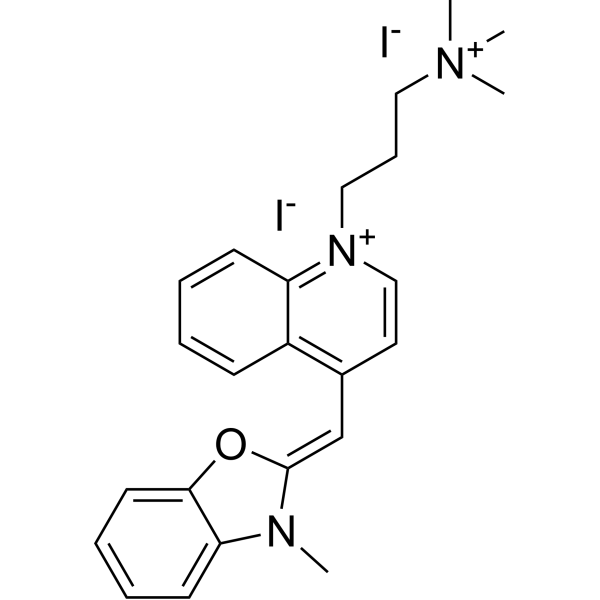
YO-PRO-1
Based on 1 publication(s) in Google Scholar
YO-PRO-1 is a non-cell-membrane-penetrating green fluorescent dye. YO-PRO-1 cannot penetrate normal cell membranes but can penetrate the membranes of apoptotic and necrotic cells. YO-PRO-1 has a high affinity for DNA. When it is not bound to DNA, it emits almost no fluorescence, and when it binds to DNA, it can emit green fluorescence (Ex/Em: 488/530±30 nM). YO-PRO-1 is often used together with Propidium Iodide (HY-D0815) to analyze and identify apoptotic and necrotic cells.
Nur für Forschungszwecke. Wir verkaufen nicht an Patienten.
- Reinheit : 98.2%
- CAS. Nr.: 152068-09-2
- Formel: C24H29I2N3O
- Molecular Weight:629.32
-
Speicherung:
4°C, sealed storage, away from moisture and light
* In solvent : -80°C, 6 months; -20°C, 1 month (sealed storage, away from moisture and light)

To place orders, for customer services and technical support, please contact: MedChemExpress USA
Tel: 609-228-6898 E-mail: [email protected] [email protected]


-
 Biologische Aktivität
-
Chemical Information
- Protokoll
- Reinheit & Dokumentation
- Verweise
-
 Help & FAQs


Publications Citing Use of MedChemExpress (MCE) YO-PRO-1
More

Biologische Aktivität
Beschreibung
In Vitro
Simple steps for YO-PRO-1 staining: Prepare cell/tissue samples; Wash the samples after incubating with YO-PRO-1 for 30 minutes to 1 hour. It can be co-stained with Hoechst 33342 (HY-15559), Propidium Iodide (HY-D0815), etc.; Microscopic observation and detection: Image by excitation with a 488 nm laser line[1][2].
MedChemExpress (MCE) has not independently confirmed the accuracy of these methods. They are for reference only.


Chemical Information
-
CAS. Nr. 152068-09-2
-
Appearance Solid
-
Molecular Weight 629.32
-
Formel C24H29I2N3O
-
Color Yellow to orange
-
SMILES
C[N+](CCC[N+]1=C2C=CC=CC2=C(/C=C3OC4=CC=CC=C4N\3C)C=C1)(C)C.[I-].[I-]
-
Versand
Room temperature in continental US; may vary elsewhere.
-
Speicherung
4°C, sealed storage, away from moisture and light
* In solvent : -80°C, 6 months; -20°C, 1 month (sealed storage, away from moisture and light)


Publications (1)
-
Journal Impact Factor
-
Most Recent


Reinheit & Dokumentation
-
Data Sheet (269 KB)
-
SDS (480 KB)
- English - EN (480 KB)
- Français - FR (480 KB)
- Deutsch - DE (480 KB)
- Norwegian - NO (480 KB)
- Español - ES (480 KB)
- Swedish - SV (480 KB)
- Italian - IT (480 KB)
- Korean - KR (480 KB)
- Portuguese - PT (480 KB)
-
Handling Instructions (2659 KB)
Verweise
[1]. Boffa DJ, et al. Measurement of apoptosis of intact human islets by confocal optical sectioning and stereologic analysis of YO-PRO-1-stained islets. Transplantation. 2005 Apr 15;79(7):842-5. [Content Brief]
[2]. Fujisawa S, et al. Evaluation of YO-PRO-1 as an early marker of apoptosis following radiofrequency ablation of colon cancer liver metastases. Cytotechnology. 2014 Mar;66(2):259-73. [Content Brief]

Calculators
Konzentration (Stammlösung) × Volumen (Stammlösung) = Konzentration (Ziellösung) × Volumen (Ziellösung)


YO-PRO-1 Related Classifications
HY-D0918 Related Classifications
 Powered by Bioz