



CHD1Li 6.11

Based on 1 Customer Validation
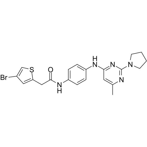
CHD1Li 6.11 is a potent and orally active Chromodomain Helicase DNA Binding Protein 1 Like (CHD1L) (oncogenic gene) inhibitor (IC50 = 3.3 µM for cat-CHD1L recombinant protein). CHD1Li 6.11 can inhibit EMT, induce mesenchymal-epithelial transition (reverse EMT) and promote apoptosis in tumor organoid models. CHD1Li 6.11 can be used for the research of cancer, such as colorectal cancer.
연구목적의 판매만을 진행합니다. 환자를 대상으로 한 판매는 하지 않습니다.
- Purity : 99.54%
- CAS No.: 2716890-91-2
- 화학식: C21H22BrN5OS
- 분자량:472.40
-
보관:Powder -20°C, 3 years , 4°C, 2 years ; In solvent -80°C, 6 months , -20°C, 1 month

To place orders, for customer services and technical support, please contact: MedChemExpress USA
Tel: 609-228-6898 E-mail: [email protected] [email protected]


-
 Biological Activity
-
Chemical Information
-
용액&용해도
- Protocol
- 순도&문서
- References
-
 Help & FAQs


All DNA/RNA Synthesis Isoforms
More

Biological Activity
제품 설명
IC50 & Target
|
Helicase |
Cellular Effect
|
Cell Line

|
Type | Value | Description | References |
|---|---|---|---|---|
| HCT-116 | IC50 |
0.8 μM
Compound: 6.11
|
Inhibition of cell stemness in human HCT-116 M-phenotype cells assessed as reduction in colony formation
Inhibition of cell stemness in human HCT-116 M-phenotype cells assessed as reduction in colony formation
|
[PMID: 35192363] |
| HCT-116 | IC50 |
4.3 μM
Compound: 6.11
|
Cytotoxicity against human HCT-116 M-phenotype tumor organoid assessed as reduction in cell viability incubated for 72 hrs by CellTiter Glo assay
Cytotoxicity against human HCT-116 M-phenotype tumor organoid assessed as reduction in cell viability incubated for 72 hrs by CellTiter Glo assay
|
[PMID: 35192363] |
| HCT-116 | IC50 |
5 μM
Compound: 6.11
|
Induction of mesenchymal-epithelial transition in human HCT-116 M-phenotype tumor organoid assessed as downregulation of vimentin expression by fluorescence analysis
Induction of mesenchymal-epithelial transition in human HCT-116 M-phenotype tumor organoid assessed as downregulation of vimentin expression by fluorescence analysis
|
[PMID: 35192363] |
| SW-620 | IC50 |
1.6 μM
Compound: 6.11
|
Inhibition of cell stemness in human SW-620 M-phenotype cells assessed as reduction in colony formation
Inhibition of cell stemness in human SW-620 M-phenotype cells assessed as reduction in colony formation
|
[PMID: 35192363] |
| SW-620 | IC50 |
2.6 μM
Compound: 6.11
|
Cytotoxicity against human SW-620 M-phenotype tumor organoid assessed as reduction in cell viability incubated for 72 hrs by CellTiter Glo assay
Cytotoxicity against human SW-620 M-phenotype tumor organoid assessed as reduction in cell viability incubated for 72 hrs by CellTiter Glo assay
|
[PMID: 35192363] |
| SW-620 | IC50 |
2.6 μM
Compound: 6.11
|
Cytotoxicity against human SW-620 tumor organoid assessed as reduction in cell viability incubated for 72 hrs by CellTiter Glo assay
Cytotoxicity against human SW-620 tumor organoid assessed as reduction in cell viability incubated for 72 hrs by CellTiter Glo assay
|
[PMID: 35192363] |
| SW-620 | IC50 |
3.3 μM
Compound: 6.11
|
Induction of mesenchymal-epithelial transition in human SW-620 M-phenotype tumor organoid assessed as downregulation of vimentin expression by fluorescence analysis
Induction of mesenchymal-epithelial transition in human SW-620 M-phenotype tumor organoid assessed as downregulation of vimentin expression by fluorescence analysis
|
[PMID: 35192363] |
In Vitro
CHD1Li 6.11 shows an IC50 of 2.6 μM against SW620 tumor organoid[1].
CHD1Li 6.11 (1-100 μM, 24 h) reduces TCF-transcription in M-Phenotype SW620 and HCT116 cells[1].
CHD1Li 6.11 (1-100 μM, 72 h) reduces viabilities in M-Phenotype SW620 and HCT116 cells[1].
MedChemExpress (MCE) has not independently confirmed the accuracy of these methods. They are for reference only. Further protocols information, click here.
In Vivo
MedChemExpress (MCE) has not independently confirmed the accuracy of these methods. They are for reference only.
-
Animal Model:Athymic nude mouse xenografts baring SW620 isolated M-Phenotype flank tumors[1]
-
Dosage:75 and 125 mg/kg
-
Administration:Orally administration, 5 times a week for 28 days
-
Result:Reduced tumor volume.
Had no abnormal observations associated with gross toxicity.


Chemical Information
-
CAS No. 2716890-91-2
-
Appearance Solid
-
분자량 472.40
-
화학식 C21H22BrN5OS
-
Color Off-white to gray
-
SMILES
O=C(NC1=CC=C(NC2=NC(N3CCCC3)=NC(C)=C2)C=C1)CC4=CC(Br)=CS4
-
선적
Room temperature in continental US; may vary elsewhere.
-
보관
Powder -20°C 3 years 4°C 2 years In solvent -80°C 6 months -20°C 1 month


용액&용해도
In Vitro:
DMSO : 100 mg/mL (211.69 mM; Need ultrasonic; Hygroscopic DMSO has a significant impact on the solubility of product, please use newly opened DMSO)
Please refer to the solubility information to select the appropriate solvent. Once prepared, please aliquot and store the solution to prevent product inactivation from repeated freeze-thaw cycles.
Storage method and period of stock solution: -80°C, 6 months; -20°C, 1 month. When stored at -80°C, please use it within 6 months. When stored at -20°C, please use it within 1 month.
Please refer to the solubility information to select the appropriate solvent. Once prepared, please aliquot and store the solution to prevent product inactivation from repeated freeze-thaw cycles.
Storage method and period of stock solution: -80°C, 6 months; -20°C, 1 month. When stored at -80°C, please use it within 6 months. When stored at -20°C, please use it within 1 month.
Concentration (start) × Volume (start) = Concentration (final) × Volume (final)
In Vivo:
Select the appropriate dissolution method based on your experimental animal and administration route.
- For the following dissolution methods, please ensure to first prepare a clear stock solution using an In Vitro approach and then sequentially add co-solvents:
- To ensure reliable experimental results, the clarified stock solution can be appropriately stored based on storage conditions. As for the working solution for In Vivo experiments, it is recommended to prepare freshly and use it on the same day.
- The percentages shown for the solvents indicate their volumetric ratio in the final prepared solution. If precipitation or phase separation occurs during preparation, heat and/or sonication can be used to aid dissolution.
Add each solvent one by one: 10% DMSO 40% PEG300 5% Tween-80 45% Saline
Solubility: ≥ 2.5 mg/mL (5.29 mM); Clear solution
This protocol yields a clear solution of ≥ 2.5 mg/mL (saturation unknown).
Taking 1 mL working solution as an example, add 100 μL DMSO stock solution (25.0 mg/mL) to 400 μL PEG300, and mix evenly; then add 50 μL Tween-80 and mix evenly; then add 450 μL Saline to adjust the volume to 1 mL.
Preparation of Saline: Dissolve 0.9 g sodium chloride in ddH₂O and dilute to 100 mL to obtain a clear Saline solution.
In Vivo Dissolution Calculator
Please enter the basic information of animal experiments:
-
-
-
-
Recommended: Prepare an additional quantity of animals to account for potential losses during experiments.
Please enter your animal formula composition:
-
%DMSO +
Recommended: Keep the proportion of DMSO in working solution below 2% if your animal is weak.
-
%+
-
+%Tween-80 + +
-
%Saline +
The co-solvents required include: DMSO, . All of co-solvents are available by MedChemExpress (MCE). , Tween 80. All of co-solvents are available by MedChemExpress (MCE).
Working solution concentration: 0.22 mg/mL
Method for preparing stock solution: mg drug dissolved in μL DMSO. Stock solution concentration: mg/mL.
1. Take μL DMSO stock solution;
2. Add μL .
μL , mix evenly;
3. Then add μL Tween 80, mix evenly;
4. Then add μL
Please ensure that the stock solution in the first step is dissolved to a clear state, and add co-solvents in sequence. You can use ultrasonic heating (ultrasonic cleaner, recommended frequency 20-40 kHz), vortexing, etc. to assist dissolution.

Protocol
-
Protocol for Electrophoretic Mobility Shift Assay (EMSA)
Electrophoretic mobility shift assay detects protein-nucleic acid binding by incubating a labeled DNA or RNA probe with purified protein or cell extract, then separating free probe from slower-migrating protein-probe complexes on a native gel. For cancer cells, primary neurons, mouse tumor samples, intestinal organoids, inflammatory macrophages, or drug-treated samples, EMSA can measure transcription-factor DNA binding or RNA-binding protein activity in extracts, but it does not directly measure transcription, protein expression, or chromatin occupancy in intact cells. Specificity is judged by competition with unlabeled wild-type probe, failure of mutated or unrelated competitors to compete, and antibody supershift or disruption when the binding protein identity must be confirmed.
-
Baculovirus-insect cell protein expression and purification
Baculovirus-insect cell expression uses recombinant baculovirus to deliver a target gene into insect cells, where late or very-late viral transcription drives recombinant protein production; the method was classically demonstrated by expression of human β-interferon in baculovirus-infected insect cells. The readout is target protein accumulation, assessed by activity, fluorescence if a fluorescent reporter is used, SDS-PAGE, Western blot, or purified protein yield. The system can express soluble, secreted, membrane-associated, and multiprotein targets, but expression outcome depends on the construct, baculovirus vector, insect cell line, multiplicity of infection, infection cell density, harvest time, and target-specific stability.
-
Apoptosis
Apoptosis, also called programmed cell death, is generally characterized by distinct morphological characteristics.
-
TUNEL staining for apoptotic DNA fragmentation
TUNEL staining detects DNA strand breaks by using terminal deoxynucleotidyl transferase to add labeled nucleotides to exposed 3′-OH DNA termini, generating either microscopic staining in fixed cells or tissue sections, or fluorescence/cytometric signal in cell suspensions. TUNEL positivity reflects DNA fragmentation but should not be interpreted alone as definitive apoptosis, because TUNEL can also label necrotic, autolytic, mechanically damaged, or DNA-repair-associated DNA breaks.
-
Patient-Derived Organoid (PDO) Establishment and Expansion
Patient-derived organoids (PDOs) are 3D in vitro models derived from patient tumor tissues that recapitulate the histological, genetic, and functional heterogeneity of the original tumors. These models are established by isolating tumor cells or tissue fragments and culturing them in a 3D extracellular matrix (ECM), such as Matrigel or decellularized ECM, to support self-organization, proliferation, and differentiation. The culture system preserves key features of the tumor microenvironment, including cell-cell interactions, stromal components, and ECM signaling, enabling accurate modeling of tumor biology and drug response for personalized medicine applications.
-
E. coli fusion-tag soluble protein purification
The purification of soluble recombinant proteins in Escherichia coli is achieved by fusing the target protein with a solubility-enhancing affinity tag (e. g. , His-tag, GST, MBP, Fh8, CSQ-tag, or thioredoxin) to improve expression yield, prevent aggregation, and enable efficient purification via affinity chromatography. The fusion protein is expressed under inducible promoters (e. g. , IPTG-induced T7 promoter), lysed from bacterial cells, and purified using resin-based affinity chromatography (e. g. , Ni-NTA for His-tag, amylose resin for MBP, chitin resin for intein tags, or HIC for Fh8). Tags can be removed post-purification using site-specific proteases (e. g. , TEV, enterokinase) or through intracellular cleavage systems. Solubility screening using multiple fusion partners (e. g. , Expresso® system) allows optimization of expression conditions for difficult-to-express proteins.
-
Annexin V plus membrane-impermeant dye apoptosis staining
Annexin V-based apoptosis assays rely on the detection of phosphatidylserine (PS) externalization from the inner leaflet of the plasma membrane to the outer leaflet, an early biochemical hallmark of apoptosis. Fluorescently labeled Annexin V binds PS in a calcium-dependent manner, enabling identification of early apoptotic cells by flow cytometry or fluorescence microscopy. When combined with a membrane-impermeant DNA-binding dye (e. g. , propidium iodide), this approach allows discrimination between viable (Annexin V−/dye−), early apoptotic (Annexin V+/dye−), and late apoptotic or necrotic (Annexin V+/dye+) cell populations by assessing membrane integrity and PS exposure.
-
Apoptosis Solutions
Apoptosis is a regulated, generally non-lytic cell-death pathway that removes unwanted, damaged, infected, or abnormal cells through coordinated morphological changes, caspase activation, DNA fragmentation, and membrane remodeling. The intrinsic apoptosis pathway is controlled mainly by mitochondrial outer membrane permeabilization, BCL-2 family proteins, cytochrome c release, apoptosome formation, caspase-9 activation, and downstream executioner caspase-3/7 activation. The extrinsic apoptosis pathway is initiated by death receptors such as Fas, TNFR, and TRAIL receptors, which recruit adaptor proteins and activate caspase-8 before engaging executioner caspases or mitochondrial amplification through BID cleavage. Apoptosis is linked to many phenotypes, including cancer cell killing, tissue homeostasis, immune regulation, neurodegeneration, infection response, and treatment-induced cytotoxicity; unresolved questions include how apoptosis interacts with necroptosis, pyroptosis, ferroptos
-
Inclusion-body expression, solubilization, refolding and purification
Inclusion-body recovery uses insoluble recombinant protein aggregates from E. coli as a starting material; the workflow is cell disruption, inclusion-body isolation/washing, denaturant or mild solubilization, refolding into soluble protein, and final chromatographic purification. The readouts are soluble protein recovery, purity by SDS-PAGE/chromatography, structural recovery by methods such as circular dichroism when used, and biological activity when an assay is available.
-
Protocol For Protein Expression And Purification
Recombinant protein expression in Escherichia coli followed by purification of a His-tagged soluble protein by immobilized metal affinity chromatography (IMAC), with optional MBP fusion and TEV tag removal when the construct includes these elements. The biological readout is production of the encoded target protein, detected as an inducible band at the expected molecular mass by SDS-PAGE and quantified by total protein assay or chromatographic absorbance; the purification readout is enrichment of the target protein in elution fractions after selective binding of polyhistidine residues to immobilized Ni2+/metal-chelate resin and elution by imidazole-containing buffer. Expression is driven by an inducible bacterial expression system, commonly T7/lac-based, in which IPTG or lactose/auto-induction activates transcription and translation of the cloned gene; lower induction temperature, lower inducer concentration, induction timing, and solubility-enhancing fusion tags can influence the frac
-
Mammalian transient protein expression and purification
Mammalian transient protein expression introduces plasmid DNA into HEK293 or CHO cells for short-term recombinant protein production, allowing secreted, glycosylated, Fc-tagged, His-tagged, or membrane proteins to be produced without stable clone generation.


순도&문서
-
Data Sheet (271 KB)
-
SDS (254 KB)
- English - EN (254 KB)
- Français - FR (254 KB)
- Deutsch - DE (254 KB)
- Norwegian - NO (254 KB)
- Español - ES (254 KB)
- Swedish - SV (254 KB)
- Italian - IT (254 KB)
- Korean - KR (254 KB)
- Portuguese - PT (254 KB)
-
Handling Instructions (2659 KB)
References


Complete Stock Solution Preparation Table
Please refer to the solubility information to select the appropriate solvent. Once prepared, please aliquot and store the solution to prevent product inactivation from repeated freeze-thaw cycles.
Storage method and period of stock solution: -80°C, 6 months; -20°C, 1 month. When stored at -80°C, please use it within 6 months. When stored at -20°C, please use it within 1 month.
| Optional Solvent | Concentration Solvent Mass | 1 mg | 5 mg | 10 mg | 25 mg |
|---|---|---|---|---|---|
| DMSO | 1 mM | 2.1169 mL | 10.5843 mL | 21.1685 mL | 52.9213 mL |
| 5 mM | 0.4234 mL | 2.1169 mL | 4.2337 mL | 10.5843 mL | |
| 10 mM | 0.2117 mL | 1.0584 mL | 2.1169 mL | 5.2921 mL | |
| 15 mM | 0.1411 mL | 0.7056 mL | 1.4112 mL | 3.5281 mL | |
| 20 mM | 0.1058 mL | 0.5292 mL | 1.0584 mL | 2.6461 mL | |
| 25 mM | 0.0847 mL | 0.4234 mL | 0.8467 mL | 2.1169 mL | |
| 30 mM | 0.0706 mL | 0.3528 mL | 0.7056 mL | 1.7640 mL | |
| 40 mM | 0.0529 mL | 0.2646 mL | 0.5292 mL | 1.3230 mL | |
| 50 mM | 0.0423 mL | 0.2117 mL | 0.4234 mL | 1.0584 mL | |
| 60 mM | 0.0353 mL | 0.1764 mL | 0.3528 mL | 0.8820 mL | |
| 80 mM | 0.0265 mL | 0.1323 mL | 0.2646 mL | 0.6615 mL | |
| 100 mM | 0.0212 mL | 0.1058 mL | 0.2117 mL | 0.5292 mL |


CHD1Li 6.11 Related Classifications
HY-144256 Related Classifications
 Powered by Bioz