



Dehydrobruceine B
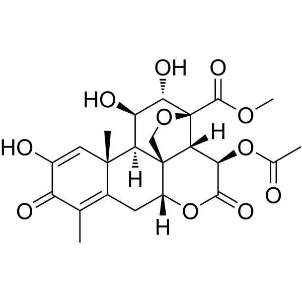
Dehydrobruceine B, a quassinoid, can be isolated from Brucea javanica. Dehydrobruceine B shows a synergistic effect with Cisplatin (HY-17394) to induce apoptosis via mitochondrial method. Dehydrobruceine B increases apoptosis-inducing factor (AIF) and Bax expression and suppresses Keap1-Nrf2.
For research use only. We do not sell to patients.
- CAS No.: 53730-90-8
- Formula: C23H26O11
- Molecular Weight:478.45
-
Storage:
Please store the product under the recommended conditions in the Certificate of Analysis.

To place orders, for customer services and technical support, please contact: MedChemExpress USA
Tel: 609-228-6898 E-mail: [email protected] [email protected]


-
 Biological Activity
-
Chemical Information
- Protocols
- Purity & Documentation
- References
-
 Help & FAQs




Biological Activity
Description
IC50 & Target
[1]|
Bax |
Bcl-xL |
Bcl-2 |
In Vitro
Dehydrobruceine B (1 μM; 48 h) and Cisplatin (3-18 μM; 48 h) combination synergizes the cytotoxic and apoptotic effects in A549 cells[1].
Dehydrobruceine B (1 μM; 24 h) induces excessive generation of intracellular ROS in Cisplatin (3 μM, 6 μM; 24 h)-treated A549 cells[1].
Dehydrobruceine B (1 μM; 24 h) increases depolarization of mitochondrial membrane potential (MMP) and translocation of cytochrome c in Cisplatin (3 μM, 6 μM; 24 h)-treated A549 cells[1].
Dehydrobruceine B (1 μM; 24 h) enhances the change of anti-apoptotic and pro-apoptotic protein levels in A549 cells[1].
MedChemExpress (MCE) has not independently confirmed the accuracy of these methods. They are for reference only.
-
Cell Line:A549 cells
-
Concentration:1 μM; with or without Cisplatin
-
Incubation Time:48 hours
-
Result:Upregulated the protein level of Bax, while downregulated the levels of Bcl-2 and Bcl-xL.
Enhanced caspase activation and PARP cleavage.
-
Cell Line:A549 cells
-
Concentration:1 μM; with or without Cisplatin
-
Incubation Time:48 hours
-
Result:Inhibited cell viability with 9 μM and 18 μM Cisplatin, respectively.
Induced cell apoptosis with 3 μM and 6 μM Cisplatin, respectively.
-
Cell Line:A549 cells
-
Concentration:1 μM; with or without 3 μM and 6 μM Cisplatin, respectively
-
Incubation Time:24 hours
-
Result:Resulted apoptosis-inducing factor (AIF) translocated from cytosol into nucleus dramatically in the co-treatment condition.


Chemical Information
-
CAS No. 53730-90-8
-
Molecular Weight 478.45
-
Formula C23H26O11
-
SMILES
COC([C@]12[C@@]3([H])[C@@]4(CO2)[C@@]([C@H]([C@@H]1O)O)([H])[C@@]5(C(C[C@@]4([H])OC([C@@H]3OC(C)=O)=O)=C(C(C(O)=C5)=O)C)C)=O
-
Structure Classification
-
Shipping
Room temperature in continental US; may vary elsewhere.
-
Storage
Please store the product under the recommended conditions in the Certificate of Analysis.


Purity & Documentation
References

Calculators
Concentration (start) × Volume (start) = Concentration (final) × Volume (final)


Dehydrobruceine B Related Classifications
HY-N10313 Related Classifications
 Powered by Bioz