



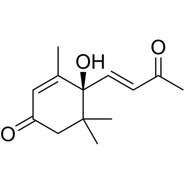
Dehydrovomifoliol

Based on 1 Customer Validation
Dehydrovomifoliol is a AKT/mTOR dual inhibitor. Dehydrovomifoliol reduces lipid accumulation and lipogenesis by inhibiting the AKT/mTOR signaling pathway. Dehydrovomifoliol is used in nonalcoholic fatty liver disease research (NAFLD) .
Nur für Forschungszwecke. Wir verkaufen nicht an Patienten.
- Reinheit : 98.88%
- CAS. Nr.: 39763-33-2
- Formel: C13H18O3
- Molecular Weight:222.28
-
Speicherung:
4°C, sealed storage, away from moisture and light
* In solvent : -80°C, 6 months; -20°C, 1 month (sealed storage, away from moisture and light)

To place orders, for customer services and technical support, please contact: MedChemExpress USA
Tel: 609-228-6898 E-mail: [email protected] [email protected]


-
 Biologische Aktivität
-
Chemical Information
- Protokoll
- Reinheit & Dokumentation
- Verweise
-
 Help & FAQs




Biologische Aktivität
Beschreibung
Cellular Effect
|
Cell Line

|
Type | Value | Description | References |
|---|---|---|---|---|
| A549 | IC50 |
>64.4 μM
Compound: 12
|
Cytostatic activity against human A549 cells after 48 cells by MTT assay
Cytostatic activity against human A549 cells after 48 cells by MTT assay
|
[PMID: 21561060] |
| C8166 | CC50 |
>450.5 μM
Compound: 12
|
Cytotoxicity against human C8166 cells after 3 days by MTT assay
Cytotoxicity against human C8166 cells after 3 days by MTT assay
|
[PMID: 21561060] |
| C8166 | EC50 |
>450.5 μM
Compound: 12
|
Antiviral activity against Human immunodeficiency virus 1 3B infected in human C8166 cells assessed as numbers of syncytia after 3 days by MTT assay
Antiviral activity against Human immunodeficiency virus 1 3B infected in human C8166 cells assessed as numbers of syncytia after 3 days by MTT assay
|
[PMID: 21561060] |
| HL-60 | IC50 |
>64.4 μM
Compound: 12
|
Cytostatic activity against human HL60 cells after 48 cells by MTT assay
Cytostatic activity against human HL60 cells after 48 cells by MTT assay
|
[PMID: 21561060] |
| HT-29 | IC50 |
>10 μM
Compound: S7
|
Cytotoxicity against human HT-29 cells assessed as reduction in cell viability after 72 hrs by CellTiter 96 aqueous one solution assay
Cytotoxicity against human HT-29 cells assessed as reduction in cell viability after 72 hrs by CellTiter 96 aqueous one solution assay
|
[PMID: 30057155] |
| HT-29 | IC50 |
>10 μM
Compound: S7
|
Inhibition of mitochondrial membrane potential in human HT-29 cells after 3 hrs by JC-1 staining based fluorescence assay
Inhibition of mitochondrial membrane potential in human HT-29 cells after 3 hrs by JC-1 staining based fluorescence assay
|
[PMID: 30057155] |
| MCF7 | IC50 |
>64.4 μM
Compound: 12
|
Cytostatic activity against human MCF7 cells after 48 cells by MTT assay
Cytostatic activity against human MCF7 cells after 48 cells by MTT assay
|
[PMID: 21561060] |
| MDA-MB-435 | IC50 |
>10 μM
Compound: S7
|
Cytotoxicity against human MDA-MB-435 cells assessed as reduction in cell viability after 72 hrs by CellTiter 96 aqueous one solution assay
Cytotoxicity against human MDA-MB-435 cells assessed as reduction in cell viability after 72 hrs by CellTiter 96 aqueous one solution assay
|
[PMID: 30057155] |
| OVCAR-3 | IC50 |
>10 μM
Compound: S7
|
Cytotoxicity against human OVCAR3 cells assessed as reduction in cell viability after 72 hrs by CellTiter 96 aqueous one solution assay
Cytotoxicity against human OVCAR3 cells assessed as reduction in cell viability after 72 hrs by CellTiter 96 aqueous one solution assay
|
[PMID: 30057155] |
| RAW264.7 | IC50 |
>50 μM
Compound: 10
|
Inhibition of LPS-induced IL-1beta production in mouse RAW264.7 cells after 24 hrs by ELISA
Inhibition of LPS-induced IL-1beta production in mouse RAW264.7 cells after 24 hrs by ELISA
|
[PMID: 24963714] |
| RAW264.7 | IC50 |
>64.4 μM
Compound: 12
|
Antiinflammatory against mouse RAW264.7 cells assessed as inhibition of LPS-induced nitric oxide production
Antiinflammatory against mouse RAW264.7 cells assessed as inhibition of LPS-induced nitric oxide production
|
[PMID: 21561060] |
| RAW264.7 | IC50 |
38.5 μM
Compound: 10
|
Inhibition of LPS-induced NO production in mouse RAW264.7 cells after 24 hrs by Griess reagent based assay
Inhibition of LPS-induced NO production in mouse RAW264.7 cells after 24 hrs by Griess reagent based assay
|
[PMID: 24963714] |
| SMMC-7721 | IC50 |
>64.4 μM
Compound: 12
|
Cytostatic activity against human SMMC7721 cells after 48 cells by MTT assay
Cytostatic activity against human SMMC7721 cells after 48 cells by MTT assay
|
[PMID: 21561060] |
| SW480 | IC50 |
>64.4 μM
Compound: 12
|
Cytostatic activity against human SW480 cells after 48 cells by MTT assay
Cytostatic activity against human SW480 cells after 48 cells by MTT assay
|
[PMID: 21561060] |


Chemical Information
-
CAS. Nr. 39763-33-2
-
Appearance Oil
-
Molecular Weight 222.28
-
Formel C13H18O3
-
Color Colorless to light yellow
-
SMILES
CC(/C=C/[C@](C(C)(C1)C)(C(C)=CC1=O)O)=O
-
Structure Classification
-
Initial Source
-
Versand
Room temperature in continental US; may vary elsewhere.
-
Speicherung
4°C, sealed storage, away from moisture and light
* In solvent : -80°C, 6 months; -20°C, 1 month (sealed storage, away from moisture and light)


Reinheit & Dokumentation
-
Data Sheet (267 KB)
-
SDS (252 KB)
- English - EN (252 KB)
- Français - FR (252 KB)
- Deutsch - DE (252 KB)
- Norwegian - NO (252 KB)
- Español - ES (252 KB)
- Swedish - SV (252 KB)
- Italian - IT (252 KB)
- Korean - KR (252 KB)
- Portuguese - PT (252 KB)
-
Handling Instructions (2659 KB)
Verweise

Calculators
Konzentration (Stammlösung) × Volumen (Stammlösung) = Konzentration (Ziellösung) × Volumen (Ziellösung)


Dehydrovomifoliol Related Classifications
HY-137996 Related Classifications
 Powered by Bioz