



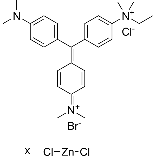
- Formula: C27H35N3.Br.xCl2Zn.Cl
Methyl Green zinc chloride is a potent fluorescent dye. Methyl Green zinc chloride is a DNA stains of cells and electrophoretic gels. Methyl Green zinc chloride can be used as direct measuring of viability by both microscopy and flow cytometry, with peaks at 633 and 677 nm.
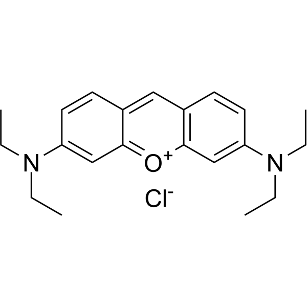

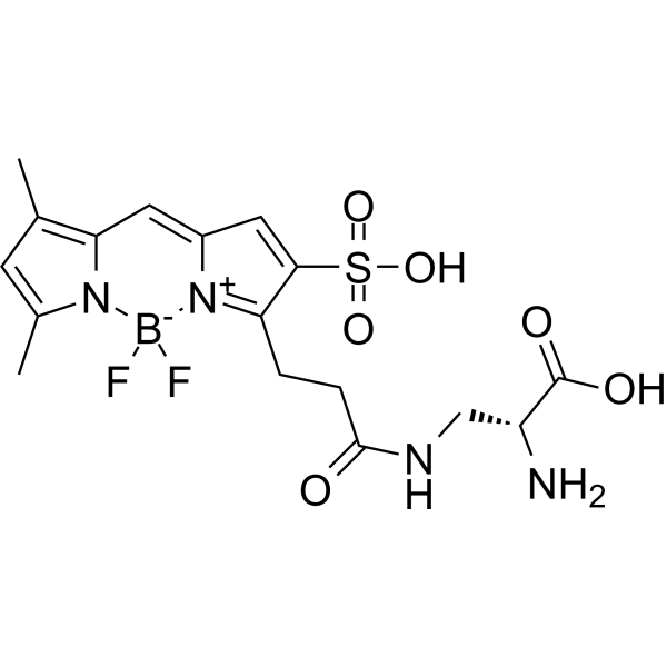
- Formula: C17H21BF2N4O6S
- Molecular Weight: 458.24
sBADA is a potent green fluorescent dye that targets and labels peptidoglycan in the cell walls of live bacteria(Ex/Em = 490/510 nm).
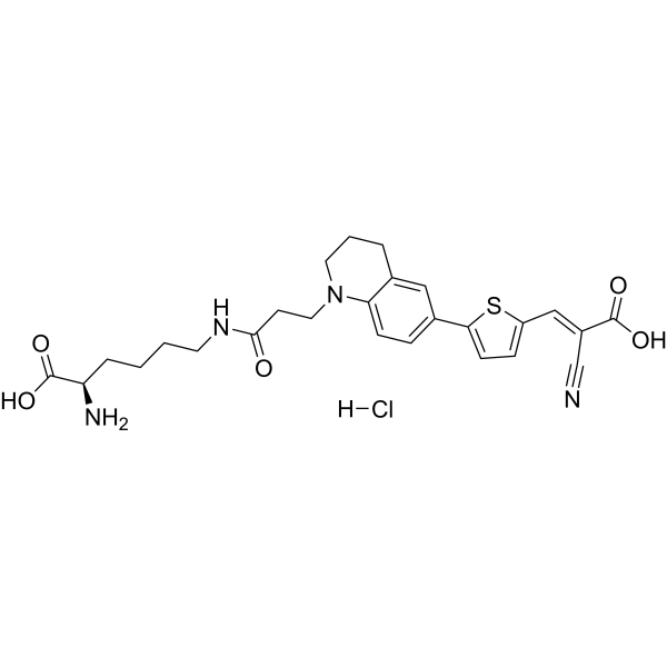
- Formula: C26H31ClN4O5S
- Molecular Weight: 547.07
Rf470DL is a rotor-fluorogenic D-amino acid (RfDAA). Rf470DL can be used for labeling bacteria (Ex=470 nm, Em=640 nm).
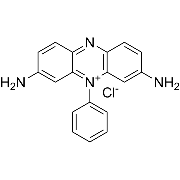
- Formula: C18H15ClN4
- Molecular Weight: 322.80
Phenosafranine is a phenazine dye. Phenosafranine exhibits higher binding affinity for triple-stranded RNA than for double-stranded RNA, and binds to both types of RNA via intercalation. Phenosafranine interacts with hemoglobin. Phenosafranine can be used for plant cell staining, as well as the detection of hemoglobin, dopamine, serotonin, etc.
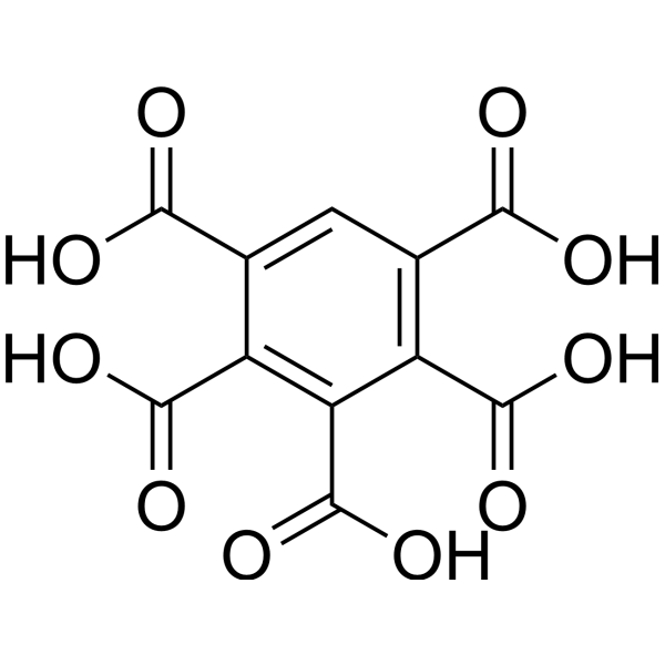
- Formula: C11H6O10
- Molecular Weight: 298.16
Benzenepentacarboxylic acid is a fluorescent dye that detects and scavenge HO radicals.

- Formula: C35H44N4O9S2
- Molecular Weight: 728.88
sCy5DA is a is a fluorescent D-amino acid. sCy5DA incorporates into bacterial peptidoglycan layers and cross-links via transpeptidase action, replacing peptidoglycan stem peptide D-amino acids. sCy5DA labels live bacterial cell walls, nascent peptidoglycan biosynthesis sites, and diverse bacterial species including Gram-negative, Gram-positive, and mycobacteria (Ex/Em = 646/665 nm).

- Formula: C38H51N5O8S2
- Molecular Weight: 769.97
sCy5DL-amide is an amidated fluorescent D-amino acid conjugated to Sulfo-Cyanine 5, and a peptidoglycan labeling agent. sCy5DL-amide incorporates into bacterial peptidoglycan cell wall via transpeptidase-mediated reactions, replacing the fourth or fifth D-amino acid of the peptidoglycan stem peptide for visualization of nascent peptidoglycan biosynthesis. sCy5DL-amide produces clear cell outline, septum labeling, and high localization density in Bacillus subtilis, including a 'V-shape' pattern at cell-cell contact areas. sCy5DL-amide exhibits robust incorporation into Gram-positive bacteria and mycobacteria, with reduced incorporation into Gram-negative bacteria (Ex/Em = 646/666 nm).

- Formula: C28H28N4O6
- Molecular Weight: 516.55
RADA is a fluorescent D-amino acid (FDAA) with high photostability and thermostability, which emits yellow-to-orange fluorescence. RADA shows low outer membrane permeability in wild-type Gram-negative Escherichia coli, but it targets penicillin-binding proteins and L,D-transpeptidases, mimics the interaction between acyl acceptors and enzyme intermediates, and integrates into peptidoglycan during biosynthesis. As a peptidoglycan labeling reagent, RADA metabolically integrates into the nascent peptidoglycan of live bacterial cells, labels the peptidoglycan at the poles and lateral walls of mycobacteria, and enables visualization of peptidoglycan synthesis and remodeling processes. RADA serves as a non-specific stain for fixed cells, is non-toxic to bacterial cells, and its red-shifted excitation/emission spectra reduce phototoxicity. RADA also supports virtual pulse-chase labeling experiments and stochastic optical reconstruction microscopy for sub-diffraction-limited imaging of bacterial cell walls.

- Formula: C32H38N4O11S
- Molecular Weight: 686.73
RMR-Tre is a fluorescent probe targeting the mycobacterial acyltransferase Ag85. Under the catalysis of Ag85, RMR-Tre undergoes 6-position mycoloylation and anchors to the mycobacterial membrane, while achieving fluorescence activation by inhibiting the intramolecular twisted charge transfer state transition. RMR-Tre can distinguish live mycobacteria from dead ones through metabolism-driven labeling, enabling rapid, wash-free, low-background detection of viable bacteria. RMR-Tre reports the drug resistance of Mycobacterium tuberculosis via the trehalose catalytic shift activity readout associated with TreS. In addition, RMR-Tre can be combined with flow cytometry or high-content imaging techniques to visualize and quantitatively analyze the metabolic heterogeneity of Mycobacterium tuberculosis related to persistence and drug resistance. RMR-Tre is widely used in tuberculosis-related research.

- Formula: C27H23BF3N3O2
- Molecular Weight: 489.30
CDy11 is a fluorescent probe and amyloid-binding dye (λex=590 nm; λem=612 nm), with a Ka of 29 μM for Pseudomonas aeruginosa Fap. CDy11 specifically recognizes amyloid fibrils in bacterial biofilms and exhibits significantly enhanced fluorescence upon binding to the target. CDy11 shows no staining effect on amyloid-deficient mutant strains, planktonic cells or protein monomers. CDy11 supports in vivo imaging of Pseudomonas aeruginosa biofilms in mouse implant and corneal infection models. CDy11 is widely used in studies of Staphylococcus aureus biofilm infections, dental caries, and Pseudomonas aeruginosa-associated implant and corneal infections.

- Formula: C27H27BF2N4O
- Molecular Weight: 472.34
CDr15 is a deep-red fluorescent probe (Ex=733 nm) that can selectively intercalate into and label bacterial extracellular DNA (eDNA). CDr15 exhibits extremely high specificity for biofilm imaging and cannot effectively bind to mammalian nuclear DNA. CDr15 enables real-time visualization of the microcolony structure and developmental process of three-dimensional *Pseudomonas aeruginosa* biofilms, and accurately localizes biofilm-forming regions of microorganisms in a mouse corneal infection model. With low background interference signals, CDr15 serves as an ideal diagnostic tool for research fields including bacterial biofilms and corneal infections.

- Formula: C27H26BF2N3O3
- Molecular Weight: 489.32
CDy14 is a fluorescent probe targeting the extracellular polysaccharide Psl. CDy14 enables the detection of biofilms. CDy14 can be used for research on chronic bacterial infections.

- Formula: C18H16BrN5S
- Molecular Weight: 414.32
Thiazolyl Blue (solution) (MTT (solution)) is a cell-permeable and positively charged tetrazolium dye that is used to detect reductive metabolism in cells. Thiazolyl Blue is taken up by cells through the plasma membrane and then reduced to formazan by intracellular NAD (P) H-oxidoreductases. Thiazolyl Blue is frequently used in colorimetric assays to measure cell proliferation, cytotoxicity, and apoptosis.
Solvent and concentration: PBS: 5 mg/mL

- Formula: C55H45N5O15S
- Molecular Weight: 1048.04
CDG-DNB3 is a selective fluorescent probe for Mycobacterium tuberculosis. CDG-DNB3 provides rapid and specific labeling of live Mycobacterium tuberculosis. CDG-DNB3 can image Bacillus Calmette-Guérin phagocytosis in real time.

- Formula: C52H65IN8O11S2
- Molecular Weight: 1169.15

- Formula: C26H23B2F2N3O3
- Molecular Weight: 485.10
BacGO is a highly selective, wash free fluorescent probe for Gram positive bacteria. BacGO binds to the carbohydrate structure in peptidoglycan through boric acid and exhibits depolymerization induced luminescence (DIE) properties. BacGO can be used for imaging complex environmental samples (such as activated sludge) and flat plate bacteria without affecting bacterial activity.

- Formula: C29H35N3O3
- Molecular Weight: 473.61
ARHB is a highly selective and sensitive NAT2 fluorescent probe suitable for real-time detection of NAT2 activity in various bacteria. ARHB can successfully penetrate the bacterial cells, and the fluorescence intensity is positively correlated with the expression level of NAT2. ARHB is used for high-throughput screening of natural inhibitors for tuberculosis.

- Formula: C76H103ClFeN11O133+
- Molecular Weight: 1469.99
N14G-Fe, the Fe3+-chelated form of N14G, identifies Mtb in sputum samples with tuberculosis, exhibiting exceptional fluorescence. N14G-Fe can effectively traverse the cell wall and inner membrane region where IrtAB is located.



