



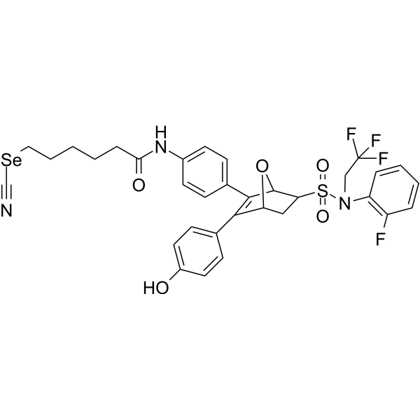
ER degrader 7
ER degrader 7 is an estrogen receptor α (ERα) degrader and a tubulin (tubulin) inhibitor. ER degrader 7 inhibits the cell viability of various cancer cell lines. ER degrader 7 disrupts the microtubule network in breast cancer cells, induces G2/M phase cell cycle arrest, and exhibits concentration-dependent accumulation in Tamoxifen-resistant LCC2 cells. ER degrader 7 suppresses tumor growth in vivo without causing body weight loss. ER degrader 7 can be applied in studies related to breast cancer.
Nos produits utilisent uniquement pour la recherche. Nous ne vendons pas aux patients.
- CAS No.: 2922929-63-1
- Formule: C33H31F4N3O5SSe
- Masse moléculaire:736.63
-
Stockage:
Please store the product under the recommended conditions in the Certificate of Analysis.

To place orders, for customer services and technical support, please contact: MedChemExpress USA
Tel: 609-228-6898 E-mail: [email protected] [email protected]


-
 Activité biologique
-
Chemical Information
- Protocole
- Pureté et documentation
- Références
-
 Help & FAQs




Activité biologique
Description
Cellular Effect
|
Cell Line

|
Type | Value | Description | References |
|---|---|---|---|---|
| MCF7 | IC50 |
0.06 μM
|
Antiproliferative activity against human MCF-7 ERα-positive breast cancer cells assessed as reduction in cell viability.
Antiproliferative activity against human MCF-7 ERα-positive breast cancer cells assessed as reduction in cell viability.
|
37584263 |
| T47D | IC50 |
2.56 μM
|
Antiproliferative activity against human T47D ERα-positive breast cancer cells assessed as reduction in cell viability.
Antiproliferative activity against human T47D ERα-positive breast cancer cells assessed as reduction in cell viability.
|
37584263 |
| MCF-10A | IC50 |
15.84 μM
|
Cytotoxicity against human MCF-10A normal breast cells assessed as reduction in cell viability.
Cytotoxicity against human MCF-10A normal breast cells assessed as reduction in cell viability.
|
37584263 |
| MDA-MB-231 | IC50 |
3.75 μM
|
Antiproliferative activity against human MDA-MB-231 ERα-negative breast cancer cells assessed as reduction in cell viability.
Antiproliferative activity against human MDA-MB-231 ERα-negative breast cancer cells assessed as reduction in cell viability.
|
37584263 |
In Vitro
ER degrader 7 (compound 35t) potently inhibits the proliferation of MCF-7 and T47D ERα-positive breast cancer cells, with IC50 values of 0.06 μM and 2.56 μM, respectively; it exhibits high cancer cell selectivity compared to normal MCF-10A breast cells, with a selectivity index (SI) value of 264.00[1].
ER degrader 7 potently inhibits the proliferation of endocrine-resistant ERα-positive breast cancer cell lines LCC2, T47D538G and T47DY537S, with IC50 values of 1.59 μM, 1.67 μM and 1.37 μM, respectively[1].
ER degrader 7 inhibits the proliferation of ERα-negative MDA-MB-231 breast cancer cells with an IC50 of 3.75 μM[1].
ER degrader 7 (0.5-5 μM; 6-24) induces proteasome-dependent ERα degradation in a dose-dependent manner in MCF-7 cells, with activity comparable to that of Fulvestrant (HY-13636)[1].
ER degrader 7 (0.5-5 μM; 6-24 h) exhibits weak degradation activity in three ERα+ drug-resistant breast cancer cell lines[1].
ER degrader 7 (2-4 μM; 48 h) disrupts the microtubule network in MCF-7 and LCC2 breast cancer cells, causing fragmentation and disorganization of microtubule structures, an effect similar to that induced by Colchicine treatment[1].
ER degrader 7 (1-12 μM; 48 h) induces G2/M phase arrest in MCF-7 and LCC2 cells, with LCC2 cells showing a concentration-dependent accumulation in the G2/M phase[1].
ER degrader 7 exhibits selective binding to ERα over ERβ, with an ERα RBA of 8.57% in a competitive fluorescent receptor binding assay[1].
ER degrader 7 (30 μM) significantly inhibits tubulin polymerization in a cell-free assay, with activity comparable to that of Colchicine (HY-16569) [1].
MedChemExpress (MCE) has not independently confirmed the accuracy of these methods. They are for reference only. Further protocols information, click here.
-
Cell Line:MCF-7 cells
-
Concentration:0.5, 1 and 5 μM
-
Incubation Time:6, 12 and 24 h
-
Result:Induced ERα degradation in a concentration-dependent manner.
Exhibited good degradation activity at 1 μM, comparable to that of Fulvestrant.
-
Cell Line:MCF-7 and LCC2 breast cancer cells
-
Concentration:2 μM (MCF-7 cells); 4 μM (LCC2 cells)
-
Incubation Time:48 h
-
Result:Disrupted microtubule networks in MCF-7 and LCC2 breast cancer cells, causing microtubule fragmentation and disorganization, similar to the effect observed with Colchicine treatment.
-
Cell Line:MCF-7 and LCC2 breast cancer cells
-
Concentration:1, 4 and 8 μM (MCF-7 cells); 3, 6 and 12 μM (LCC2 cells)
-
Incubation Time:48 h
-
Result:Induced G2/M phase arrest in MCF-7 and LCC2 cells, with the percentage of MCF-7 cells at G2/M increasing from 17.45% to 40.88%, and LCC2 cells showing a concentration-dependent accumulation in G2/M phase.
In Vivo
MedChemExpress (MCE) has not independently confirmed the accuracy of these methods. They are for reference only.
-
Animal Model:Nude mice were subcutaneously inoculated with MCF-7 cells and Tamoxifen-resistant LCC2 cells to establish an ERα+ breast cancer xenograft mode[1]
-
Dosage:2 and 4 mg/kg
-
Administration:i.p.; once every two days
-
Result:Achieved tumor growth inhibition (TGI) of 51.39% at 2 mg/kg, without body-weight loss, accompanied by tumor-tissue ERα degradation and reduced Ki67 positivity[.


Chemical Information
-
CAS No. 2922929-63-1
-
Masse moléculaire 736.63
-
Formule C33H31F4N3O5SSe
-
SMILES
O=C(CCCCC[Se]C#N)NC1=CC=C(C2=C(C3=CC=C(O)C=C3)[C@H]4CC(S(N(CC(F)(F)F)C5=C(F)C=CC=C5)(=O)=O)[C@@H]2O4)C=C1
-
Livraison
Room temperature in continental US; may vary elsewhere.
-
Stockage
Please store the product under the recommended conditions in the Certificate of Analysis.

Protocole
-
Mammalian live/dead viability and cytotoxicity staining
Live/dead viability and cytotoxicity staining assays are based on the simultaneous detection of intracellular esterase activity in metabolically active (viable) cells and membrane integrity loss in non-viable cells. In commonly used dual-staining approaches, membrane-permeant fluorogenic substrates are converted by intracellular esterases into fluorescent products in live cells, while impermeant DNA-binding dyes selectively enter cells with compromised plasma membranes and label nucleic acids in dead or dying cells, enabling discrimination between viable and non-viable populations by fluorescence microscopy or flow cytometry.
-
BrdU Incorporation Assay
Bromodeoxyuridine (BrdU) incorporation assay is based on the principle that BrdU, a thymidine analog, is incorporated into newly synthesized DNA during the S phase of the cell cycle, thereby serving as a marker of DNA replication and cellular proliferation. Incorporated BrdU can be detected using anti-BrdU antibodies following DNA denaturation, enabling visualization or quantification of proliferating cells through immunochemical detection methods such as immunofluorescence or immunohistochemistry.
-
Patient-Derived Orthotopic Xenograft (PDOX)
Patient-derived orthotopic xenograft (PDOX) modeling implants fresh patient tumor tissue or patient-derived tumor cells into the anatomically corresponding organ or tissue site of immunodeficient mice, usually by surgical orthotopic implantation, to preserve patient tumor histology, local microenvironmental context, invasion, metastatic behavior, and treatment-response features better than subcutaneous implantation. PDOX readouts include tumor engraftment, orthotopic tumor growth, local invasion, metastasis, recurrence after resection, histologic similarity to the donor tumor, biomarker retention, molecular concordance, survival, and response or resistance to therapy. PDOX models are used for preclinical drug testing and individualized therapy evaluation, but engraftment success varies by tumor type and specimen quality.
-
CCK-8/WST-8 Cell Proliferation Assay
The CCK-8/WST-8 assay is based on the reduction of the water-soluble tetrazolium salt WST-8 to a water-soluble formazan product by cellular dehydrogenases in metabolically active cells, where the generated formazan amount is proportional to the number of living cells and is quantified by measuring absorbance in the visible range, providing a colorimetric readout for cell viability and proliferation assessment. This class of tetrazolium-based assays improves upon earlier MTT-based systems by producing a water-soluble formazan, eliminating the need for organic solubilization steps and enabling direct spectrophotometric measurement in culture medium.
-
Protocol for Cell Cycle
Cell-cycle analysis by flow cytometry measures DNA content in single cells to estimate the fraction of cells in G0/G1, S, and G2/M phases. Propidium iodide intercalates into DNA, and after RNA removal with RNase, fluorescence intensity reflects cellular DNA content: 2N cells are assigned to G0/G1, cells between 2N and 4N to S phase, and 4N cells to G2/M. DNA-content analysis alone cannot reliably separate G0 from G1 or G2 from M. Ki-67 can distinguish quiescent G0 cells from cycling cells, EdU or BrdU incorporation marks active DNA synthesis in S phase, and phospho-histone H3 staining identifies mitotic cells within the 4N population.
-
Breast Cancer Modeling
Breast cancer is a heterogeneous cancer, and it has been distinguished into four subtypes: luminal A, luminal B, HER2-positive and basal-like. Molecular mutations, epigenetic alterations, hormone exposure and immune microenvironment are related to the progression of breast cancer.
-
Cell Viability Determination by MTT Colorimetric Assay
The following protocol uses the MTT colorimetric assay as a classic literature-established method for assessing cell viability/metabolic activity in cultured mammalian cells. MTT[3-(4,5-dimethylthiazol-2-yl)-2,5-diphenyltetrazolium bromide] is reduced by metabolically active cells to a colored formazan product; the amount of formazan is quantified spectrophotometrically and provides an indirect measure of metabolically active viable cells. Importantly, MTT reduction reflects cellular oxidoreductase/metabolic activity rather than an absolute direct count of living cells, so changes in cellular metabolism can alter the signal independently of cell number.
-
Cell Cytotoxicity Assay
Cytotoxicity assays are usually based on the assessment of cell membrane damage, which can also be indirectly detected by measuring cell viability. Detection methods include MTT assay, CKK-8 assay, LDH assay and ATP assay, etc.
-
Flow cytometric DNA-content cell-cycle staining
Flow cytometric DNA-content cell-cycle staining measures the fluorescence intensity of DNA-bound fluorochromes in single cells or nuclei to estimate DNA content distributions, allowing assignment of populations to G0/G1, S, and G2/M phases by DNA histogram deconvolution. Propidium iodide (PI) intercalates into DNA, and PI fluorescence is proportional to cellular DNA content when staining is performed under conditions that make DNA accessible and minimize non-DNA signal. Cells with G2/M DNA content are expected to show approximately twice the fluorescence intensity of G0/G1 cells, while S-phase cells occupy intermediate fluorescence values. PI-based DNA-content analysis can also detect cells with fractional DNA content, often reported as sub-G1, when DNA fragmentation and extraction during staining reduce retained DNA signal in apoptotic cells. DAPI is an alternative DNA fluorochrome for univariate DNA-content analysis, while bivariate approaches combining DNA content with proliferation
-
Research Protocol for Endocrine Diseases
Endocrine diseases often arise from disrupted hormone production, hormone signaling, or target-tissue responsiveness; for diabetes-focused endocrine disease models, insulin signaling regulates glucose uptake, hepatic glucose output, lipid metabolism, and β-cell compensation. Type 2 diabetes develops through interacting defects in insulin resistance, β-cell dysfunction, adipose inflammation, hepatic glucose overproduction, altered incretin signaling, and ectopic lipid metabolism. A major unresolved question is whether endocrine dysfunction is driven primarily by target-tissue insulin resistance, intrinsic β-cell failure, immune/inflammatory stress, or combined multi-organ failure that differs by disease stage.
-
Patient-Derived Xenograft (PDX)
Patient-derived xenograft (PDX) models are generated by engrafting primary human tumor tissue directly into immunodeficient mice, allowing in vivo propagation of patient tumor biology without initial in vitro adaptation. These models are used to preserve key histopathological and molecular characteristics of the original tumor and enable assessment of tumor growth dynamics and therapeutic response in a living organism. The biological readout is tumor engraftment and subsequent growth in the murine host, which reflects the ability of human tumor cells to survive, vascularize, and expand in an immunocompromised microenvironment.
-
MTT Cell Proliferation Assay
The MTT assay is a colorimetric endpoint assay for estimating viable cell number, cell growth, cytotoxicity, or cell activation in cultured mammalian cells. Living cells reduce the yellow tetrazolium salt MTT into purple/blue formazan, while dead cells do not generate the same signal; the resulting color can be quantified with a multiwell spectrophotometer. MTT reduction is commonly interpreted as a readout of metabolic activity that often correlates with viable cell number, but it should not be treated as a direct cell-counting method unless the assay is optimized for the cell type and experimental condition. Studies show that MTT reduction can involve mitochondrial and non-mitochondrial reducing systems, and formazan may accumulate in intracellular lipid droplets rather than simply marking mitochondria.
-
Subcutaneous Cell-Line-Derived Xenograft
Subcutaneous cell-line-derived xenograft (CDX) models are established by implanting cultured human cancer cell lines into immunodeficient mice, where the injected cells form localized tumors that can be monitored in vivo as a measure of tumorigenic potential, growth kinetics, and treatment response. These models are widely used in oncology research because they allow reproducible tumor formation and enable comparative assessment of tumor growth between different cell lines or genetic manipulations in a controlled in vivo microenvironment. Subcutaneous implantation of cancer cells in immunodeficient mice is a standard approach for evaluating tumor growth behavior and therapeutic response across multiple cancer types, including prostate, esophageal, pancreatic, and colon cancer models.
-
Orthotopic Cell-Line Xenograft
Orthotopic cell-line xenograft models involve implantation of human cancer cell lines into the anatomically corresponding organ of immunodeficient mice to reproduce tumor growth within a native microenvironment, enabling more clinically relevant tumor behavior compared with subcutaneous models. These models are widely used because orthotopic placement better recapitulates tumor progression, including invasion and metastatic spread, which are often underrepresented in heterotopic implantation systems. Compared with conventional xenografts, orthotopic implantation is described as more technically complex but provides improved simulation of tumor-microenvironment interactions and metastatic behavior, making it particularly valuable for translational oncology research. Surgical orthotopic implantation approaches have been emphasized as enabling faithful reproduction of clinical cancer features, including metastasis and disease progression patterns that align with the tumor’s organ of origi


Pureté et documentation
Références

Calculators
Concentration (start) × Volume (start) = Concentration (final) × Volume (final)


ER degrader 7 Related Classifications
HY-155197 Related Classifications
 Powered by Bioz