



Avadomide
Based on 6 publication(s) in Google Scholar
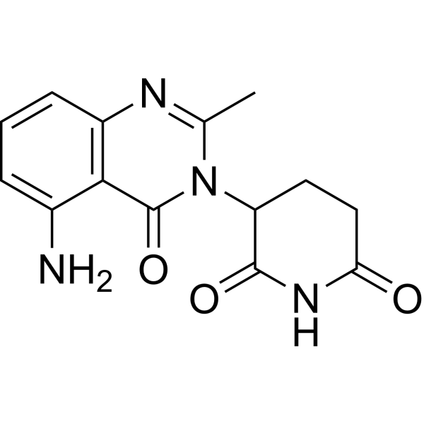
Avadomide is an orally active cereblon modulator. Avadomide modulates cereblon E3 ligase activity, inhibits NF-κB pathway, arrests the cell cycle at G1 phase, and thus induces apoptosis in cancer cell PDAC. Avadomide exhibits potent antitumor and immunomodulatory activities.
For research use only. We do not sell to patients.
- Purity : 99.56%
- CAS No.: 1015474-32-4
- Formula: C14H14N4O3
- Molecular Weight:286.29
-
Storage:Powder -20°C, 3 years , 4°C, 2 years ; In solvent -80°C, 1 year , -20°C, 6 months

To place orders, for customer services and technical support, please contact: MedChemExpress USA
Tel: 609-228-6898 E-mail: [email protected] [email protected]


-
 Biological Activity
-
Chemical Information
-
Solvent & Solubility
- Protocols
- Purity & Documentation
- References
-
 Help & FAQs


-
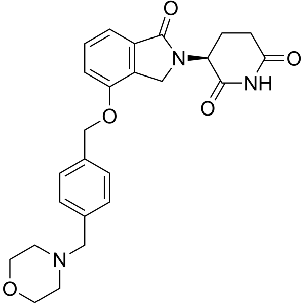
HY-101291
IberdomideCELMoD -
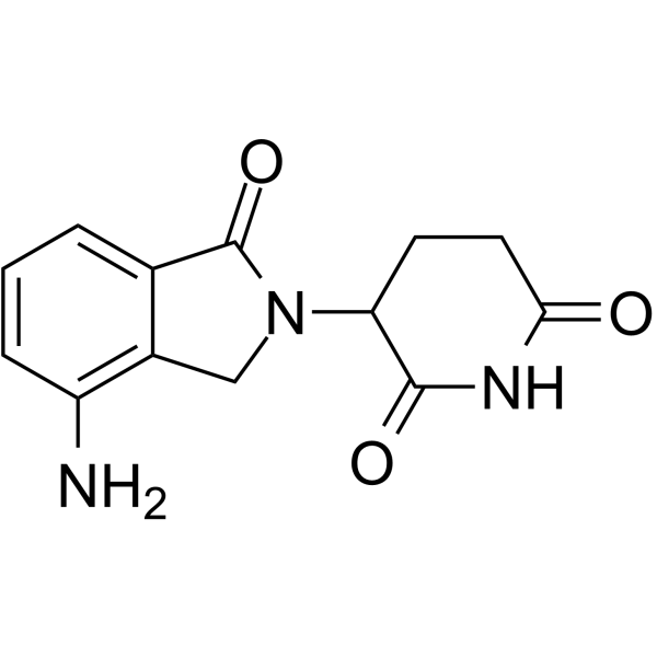
HY-A0003
LenalidomideImmunomodulator -
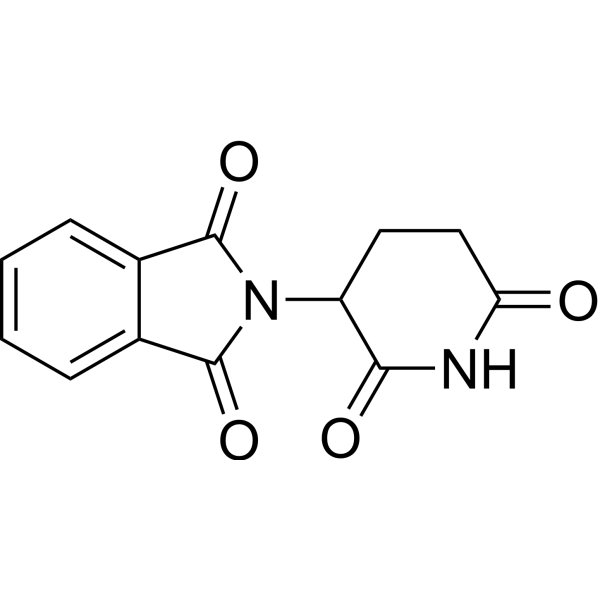
HY-14658
ThalidomideCereblon Inhibitor -
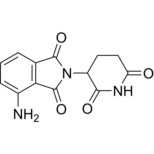
HY-10984
PomalidomideImmunomodulatory Agent -
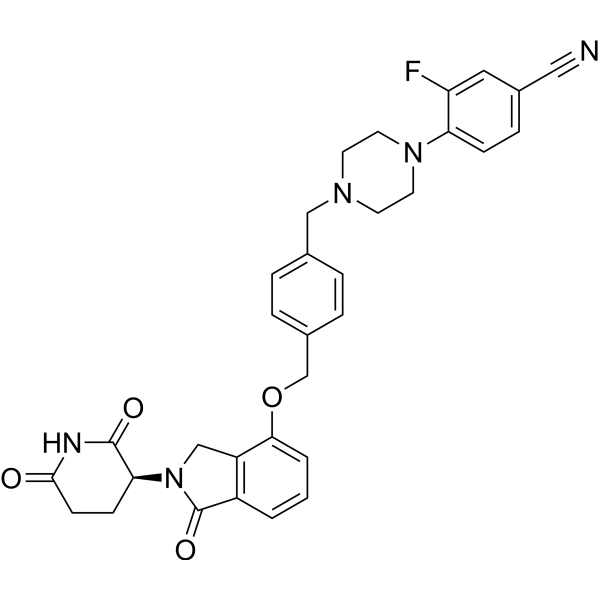
HY-129395
MezigdomideCereblon E3 Ubiquitin Ligase Modulating Agent -
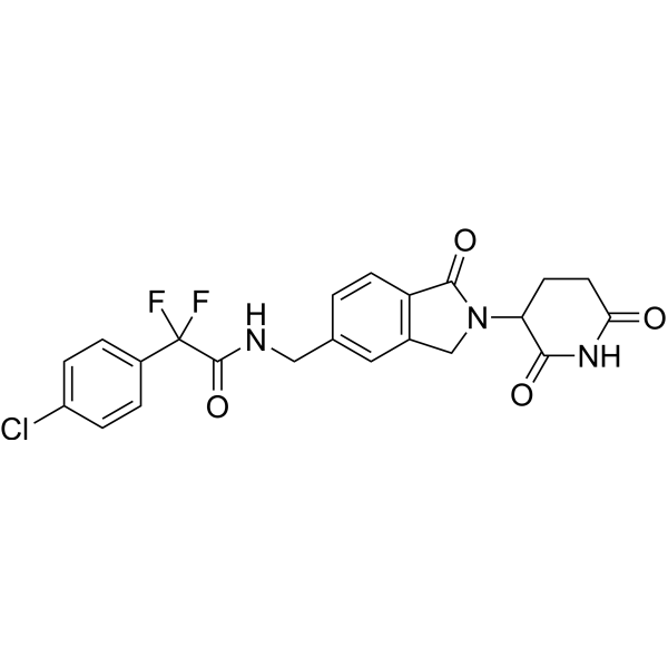
HY-130800
EragidomideGSPT1 Molecular Glue Degrader -
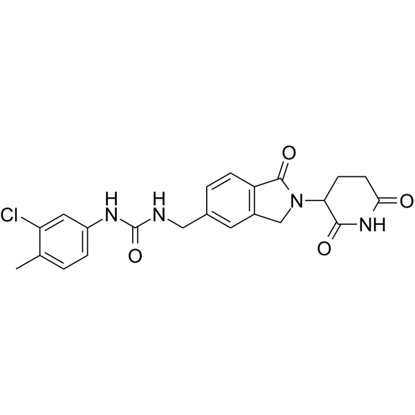
HY-101488
CC-885CRBN Modulator/GSPT1 Molecular Glue Degrader -
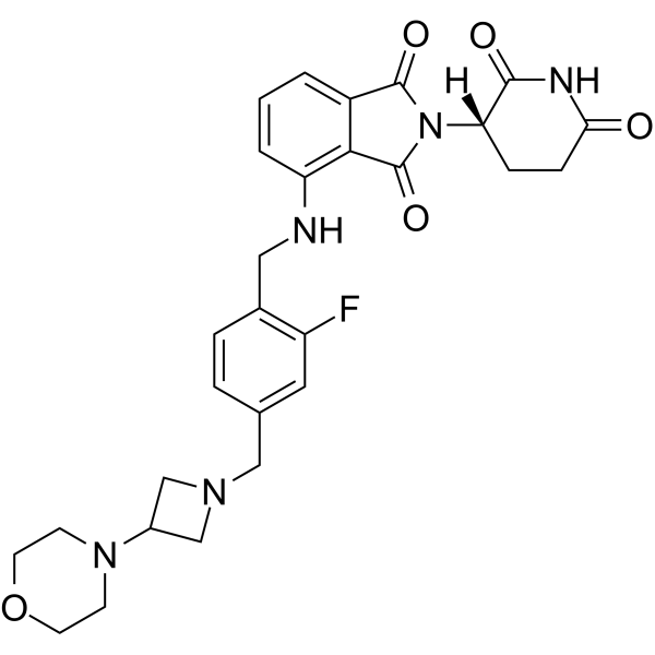
HY-146237
GolcadomideCereblon Modulator -
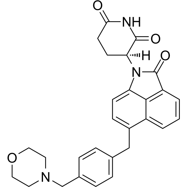
HY-144841
CemsidomidePROTAC IKZF1/3 Degrader -
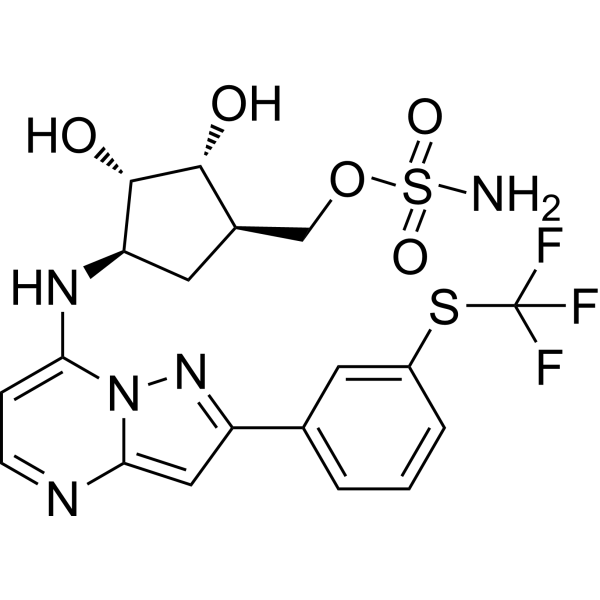
HY-100487
TAK-243UAE/E1 Inhibitor -
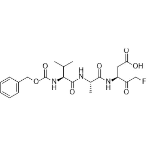
HY-16658B
Z-VAD-FMKPan-Caspase Inhibitor -
HY-19817
PRT4165E1/E2/E3 Enzyme Inhibitor
Publications Citing Use of MedChemExpress (MCE) Avadomide
More- Nat Commun. 2020 Jul 14;11(1):3508. [Abstract]
- Cell Rep Med. 2024 Mar 19;5(3):101472. [Abstract]
- Cell Chem Biol. 2020 Jul 16;27(7):866-876.e8. [Abstract]
- J Dermatol Sci. 2025 Sep 26:S0923-1811(25)00177-X. [Abstract]
- iScience. 2023 Jun 7;26(7):107059. [Abstract]
- Cancer Chemother Pharmacol. 2023 Oct;92(4):303-314. [Abstract]
 Customer Validation & Images

-
Cell Proliferation/Viability Assay
-
Flow Cytometry
-
WB
-
In Vivo Efficacy Study
-
Histological Imaging/Staining


Biological Activity
Description
Cellular Effect
|
Cell Line

|
Type | Value | Description | References |
|---|---|---|---|---|
| Daudi | EC50 |
182.3 nM
Compound: CC-122
|
Antiproliferative activity against human Daudi cells assessed as reduction in cell viability incubated for 2 days by CCK8 assay
Antiproliferative activity against human Daudi cells assessed as reduction in cell viability incubated for 2 days by CCK8 assay
|
[PMID: 36577219] |
| HEK-293T | EC50 |
>10 μM
Compound: CC-122
|
Antiproliferative activity against human HEK-293T cells assessed as reduction in cell viability incubated for 2 days by CCK8 assay
Antiproliferative activity against human HEK-293T cells assessed as reduction in cell viability incubated for 2 days by CCK8 assay
|
[PMID: 36577219] |
| MM1.S | EC50 |
21.91 nM
Compound: CC-122
|
Antiproliferative activity against human MM1.S cells assessed as reduction in cell viability incubated for 2 days by CCK8 assay
Antiproliferative activity against human MM1.S cells assessed as reduction in cell viability incubated for 2 days by CCK8 assay
|
[PMID: 36577219] |
In Vitro
Avadomide inhibits proliferation and induces apoptosis in ABC and GCB DLBCL. In DLBCL cell lines, Avadomide-induced degradation or short hairpin RNA-mediated knockdown of Aiolos and Ikaros correlates with increased transcription of IFN-stimulated genes independent of IFN-α, -β, and -γ production and/or secretion and results in apoptosis in both activated B-cell (ABC) and germinal center B-cell DLBCL[1].
MedChemExpress (MCE) has not independently confirmed the accuracy of these methods. They are for reference only.
In Vivo
MedChemExpress (MCE) has not independently confirmed the accuracy of these methods. They are for reference only.
Clinical Trial
| NCT Number | Sponsor | Condition | Start Date |
Phase
|
|---|---|---|---|---|
| NCT01329991 | 2011-05 | PHASE1 |


Chemical Information
-
CAS No. 1015474-32-4
-
Appearance Solid
-
Molecular Weight 286.29
-
Formula C14H14N4O3
-
Color White to gray
-
SMILES
O=C(C(N1C(C)=NC2=C(C(N)=CC=C2)C1=O)CC3)NC3=O
-
Synonyms
CC 122
-
Shipping
Room temperature in continental US; may vary elsewhere.
-
Storage
Powder -20°C 3 years 4°C 2 years In solvent -80°C 1 year -20°C 6 months


Publications (6)
-
Journal Impact Factor
-
Most Recent
-
Nat Commun
2020 Jul 14;11(1):3508. PMID: 32665554 -
Cell Rep Med
STAT3 couples activated tyrosine kinase signaling to the oncogenic core transcriptional regulatory circuitry of anaplastic large cell lymphoma. [Abstract]2024 Mar 19;5(3):101472. PMID: 38508140 -
Cell Chem Biol
A Cell-Based Target Engagement Assay for the Identification of Cereblon E3 Ubiquitin Ligase Ligands and Their Application in HDAC6 Degraders. [Abstract]2020 Jul 16;27(7):866-876.e8. PMID: 32413286 -
J Dermatol Sci
Regulation of melanogenesis via ubiquitin-proteasome system and autophagy by 3,3,5-trimethylcyclohexyl succinate dimethylamide and tranexamic acid. [Abstract]2025 Sep 26:S0923-1811(25)00177-X. PMID: 41046159 -
iScience
iTAG an optimized IMiD-induced degron for targeted protein degradation in human and murine cells. [Abstract]2023 Jun 7;26(7):107059. PMID: 37360684 -
Cancer Chemother Pharmacol
Anti-tumor effect of avadomide in gemcitabine-resistant pancreatic ductal adenocarcinoma. [Abstract]2023 Oct;92(4):303-314. PMID: 37491611
Avadomide purchased from MedChemExpress. Usage Cited in: Cancer Chemother Pharmacol. 2023 Oct;92(4):303-314. [Abstract]
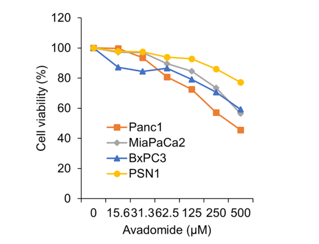
MTT assay showing the anti-tumor effects of Avadomide in four PDAC cell lines. Data are represented as mean of triplicate independent experiments.
Avadomide purchased from MedChemExpress. Usage Cited in: Cancer Chemother Pharmacol. 2023 Oct;92(4):303-314. [Abstract]
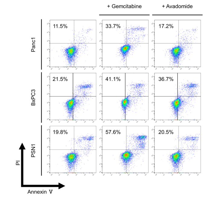
Representative dot plots from flow cytometry analysis illustrating apoptotic status by Annexin V assay. Panc1, BcPX3, and PSN1 after treatment with placebo, 10 ng/mL gemcitabine, and 200 μM avadomide for 36 h, respectively.
Avadomide purchased from MedChemExpress. Usage Cited in: Cancer Chemother Pharmacol. 2023 Oct;92(4):303-314. [Abstract]
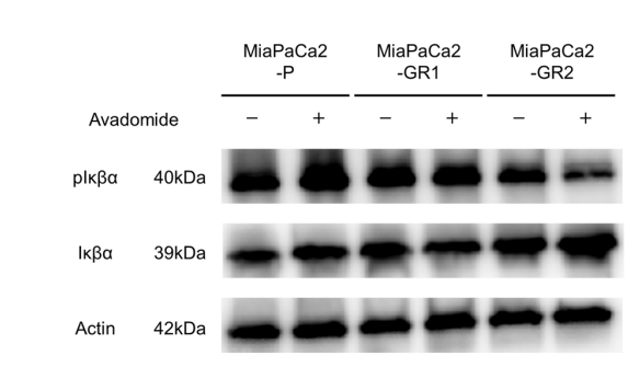
The effect of Avadomide (200 μM; 36 h) on the protein levels of pIκBα and IκBα in PDAC cells.
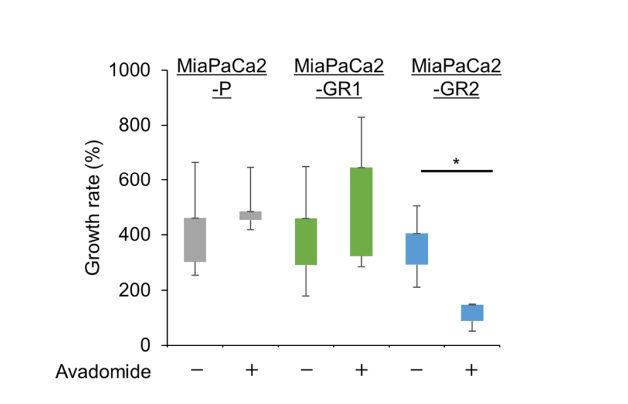
Avadomide purchased from MedChemExpress. Usage Cited in: Cancer Chemother Pharmacol. 2023 Oct;92(4):303-314. [Abstract]
Avadomide (0.5 mg/kg). Tumor growth rates varied among different cell lines. MiaPaCa2-GR2 showed the strongest inhibitory effect.
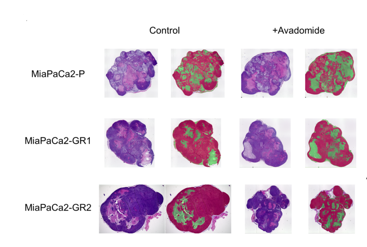
Avadomide purchased from MedChemExpress. Usage Cited in: Cancer Chemother Pharmacol. 2023 Oct;92(4):303-314. [Abstract]
Avadomide (0.5 mg/kg). Hematoxylin-eosin staining of tumor tissue.


Solvent & Solubility
In Vitro:
DMSO : 33.33 mg/mL (116.42 mM; Need ultrasonic; Hygroscopic DMSO has a significant impact on the solubility of product, please use newly opened DMSO)
Please refer to the solubility information to select the appropriate solvent. Once prepared, please aliquot and store the solution to prevent product inactivation from repeated freeze-thaw cycles.
Storage method and period of stock solution: -80°C, 1 year; -20°C, 6 months. When stored at -80°C, please use it within 1 year. When stored at -20°C, please use it within 6 months.
Please refer to the solubility information to select the appropriate solvent. Once prepared, please aliquot and store the solution to prevent product inactivation from repeated freeze-thaw cycles.
Storage method and period of stock solution: -80°C, 1 year; -20°C, 6 months. When stored at -80°C, please use it within 1 year. When stored at -20°C, please use it within 6 months.
Concentration (start) × Volume (start) = Concentration (final) × Volume (final)
In Vivo:
Select the appropriate dissolution method based on your experimental animal and administration route.
- For the following dissolution methods, please ensure to first prepare a clear stock solution using an In Vitro approach and then sequentially add co-solvents:
- To ensure reliable experimental results, the clarified stock solution can be appropriately stored based on storage conditions. As for the working solution for In Vivo experiments, it is recommended to prepare freshly and use it on the same day.
- The percentages shown for the solvents indicate their volumetric ratio in the final prepared solution. If precipitation or phase separation occurs during preparation, heat and/or sonication can be used to aid dissolution.
Add each solvent one by one: 10% DMSO 40% PEG300 5% Tween-80 45% Saline
Solubility: ≥ 2.5 mg/mL (8.73 mM); Clear solution
This protocol yields a clear solution of ≥ 2.5 mg/mL (saturation unknown).
Taking 1 mL working solution as an example, add 100 μL DMSO stock solution (25.0 mg/mL) to 400 μL PEG300, and mix evenly; then add 50 μL Tween-80 and mix evenly; then add 450 μL Saline to adjust the volume to 1 mL.
Preparation of Saline: Dissolve 0.9 g sodium chloride in ddH₂O and dilute to 100 mL to obtain a clear Saline solution.
In Vivo Dissolution Calculator
Please enter the basic information of animal experiments:
-
-
-
-
Recommended: Prepare an additional quantity of animals to account for potential losses during experiments.
Please enter your animal formula composition:
-
%DMSO +
Recommended: Keep the proportion of DMSO in working solution below 2% if your animal is weak.
-
%+
-
+%Tween-80 + +
-
%Saline +
The co-solvents required include: DMSO, . All of co-solvents are available by MedChemExpress (MCE). , Tween 80. All of co-solvents are available by MedChemExpress (MCE).
Working solution concentration: 0.22 mg/mL
Method for preparing stock solution: mg drug dissolved in μL DMSO. Stock solution concentration: mg/mL.
1. Take μL DMSO stock solution;
2. Add μL .
μL , mix evenly;
3. Then add μL Tween 80, mix evenly;
4. Then add μL
Please ensure that the stock solution in the first step is dissolved to a clear state, and add co-solvents in sequence. You can use ultrasonic heating (ultrasonic cleaner, recommended frequency 20-40 kHz), vortexing, etc. to assist dissolution.


Purity & Documentation
-
Data Sheet (282 KB)
-
SDS (393 KB)
- English - EN (393 KB)
- Français - FR (393 KB)
- Deutsch - DE (393 KB)
- Norwegian - NO (393 KB)
- Español - ES (393 KB)
- Swedish - SV (393 KB)
- Italian - IT (393 KB)
- Korean - KR (393 KB)
- Portuguese - PT (393 KB)
-
Handling Instructions (2659 KB)
References
[1]. Rasco DW, et al. A First-in-Human Study of Novel Cereblon Modulator Avadomide (CC-122) in Advanced Malignancies. Clin Cancer Res. 2019 Jan 1;25(1):90-98. [Content Brief]
[2]. Hagner, P.R.et al.CC-122, a pleiotropic pathway modifier, mimics an IFN response and has antitumor activity in DLBCL.Blood.Aug 6;126(6):779-89. [Content Brief]
[3]. Nishi H, et al., Anti-tumor effect of avadomide in gemcitabine-resistant pancreatic ductal adenocarcinoma. Cancer Chemother Pharmacol. 2023 Oct;92(4):303-314. [Content Brief]


Complete Stock Solution Preparation Table
Please refer to the solubility information to select the appropriate solvent. Once prepared, please aliquot and store the solution to prevent product inactivation from repeated freeze-thaw cycles.
Storage method and period of stock solution: -80°C, 1 year; -20°C, 6 months. When stored at -80°C, please use it within 1 year. When stored at -20°C, please use it within 6 months.
| Optional Solvent | Concentration Solvent Mass | 1 mg | 5 mg | 10 mg | 25 mg |
|---|---|---|---|---|---|
| DMSO | 1 mM | 3.4930 mL | 17.4648 mL | 34.9296 mL | 87.3240 mL |
| 5 mM | 0.6986 mL | 3.4930 mL | 6.9859 mL | 17.4648 mL | |
| 10 mM | 0.3493 mL | 1.7465 mL | 3.4930 mL | 8.7324 mL | |
| 15 mM | 0.2329 mL | 1.1643 mL | 2.3286 mL | 5.8216 mL | |
| 20 mM | 0.1746 mL | 0.8732 mL | 1.7465 mL | 4.3662 mL | |
| 25 mM | 0.1397 mL | 0.6986 mL | 1.3972 mL | 3.4930 mL | |
| 30 mM | 0.1164 mL | 0.5822 mL | 1.1643 mL | 2.9108 mL | |
| 40 mM | 0.0873 mL | 0.4366 mL | 0.8732 mL | 2.1831 mL | |
| 50 mM | 0.0699 mL | 0.3493 mL | 0.6986 mL | 1.7465 mL | |
| 60 mM | 0.0582 mL | 0.2911 mL | 0.5822 mL | 1.4554 mL | |
| 80 mM | 0.0437 mL | 0.2183 mL | 0.4366 mL | 1.0916 mL | |
| 100 mM | 0.0349 mL | 0.1746 mL | 0.3493 mL | 0.8732 mL |


Avadomide Related Classifications
HY-100507 Related Classifications
 Powered by Bioz