



Terbinafine
Based on 19 publication(s) in Google Scholar
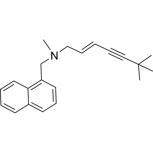
Terbinafine (TDT 067) is an orally active and potent antifungal agent. Terbinafine is a potent non-competitive inhibitor of squalene epoxidase from Candida, with a Ki of 30 nM. Terbinafine also shows antibacterial activity against certain Gram-positive and Gram-negative bacteria. Terbinafine is a click chemistry reagent, it contains an Alkyne group and can undergo copper-catalyzed azide-alkyne cycloaddition (CuAAc) with molecules containing Azide groups.
For research use only. We do not sell to patients.
- Purity : 99.96%
- CAS No.: 91161-71-6
- Formula: C21H25N
- Molecular Weight:291.43
-
Storage:Powder -20°C, 3 years , 4°C, 2 years ; In solvent -80°C, 2 years , -20°C, 1 year

To place orders, for customer services and technical support, please contact: MedChemExpress USA
Tel: 609-228-6898 E-mail: [email protected] [email protected]


-
 Biological Activity
-
Chemical Information
-
Solvent & Solubility
- Protocols
- Purity & Documentation
- References
-
 Help & FAQs


-
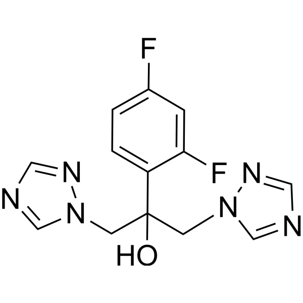
HY-B0101
FluconazoleAntifungal Agent -
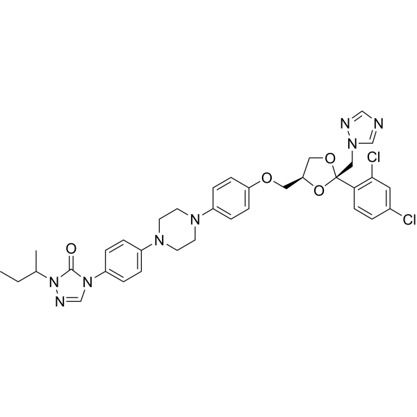
HY-17514
ItraconazoleAntifungal Agent -
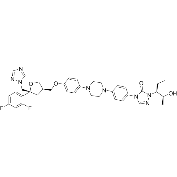
HY-17373
PosaconazoleAnti-fungal Agent -
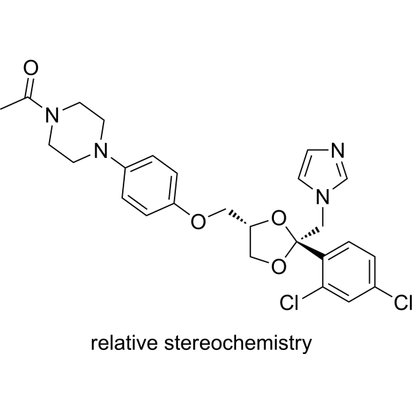
HY-B0105
KetoconazoleCYP3A4 Inhibitor -
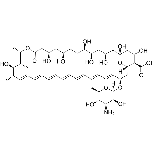
HY-B0221
Amphotericin BAntifungal Agent -
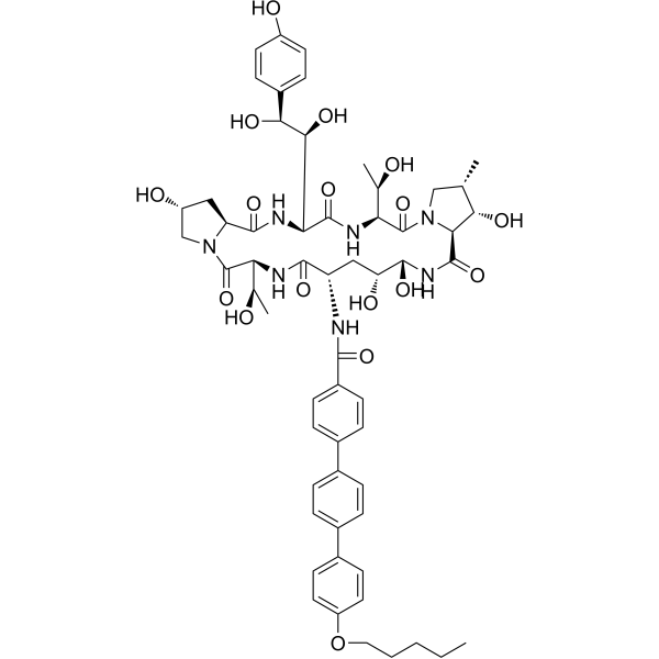
HY-13553
AnidulafunginAntifungal Agent -
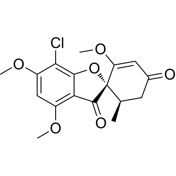
HY-17583
GriseofulvinAntifungal Antibiotic -
HY-17006
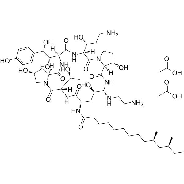
Caspofungin diacetateAntifungal Agent -
HY-B0139
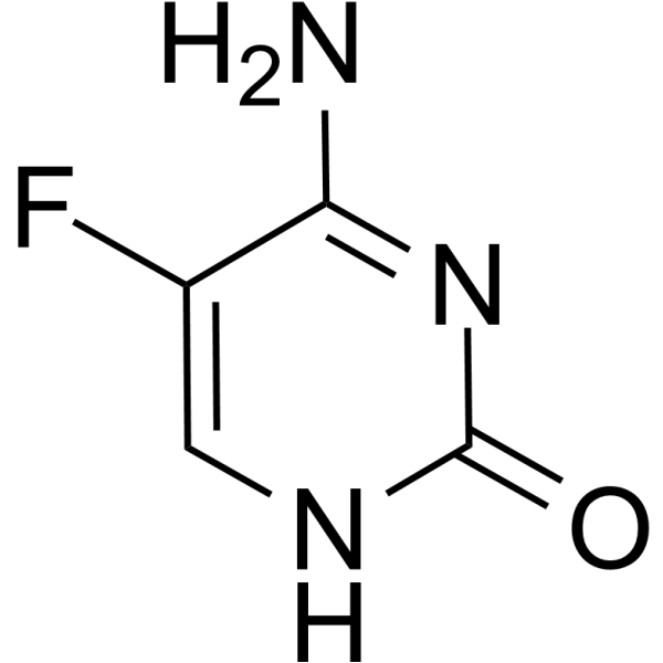
FlucytosineAntifungal Agent -
HY-76200
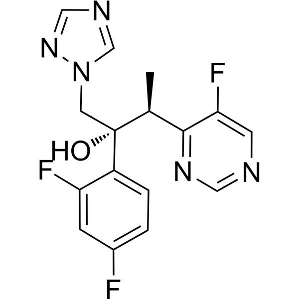
VoriconazoleAntifungal Agent -
HY-10882
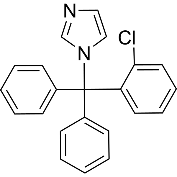
ClotrimazoleAutophagy Inhibitor -

HY-17395AR
Terbinafine (Standard)Antimicrobial Agent
Publications Citing Use of MedChemExpress (MCE) Terbinafine
More- Nature. 2026 Jul 8.
- Cancer Commun (Lond). 2021 Aug;41(8):726-746. [Abstract]
- Cancer Res. 2022 Sep 2;82(17):3032-3044. [Abstract]
- Nat Microbiol. 2024 Feb;9(2):346-358. [Abstract]
- Autophagy. 2025 Oct;21(10):2266-2283. [Abstract]
- Adv Sci (Weinh). 2026 Apr;13(19):e08588. [Abstract]
- Adv Sci (Weinh). 2023 Sep;10(27):e2206878. [Abstract]
- Cell Death Dis. 2021 May 13;12(5):482. [Abstract]
- Cancer Lett. 2025 Oct 10:630:217901. [Abstract]
- Adv Healthc Mater. 2023 Sep;12(22):e2300018. [Abstract]
- Int J Biol Macromol. 2026 Apr:354:151416. [Abstract]
- Clin Transl Med. 2024 Feb;14(2):e1586. [Abstract]
- Probiotics Antimicrob Proteins. 2026 Jan 6. [Abstract]
- J Orthop Surg Res. 2025 Oct 17;20(1):894. [Abstract]
- Infect Drug Resist. 2022 Dec 15:15:7459-7473. [Abstract]
- bioRxiv. 2026 Apr 3.
- University of Szeged. 2026.
- bioRxiv. 2024 May 14.
- Research Square Preprint. 2023 Sep 26.
 Customer Validation & Images

-
Cell Proliferation/Viability Assay
-
Cell Imaging/Staining
-
Flow Cytometry
-
Bio/Physico-chemical Assay
-
IHC
All Antibiotic Isoforms
More

Biological Activity
Description
IC50 & Target
Ki: 30 nM (squalene epoxidase)[1]
Cellular Effect
|
Cell Line

|
Type | Value | Description | References |
|---|---|---|---|---|
| J774 | IC50 |
88 μM
Compound: Tbf
|
Cytotoxicity against mouse J774 cells after 48 hrs by MTT assay
Cytotoxicity against mouse J774 cells after 48 hrs by MTT assay
|
[PMID: 19110434] |
| J774 | IC50 |
88 μM
Compound: Terbinafine, Tbf
|
Cytotoxicity against mouse J774 cells after 48 hrs by MTT assay
Cytotoxicity against mouse J774 cells after 48 hrs by MTT assay
|
[PMID: 18547811] |
| MRC5 | CC50 |
63 μM
Compound: TRB
|
Cytotoxicity against human MRC5 cells
Cytotoxicity against human MRC5 cells
|
[PMID: 20805398] |
| THP-1 | IC50 |
329.3 μM
Compound: Tbf, terbinafine
|
Cytotoxicity against human THP1 cells assessed as reduction of cell viability after 48 hrs
Cytotoxicity against human THP1 cells assessed as reduction of cell viability after 48 hrs
|
[PMID: 17287123] |
| THP-1 | IC50 |
329.3 μM
Compound: Tbf, Terbinafine
|
Cytotoxicity against human THP1 cells after 48 hrs
Cytotoxicity against human THP1 cells after 48 hrs
|
[PMID: 17960923] |
| Vero | IC50 |
30.9 μg/mL
Compound: TRB
|
Cytotoxicity against african green monkey Vero cells after 48 hrs by MTT assay
Cytotoxicity against african green monkey Vero cells after 48 hrs by MTT assay
|
[PMID: 23831506] |
In Vitro
Terbinafine has a primary fungicidal action in vitro against most fungal pathogens, including dermatophytes, and dimorphic and filamentous fungi. Terbinafine specifically inhibits fungal ergosterol biosynthesis at the point of squalene epoxidation. The treated fungal cells rapidly accumulate tlic intermediate squalene[1].
MedChemExpress (MCE) has not independently confirmed the accuracy of these methods. They are for reference only. Further protocols information, click here.
In Vivo
MedChemExpress (MCE) has not independently confirmed the accuracy of these methods. They are for reference only.


Chemical Information
-
CAS No. 91161-71-6
-
Appearance Solid
-
Molecular Weight 291.43
-
Formula C21H25N
-
Color White to yellow
-
SMILES
CN(C/C=C/C#CC(C)(C)C)CC1=C2C=CC=CC2=CC=C1
-
Synonyms
TDT 067
-
Shipping
Room temperature in continental US; may vary elsewhere.
-
Storage
Powder -20°C 3 years 4°C 2 years In solvent -80°C 2 years -20°C 1 year


Publications (19)
-
Journal Impact Factor
-
Most Recent
-
-
Cancer Commun (Lond)
Squalene epoxidase promotes colorectal cancer cell proliferation through accumulating calcitriol and activating CYP24A1-mediated MAPK signaling. [Abstract]2021 Aug;41(8):726-746. PMID: 34268906
Terbinafine purchased from MedChemExpress. Usage Cited in: Cancer Commun (Lond). 2021 Aug;41(8):726-746. [Abstract]
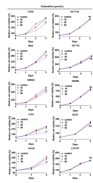
Cell viabilities of CRC cell lines treated with Terbinafine at 10, 25, 50 μmol/L were accessed by CCK8 assay. DMSO was used as control.
Terbinafine purchased from MedChemExpress. Usage Cited in: Cancer Commun (Lond). 2021 Aug;41(8):726-746. [Abstract]
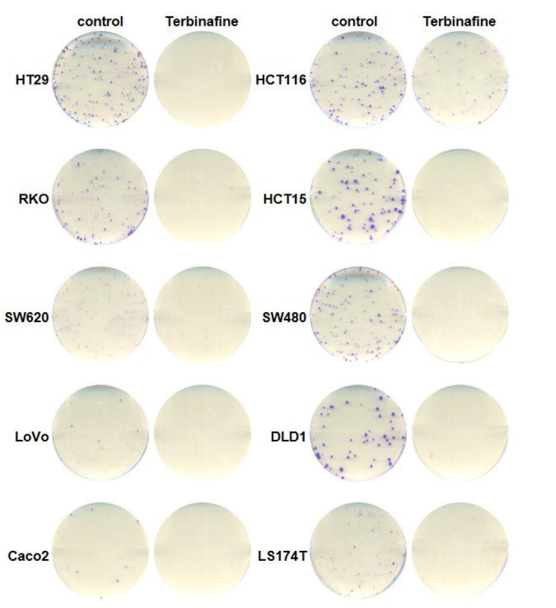
Colony formation of CRC cell lines treated with Terbinafine at 50 μmol/L. DMSO was used as control.
Terbinafine purchased from MedChemExpress. Usage Cited in: Cancer Commun (Lond). 2021 Aug;41(8):726-746. [Abstract]
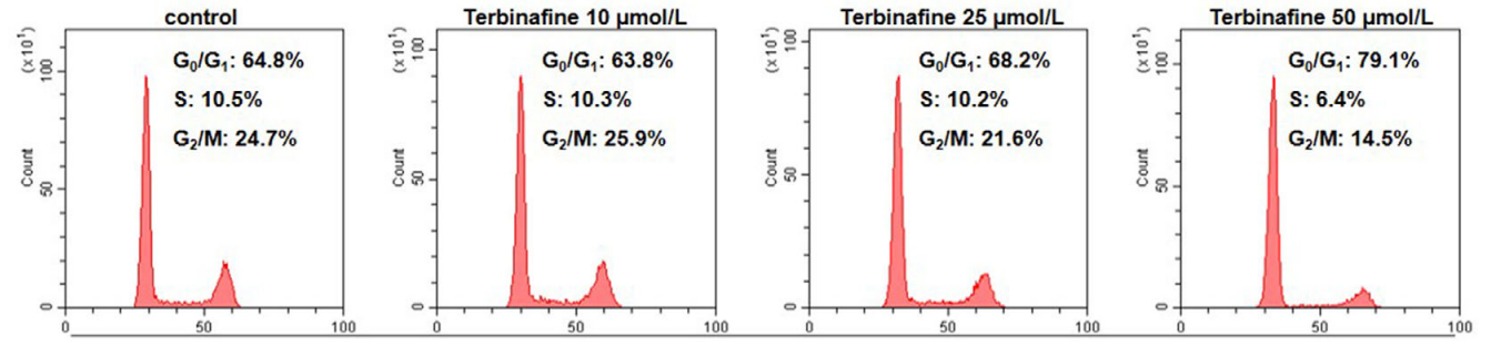
Cell cycle analysis (G0/G1, S, G2/M phase) of HT29 cells treated with terbinafine at 10, 25, 50 μmol/L. DMSO was used as control.
Terbinafine purchased from MedChemExpress. Usage Cited in: Cancer Commun (Lond). 2021 Aug;41(8):726-746. [Abstract]
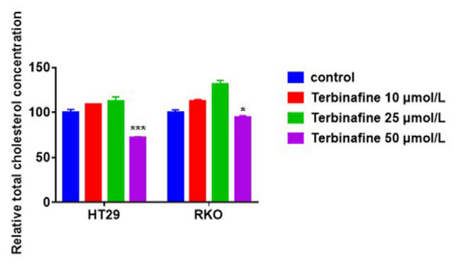
Intracellular cholesterol levels of HT29 or RKO cells treated with Terbinafine at 10, 25, 50 μmol/L were accessed by cholesterol measurement. DMSO was used as control.
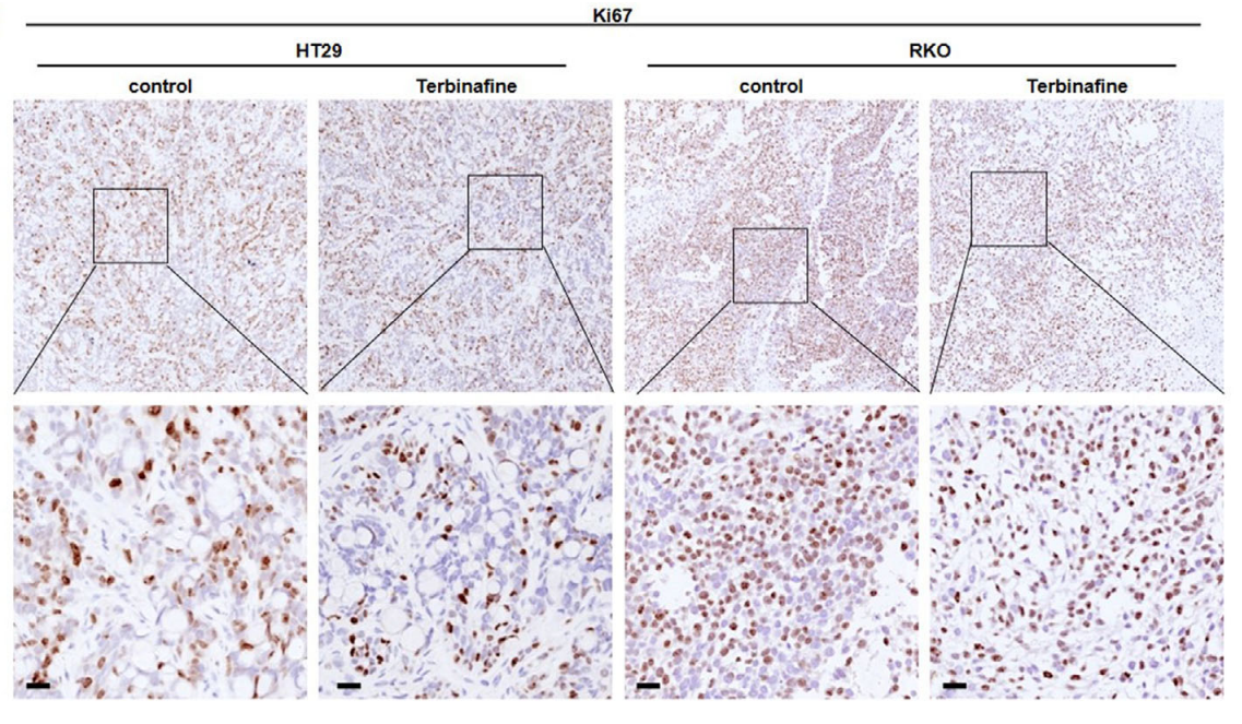
Terbinafine purchased from MedChemExpress. Usage Cited in: Cancer Commun (Lond). 2021 Aug;41(8):726-746. [Abstract]
IHC analysis of Ki67 in HT29 and RKO cells‐derived xenograft tumors and treated with terbinafine (oral, 50 mg/kg).
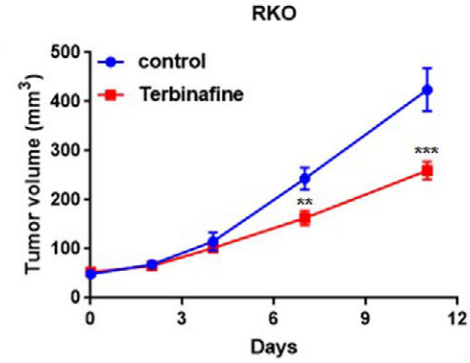
Terbinafine purchased from MedChemExpress. Usage Cited in: Cancer Commun (Lond). 2021 Aug;41(8):726-746. [Abstract]
Tumor growth curve of RKO cells treated with terbinafine (oral, 50 mg/kg) in nude mice.
-
Cancer Res
Squalene Epoxidase Metabolic Dependency Is a Targetable Vulnerability in Castration-Resistant Prostate Cancer. [Abstract]2022 Sep 2;82(17):3032-3044. PMID: 35767703 -
Nat Microbiol
Brain glucose induces tolerance of Cryptococcus neoformans to amphotericin B during meningitis. [Abstract]2024 Feb;9(2):346-358. PMID: 38225460 -
Autophagy
MoSec13 combined with MoGcn5b modulates MoAtg8 acetylation and regulates autophagy in Magnaporthe oryzae. [Abstract]2025 Oct;21(10):2266-2283. PMID: 40320672 -
Adv Sci (Weinh)
Cancer Cell-Intrinsic Cholesterol Induces Lipid-Associated Macrophage Differentiation via SP1 Palmitoylation to Promote Prostate Cancer Progression. [Abstract]2026 Apr;13(19):e08588. PMID: 41603134 -
Adv Sci (Weinh)
Targeting Squalene Epoxidase Confers Metabolic Vulnerability and Overcomes Chemoresistance in HNSCC. [Abstract]2023 Sep;10(27):e2206878. PMID: 37490552 -
Cell Death Dis
Targeting epigenetic modulation of cholesterol synthesis as a therapeutic strategy for head and neck squamous cell carcinoma. [Abstract]2021 May 13;12(5):482. PMID: 33986254 -
Cancer Lett
PD-L1 delactylation-promoted nuclear translocation accelerates liver cancer growth through elevating SQLE transcription activity. [Abstract]2025 Oct 10:630:217901. PMID: 40614853 -
Adv Healthc Mater
Cholesterol Metabolism Modulation Nanoplatform Improves Photo-Immunotherapeutic Effect In Oral Squamous Cell Carcinoma. [Abstract]2023 Sep;12(22):e2300018. PMID: 37209373 -
Int J Biol Macromol
Bletilla striata polysaccharide alleviates obesity by remodeling the gut microbiota-metabolite-liver axis and suppressing the hepatic AMPK-SREBP2/SQLE signaling pathway. [Abstract]2026 Apr:354:151416. PMID: 41833674 -
Clin Transl Med
Identifying squalene epoxidase as a metabolic vulnerability in high-risk osteosarcoma using an artificial intelligence-derived prognostic index. [Abstract]2024 Feb;14(2):e1586. PMID: 38372422 -
Probiotics Antimicrob Proteins
Screening the γ-core Motif Peptides of Ascomycetous Antifungal Proteins for Antifungal Activity and Potential Therapeutic Applicability. [Abstract]2026 Jan 6. PMID: 41493708 -
J Orthop Surg Res
Identification and validation of SQLE in steroid-induced osteonecrosis of the femoral head: a bioinformatics and experimental study. [Abstract]2025 Oct 17;20(1):894. PMID: 41107894 -
Infect Drug Resist
Correlation Between Drug Resistance and Virulence of Candida Isolates from Patients with Candidiasis. [Abstract]2022 Dec 15:15:7459-7473. PMID: 36544991 -
-
-
-


Solvent & Solubility
In Vitro:
DMSO : ≥ 100 mg/mL (343.14 mM; Hygroscopic DMSO has a significant impact on the solubility of product, please use newly opened DMSO)
* "≥" means soluble, but saturation unknown.
Please refer to the solubility information to select the appropriate solvent. Once prepared, please aliquot and store the solution to prevent product inactivation from repeated freeze-thaw cycles.
Storage method and period of stock solution: -80°C, 2 years; -20°C, 1 year. When stored at -80°C, please use it within 2 years. When stored at -20°C, please use it within 1 year.
Please refer to the solubility information to select the appropriate solvent. Once prepared, please aliquot and store the solution to prevent product inactivation from repeated freeze-thaw cycles.
Storage method and period of stock solution: -80°C, 2 years; -20°C, 1 year. When stored at -80°C, please use it within 2 years. When stored at -20°C, please use it within 1 year.
Concentration (start) × Volume (start) = Concentration (final) × Volume (final)
In Vivo:
Select the appropriate dissolution method based on your experimental animal and administration route.
- For the following dissolution methods, please ensure to first prepare a clear stock solution using an In Vitro approach and then sequentially add co-solvents:
- To ensure reliable experimental results, the clarified stock solution can be appropriately stored based on storage conditions. As for the working solution for In Vivo experiments, it is recommended to prepare freshly and use it on the same day.
- The percentages shown for the solvents indicate their volumetric ratio in the final prepared solution. If precipitation or phase separation occurs during preparation, heat and/or sonication can be used to aid dissolution.
Add each solvent one by one: 10% DMSO 40% PEG300 5% Tween-80 45% Saline
Solubility: ≥ 2.5 mg/mL (8.58 mM); Clear solution
This protocol yields a clear solution of ≥ 2.5 mg/mL (saturation unknown).
Taking 1 mL working solution as an example, add 100 μL DMSO stock solution (25.0 mg/mL) to 400 μL PEG300, and mix evenly; then add 50 μL Tween-80 and mix evenly; then add 450 μL Saline to adjust the volume to 1 mL.
Preparation of Saline: Dissolve 0.9 g sodium chloride in ddH₂O and dilute to 100 mL to obtain a clear Saline solution.
Add each solvent one by one: 10% DMSO 90% (20% SBE-β-CD in Saline)
Solubility: 2.5 mg/mL (8.58 mM); Suspended solution; Need ultrasonic
This protocol yields a suspended solution of 2.5 mg/mL. Suspended solution can be used for oral and intraperitoneal injection.
Taking 1 mL working solution as an example, add 100 μL DMSO stock solution (25.0 mg/mL) to 900 μL 20% SBE-β-CD in Saline, and mix evenly.
Preparation of 20% SBE-β-CD in Saline (4°C, storage for one week): 2 g SBE-β-CD powder is dissolved in 10 mL Saline, completely dissolve until clear.
In Vivo Dissolution Calculator
Please enter the basic information of animal experiments:
-
-
-
-
Recommended: Prepare an additional quantity of animals to account for potential losses during experiments.
Please enter your animal formula composition:
-
%DMSO +
Recommended: Keep the proportion of DMSO in working solution below 2% if your animal is weak.
-
%+
-
+%Tween-80 + +
-
%Saline +
The co-solvents required include: DMSO, . All of co-solvents are available by MedChemExpress (MCE). , Tween 80. All of co-solvents are available by MedChemExpress (MCE).
Working solution concentration: 0.22 mg/mL
Method for preparing stock solution: mg drug dissolved in μL DMSO. Stock solution concentration: mg/mL.
1. Take μL DMSO stock solution;
2. Add μL .
μL , mix evenly;
3. Then add μL Tween 80, mix evenly;
4. Then add μL
Please ensure that the stock solution in the first step is dissolved to a clear state, and add co-solvents in sequence. You can use ultrasonic heating (ultrasonic cleaner, recommended frequency 20-40 kHz), vortexing, etc. to assist dissolution.

Protocols
-
Research Protocol for Infectious Diseases
Infectious-disease experiments test how pathogens interact with host barriers, innate immune receptors, inflammatory signaling, pathogen replication, and tissue injury; pattern-recognition receptors such as TLRs, RIG-I-like receptors, NOD-like receptors, and inflammasomes detect microbial molecules and activate NF-κB, interferon, and cytokine responses. The central hypothesis is that infection severity reflects the balance between pathogen burden and host response: protective inflammation restricts pathogen growth, whereas excessive or mislocalized inflammation contributes to tissue damage and disease phenotype. Unresolved questions include which host pathways are protective versus pathogenic, why some infection models fail to translate to human disease, and which combined readouts best predict clinically relevant infection outcomes.
-
EdU Incorporation Assay (Click Chemistry-Based DNA Synthesis Measurement)
The EdU incorporation assay measures DNA synthesis by adding the thymidine analog 5-ethynyl-2′-deoxyuridine to cells or tissues, where it is incorporated into newly synthesized DNA during S phase. Incorporated EdU is detected by copper-catalyzed azide-alkyne cycloaddition, in which a fluorescent azide covalently reacts with the ethynyl group on EdU, allowing S-phase cells to be detected by fluorescence microscopy, flow cytometry, or high-content imaging. EdU detection does not require DNA denaturation or anti-BrdU antibody access, which preserves sample structure and improves compatibility with immunostaining and multiparameter cytometry compared with BrdU-based detection. EdU can be cytotoxic in a cell-type- and exposure-dependent manner, so pulse duration, concentration, and continuous-labeling designs should be validated for each cell type.
-
Gram Staining of Tissue Sections
Gram staining of tissue sections is a histochemical technique used to differentiate Gram-positive and Gram-negative bacteria within histological specimens based on differences in bacterial cell wall structure and dye retention, adapted from classical bacteriological Gram staining into tissue-compatible “histological Gram stain” variants. In tissue applications, modifications of the Brown-Hopps and Brown-Brenn methods are commonly used to improve differentiation of microorganisms embedded within host connective tissue and to reduce overstaining or loss of Gram-negative signal, which are known limitations of earlier approaches. The principle relies on crystal violet-iodine complex retention in Gram-positive organisms and subsequent decolorization and counterstaining steps that allow contrast visualization of Gram-negative organisms against tissue background.
-
Bacterial live/dead nucleic-acid viability staining
The LIVE/DEAD bacterial viability staining method is based on differential permeability of nucleic-acid-binding fluorescent dyes, most commonly SYTO 9 and propidium iodide (PI), which enables discrimination of bacterial populations with intact versus compromised cytoplasmic membranes. SYTO 9 penetrates both intact and damaged bacterial membranes and binds nucleic acids to produce green fluorescence, whereas propidium iodide penetrates only cells with compromised membranes and fluoresces red while also reducing SYTO 9 signal through competitive binding and fluorescence interactions. The resulting fluorescence pattern is interpreted as a proxy for membrane integrity, which is widely used as an indicator of bacterial viability in microscopy, flow cytometry, and spectroscopic platforms. However, mechanistic studies show that SYTO 9 and PI interactions involve displacement and fluorescence resonance energy transfer effects, which can influence signal interpretation depending on dye ratios a


Purity & Documentation
-
Data Sheet (276 KB)
-
SDS (393 KB)
- English - EN (393 KB)
- Français - FR (393 KB)
- Deutsch - DE (393 KB)
- Norwegian - NO (393 KB)
- Español - ES (393 KB)
- Swedish - SV (393 KB)
- Italian - IT (393 KB)
- Korean - KR (393 KB)
- Portuguese - PT (393 KB)
-
Handling Instructions (2659 KB)
References
[1]. Ryder NS, et al. Terbinafine: mode of action and properties of the squalene epoxidase inhibition. Br J Dermatol. 1992 Feb;126 Suppl 39:2-7. [Content Brief]
[2]. Mieth H, et al. Preclinical evaluation of terbinafine in vivo. Clin Exp Dermatol. 1989 Mar;14(2):104-7. [Content Brief]
[3]. Ciftci E, et al. Mupirocin vs terbinafine in impetigo.Indian J Pediatr. 2002 Aug;69(8):679-82. [Content Brief]


Complete Stock Solution Preparation Table
Please refer to the solubility information to select the appropriate solvent. Once prepared, please aliquot and store the solution to prevent product inactivation from repeated freeze-thaw cycles.
Storage method and period of stock solution: -80°C, 2 years; -20°C, 1 year. When stored at -80°C, please use it within 2 years. When stored at -20°C, please use it within 1 year.
| Optional Solvent | Concentration Solvent Mass | 1 mg | 5 mg | 10 mg | 25 mg |
|---|---|---|---|---|---|
| DMSO | 1 mM | 3.4314 mL | 17.1568 mL | 34.3136 mL | 85.7839 mL |
| 5 mM | 0.6863 mL | 3.4314 mL | 6.8627 mL | 17.1568 mL | |
| 10 mM | 0.3431 mL | 1.7157 mL | 3.4314 mL | 8.5784 mL | |
| 15 mM | 0.2288 mL | 1.1438 mL | 2.2876 mL | 5.7189 mL | |
| 20 mM | 0.1716 mL | 0.8578 mL | 1.7157 mL | 4.2892 mL | |
| 25 mM | 0.1373 mL | 0.6863 mL | 1.3725 mL | 3.4314 mL | |
| 30 mM | 0.1144 mL | 0.5719 mL | 1.1438 mL | 2.8595 mL | |
| 40 mM | 0.0858 mL | 0.4289 mL | 0.8578 mL | 2.1446 mL | |
| 50 mM | 0.0686 mL | 0.3431 mL | 0.6863 mL | 1.7157 mL | |
| 60 mM | 0.0572 mL | 0.2859 mL | 0.5719 mL | 1.4297 mL | |
| 80 mM | 0.0429 mL | 0.2145 mL | 0.4289 mL | 1.0723 mL | |
| 100 mM | 0.0343 mL | 0.1716 mL | 0.3431 mL | 0.8578 mL |


Terbinafine Related Classifications
HY-17395A Related Classifications
 Powered by Bioz