



ML604086
Based on 2 publication(s) in Google Scholar
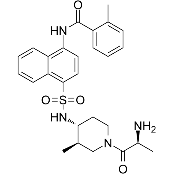
ML604086 is a selective CCR8 inhibitor, inhibiting CCL1 binding to CCR8 on circulating T-cells. ML604086 inhibits CCL1 mediated chemotaxis and increases in intracellular Ca2+ concentrations.
Para uso exclusivo en investigación. No vendemos a pacientes.
- Pureza : 99.78%
- No. CAS: 850330-18-6
- Fòrmula: C27H32N4O4S
- Peso molecular:508.63
-
Almacenamiento:Powder -20°C, 3 years , 4°C, 2 years ; In solvent -80°C, 2 years , -20°C, 1 year

To place orders, for customer services and technical support, please contact: MedChemExpress USA
Tel: 609-228-6898 E-mail: [email protected] [email protected]


-
 Actividad biológica
-
Chemical Information
-
Solvente y solubilidad
- Protocolo
- Pureza y Documentación
- Referencias
-
 Help & FAQs


-
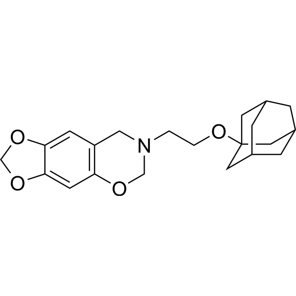
HY-122219
R243CCR8 Antagonist -
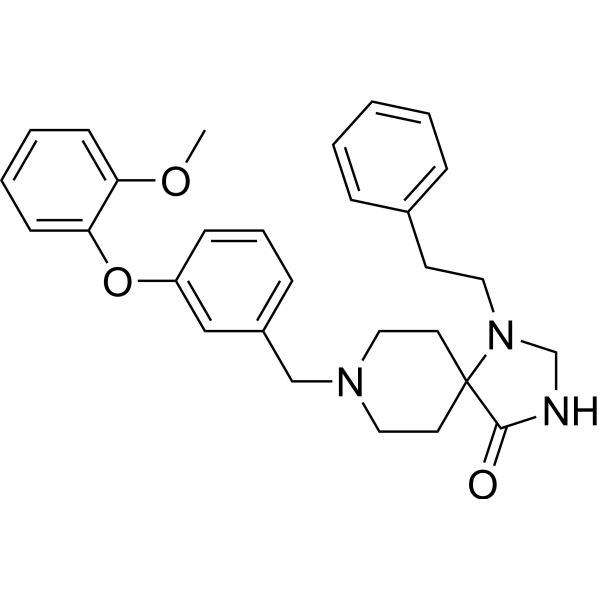
HY-121885
LMD-009CCR8 Nonpeptide Agonist -
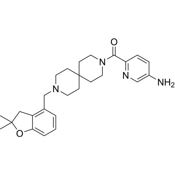
HY-119217
AZ084CCR8 Allosteric Antagonist -
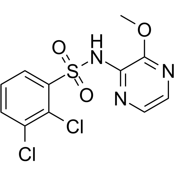
HY-U00064
AZD2098 -
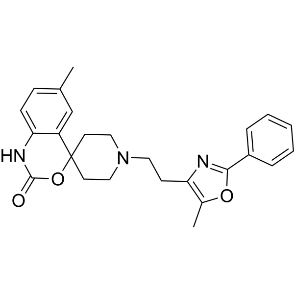
HY-15418
RS 504393CCR2 Antagonist -
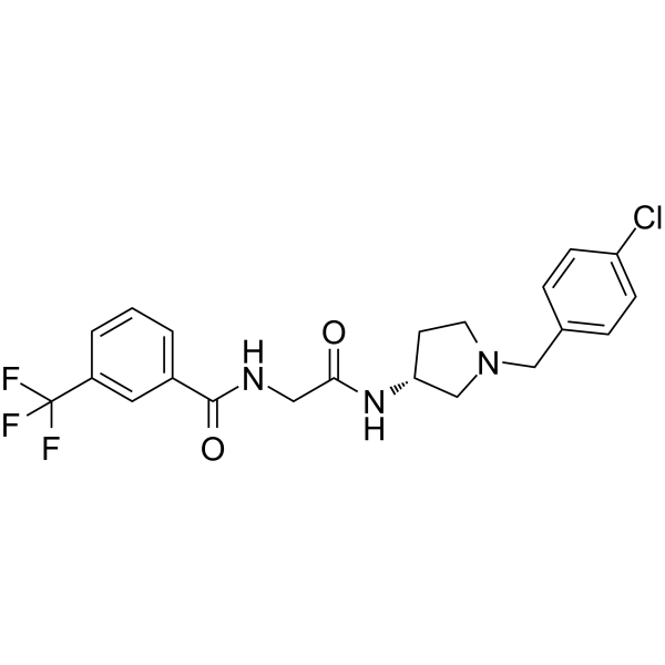
HY-108323
CCR2 antagonist 4CCR2b Antagonist -
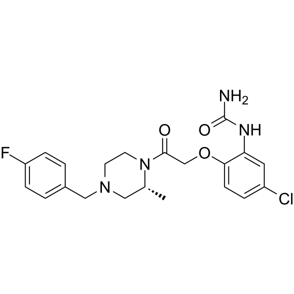
HY-12080
BX471CCR1 Antagonist -
![CCR2-RA-[R]](https://file.medchemexpress.com/product_pic/hy-50081.gif)
HY-50081
CCR2-RA-[R]CCR Antagonist -
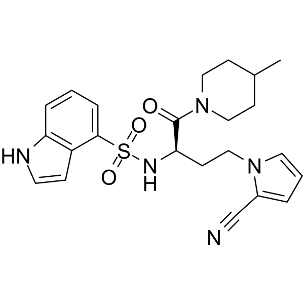
HY-116835
BI-6901CCR10 Antagonist -

HY-P7198
I-309/CCL1 Protein, Human (CHO) -
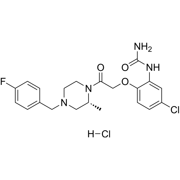
HY-12080A
BX471 hydrochlorideCCR Antagonist -
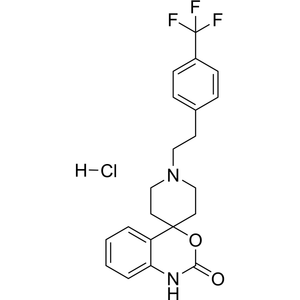
HY-18611
Abaucin hydrochlorideCCR2antagonist
Publications Citing Use of MedChemExpress (MCE) ML604086
More

Actividad biológica
Descripciòn
IC50 & Target
|
CCR8 |
In Vitro
ML604086 (0-100 μM) inhibits CCL1 mediated chemotaxis and increases in intracellular Ca2+ concentrations of cell lines stably expressing cyno CCR8 with IC50s of 1.3 μM and 1.0 μM, respectively[2].
ML604086 (10, 30 μM; 64 h) inhibits CCL1 binding to CCR8 on CD4 T-cells, and inhibits the serotonin receptor 5HT1a with the inhibition rate of 30%, 70% at 10, 30 μM respectively[2].
MedChemExpress (MCE) has not independently confirmed the accuracy of these methods. They are for reference only.
In Vivo
MedChemExpress (MCE) has not independently confirmed the accuracy of these methods. They are for reference only.
-
Animal Model:3.0-5.0 kg male and female adult Macaca fascicularis (primate model of asthma)[2]
-
Dosage:1.038 mg/kg
-
Administration:Intravenous infusion
-
Result:Did not effected the Changes in airway resistance and compliance induced by allergen provocation and increasing concentrations of methacholine.


Chemical Information
-
No. CAS 850330-18-6
-
Appearance Solid
-
Peso molecular 508.63
-
Fòrmula C27H32N4O4S
-
Color White to off-white
-
SMILES
O=C(NC1=C2C=CC=CC2=C(S(=O)(N[C@H]3[C@H](C)CN(C([C@@H](N)C)=O)CC3)=O)C=C1)C4=CC=CC=C4C
-
Envío
Room temperature in continental US; may vary elsewhere.
-
Almacenamiento
Powder -20°C 3 years 4°C 2 years In solvent -80°C 2 years -20°C 1 year


Publications (2)
-
Journal Impact Factor
-
Most Recent
-
J Clin Invest
Progranulin-dependent repair function of regulatory T cells drives bone-fracture healing. [Abstract]2024 Nov 7;135(2):e180679. PMID: 39509336 -
Cell Signal
CCL17 drives the expression of MMP9 and MMP13 expression via ERK1/2 and NF-κB signaling pathways in rheumatoid arthritis. [Abstract]2026 Aug:144:112551. PMID: 42019646


Solvente y solubilidad
In Vitro:
DMSO : 120 mg/mL (235.93 mM; Need ultrasonic; Hygroscopic DMSO has a significant impact on the solubility of product, please use newly opened DMSO)
Please refer to the solubility information to select the appropriate solvent. Once prepared, please aliquot and store the solution to prevent product inactivation from repeated freeze-thaw cycles.
Storage method and period of stock solution: -80°C, 2 years; -20°C, 1 year. When stored at -80°C, please use it within 2 years. When stored at -20°C, please use it within 1 year.
Please refer to the solubility information to select the appropriate solvent. Once prepared, please aliquot and store the solution to prevent product inactivation from repeated freeze-thaw cycles.
Storage method and period of stock solution: -80°C, 2 years; -20°C, 1 year. When stored at -80°C, please use it within 2 years. When stored at -20°C, please use it within 1 year.
Concentration (start) × Volume (start) = Concentration (final) × Volume (final)
In Vivo:
Select the appropriate dissolution method based on your experimental animal and administration route.
- For the following dissolution methods, please ensure to first prepare a clear stock solution using an In Vitro approach and then sequentially add co-solvents:
- To ensure reliable experimental results, the clarified stock solution can be appropriately stored based on storage conditions. As for the working solution for In Vivo experiments, it is recommended to prepare freshly and use it on the same day.
- The percentages shown for the solvents indicate their volumetric ratio in the final prepared solution. If precipitation or phase separation occurs during preparation, heat and/or sonication can be used to aid dissolution.
Add each solvent one by one: 10% DMSO 40% PEG300 5% Tween-80 45% Saline
Solubility: ≥ 6.25 mg/mL (12.29 mM); Clear solution
This protocol yields a clear solution of ≥ 6.25 mg/mL (saturation unknown).
Taking 1 mL working solution as an example, add 100 μL DMSO stock solution (62.5 mg/mL) to 400 μL PEG300, and mix evenly; then add 50 μL Tween-80 and mix evenly; then add 450 μL Saline to adjust the volume to 1 mL.
Preparation of Saline: Dissolve 0.9 g sodium chloride in ddH₂O and dilute to 100 mL to obtain a clear Saline solution.
In Vivo Dissolution Calculator
Please enter the basic information of animal experiments:
-
-
-
-
Recommended: Prepare an additional quantity of animals to account for potential losses during experiments.
Please enter your animal formula composition:
-
%DMSO +
Recommended: Keep the proportion of DMSO in working solution below 2% if your animal is weak.
-
%+
-
+%Tween-80 + +
-
%Saline +
The co-solvents required include: DMSO, . All of co-solvents are available by MedChemExpress (MCE). , Tween 80. All of co-solvents are available by MedChemExpress (MCE).
Working solution concentration: 0.22 mg/mL
Method for preparing stock solution: mg drug dissolved in μL DMSO. Stock solution concentration: mg/mL.
1. Take μL DMSO stock solution;
2. Add μL .
μL , mix evenly;
3. Then add μL Tween 80, mix evenly;
4. Then add μL
Please ensure that the stock solution in the first step is dissolved to a clear state, and add co-solvents in sequence. You can use ultrasonic heating (ultrasonic cleaner, recommended frequency 20-40 kHz), vortexing, etc. to assist dissolution.


Pureza y Documentación
-
Ficha de datos (278 KB)
-
SDS (251 KB)
- English - EN (251 KB)
- Français - FR (251 KB)
- Deutsch - DE (251 KB)
- Norwegian - NO (251 KB)
- Español - ES (251 KB)
- Swedish - SV (251 KB)
- Italian - IT (251 KB)
- Korean - KR (251 KB)
- Portuguese - PT (251 KB)
-
Instrucciones de manejo (2659 KB)
Referencias


Complete Stock Solution Preparation Table
Please refer to the solubility information to select the appropriate solvent. Once prepared, please aliquot and store the solution to prevent product inactivation from repeated freeze-thaw cycles.
Storage method and period of stock solution: -80°C, 2 years; -20°C, 1 year. When stored at -80°C, please use it within 2 years. When stored at -20°C, please use it within 1 year.
| Optional Solvent | Concentration Solvent Mass | 1 mg | 5 mg | 10 mg | 25 mg |
|---|---|---|---|---|---|
| DMSO | 1 mM | 1.9661 mL | 9.8303 mL | 19.6607 mL | 49.1516 mL |
| 5 mM | 0.3932 mL | 1.9661 mL | 3.9321 mL | 9.8303 mL | |
| 10 mM | 0.1966 mL | 0.9830 mL | 1.9661 mL | 4.9152 mL | |
| 15 mM | 0.1311 mL | 0.6554 mL | 1.3107 mL | 3.2768 mL | |
| 20 mM | 0.0983 mL | 0.4915 mL | 0.9830 mL | 2.4576 mL | |
| 25 mM | 0.0786 mL | 0.3932 mL | 0.7864 mL | 1.9661 mL | |
| 30 mM | 0.0655 mL | 0.3277 mL | 0.6554 mL | 1.6384 mL | |
| 40 mM | 0.0492 mL | 0.2458 mL | 0.4915 mL | 1.2288 mL | |
| 50 mM | 0.0393 mL | 0.1966 mL | 0.3932 mL | 0.9830 mL | |
| 60 mM | 0.0328 mL | 0.1638 mL | 0.3277 mL | 0.8192 mL | |
| 80 mM | 0.0246 mL | 0.1229 mL | 0.2458 mL | 0.6144 mL | |
| 100 mM | 0.0197 mL | 0.0983 mL | 0.1966 mL | 0.4915 mL |


ML604086 Related Classifications
HY-124416 Related Classifications
 Powered by Bioz