



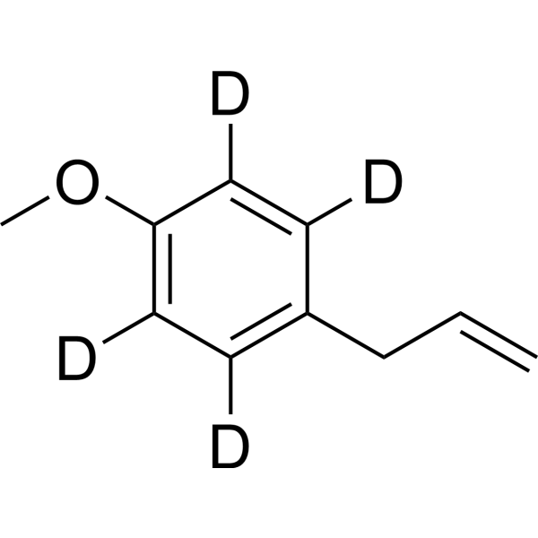
Estragole-d4

Based on 1 Customer Validation
Estragole-d4 is deuterated labeled Estragole (HY-N5060). Estragole (4-Allylanisole) is a relatively nontoxic volatile terpenoid ether and major component of the essential oil from many plants. Estragole significantly triggers Apoptosis, suppresses LPS-induced intracellular ROS production. Estragole activats Nrf-2 and regulates NF-κB. Estragole has anti-toxoplasma, anti-inflammatory, anti-edema, antioxidant and immunomodulatory properties. Estragole blocks DRG neuron excitability. Estragole has improves gastric ulcer activity[10].
For research use only. We do not sell to patients.
- Purity : 99.9%
- CAS No.: 1335401-40-5
- Formula: C10H8D4O
- Molecular Weight:152.23
-
Storage:Pure form -20°C, 3 years , 4°C, 2 years ; In solvent -80°C, 6 months , -20°C, 1 month

To place orders, for customer services and technical support, please contact: MedChemExpress USA
Tel: 609-228-6898 E-mail: [email protected] [email protected]


-
 Biological Activity
-
Chemical Information
- Protocols
- Purity & Documentation
- References
-
 Help & FAQs


All Parasite Isoforms
More

Biological Activity
Description
In Vitro
Stable heavy isotopes of hydrogen, carbon, and other elements have been incorporated into drug molecules, largely as tracers for quantitation during the drug development process. Deuteration has gained attention because of its potential to affect the pharmacokinetic and metabolic profiles of drugs[1].
MedChemExpress (MCE) has not independently confirmed the accuracy of these methods. They are for reference only. Further protocols information, click here.
Application
1. This compound can be used as a tracer
2. This compound can be used as an internal standard for quantitative analysis by NMR, GC-MS, or LC-MS.


Chemical Information
-
CAS No. 1335401-40-5
-
Unlabeled CAS 140-67-0
-
Appearance Liquid
-
Molecular Weight 152.23
-
Formula C10H8D4O
-
Color Light yellow to brown
-
SMILES
C=CCC1=C([2H])C([2H])=C(C([2H])=C1[2H])OC
-
Synonyms
4-Allylanisole-d4
-
Shipping
Room temperature in continental US; may vary elsewhere.
-
Storage
Pure form -20°C 3 years 4°C 2 years In solvent -80°C 6 months -20°C 1 month

Protocols
-
Apoptosis
Apoptosis, also called programmed cell death, is generally characterized by distinct morphological characteristics.
-
TUNEL staining for apoptotic DNA fragmentation
TUNEL staining detects DNA strand breaks by using terminal deoxynucleotidyl transferase to add labeled nucleotides to exposed 3′-OH DNA termini, generating either microscopic staining in fixed cells or tissue sections, or fluorescence/cytometric signal in cell suspensions. TUNEL positivity reflects DNA fragmentation but should not be interpreted alone as definitive apoptosis, because TUNEL can also label necrotic, autolytic, mechanically damaged, or DNA-repair-associated DNA breaks.
-
Annexin V plus membrane-impermeant dye apoptosis staining
Annexin V-based apoptosis assays rely on the detection of phosphatidylserine (PS) externalization from the inner leaflet of the plasma membrane to the outer leaflet, an early biochemical hallmark of apoptosis. Fluorescently labeled Annexin V binds PS in a calcium-dependent manner, enabling identification of early apoptotic cells by flow cytometry or fluorescence microscopy. When combined with a membrane-impermeant DNA-binding dye (e. g. , propidium iodide), this approach allows discrimination between viable (Annexin V−/dye−), early apoptotic (Annexin V+/dye−), and late apoptotic or necrotic (Annexin V+/dye+) cell populations by assessing membrane integrity and PS exposure.
-
ROS/oxidative-stress fluorescent staining
ROS/oxidative-stress fluorescent staining uses cell-permeant fluorogenic probes that become fluorescent after oxidation inside cells or tissues; commonly used examples include DCFH-DA/DCFDA for broad cellular oxidant detection, DHE for superoxide-related signal detection, MitoSOX for mitochondrial superoxide-related signal detection, and CellROX probes for oxidative-stress-associated fluorescence readouts. The assay detects probe oxidation rather than a single ROS species unless the probe and analysis method have been chemically validated for that species. DCFH-DA enters cells, is deacetylated by intracellular esterases to DCFH, and produces fluorescent DCF after oxidation, so the readout is used as an operational measure of total cellular oxidative stress rather than a species-specific ROS measurement. DHE and MitoSOX can report superoxide-related oxidation, but red fluorescence alone can include non-specific ethidium-like oxidation products; HPLC or optimized spectral approaches are
-
Apoptosis Solutions
Apoptosis is a regulated, generally non-lytic cell-death pathway that removes unwanted, damaged, infected, or abnormal cells through coordinated morphological changes, caspase activation, DNA fragmentation, and membrane remodeling. The intrinsic apoptosis pathway is controlled mainly by mitochondrial outer membrane permeabilization, BCL-2 family proteins, cytochrome c release, apoptosome formation, caspase-9 activation, and downstream executioner caspase-3/7 activation. The extrinsic apoptosis pathway is initiated by death receptors such as Fas, TNFR, and TRAIL receptors, which recruit adaptor proteins and activate caspase-8 before engaging executioner caspases or mitochondrial amplification through BID cleavage. Apoptosis is linked to many phenotypes, including cancer cell killing, tissue homeostasis, immune regulation, neurodegeneration, infection response, and treatment-induced cytotoxicity; unresolved questions include how apoptosis interacts with necroptosis, pyroptosis, ferroptos
-
Research Protocol for Inflammation-related Diseases
The NLRP3 inflammasome is a cytosolic innate immune signaling platform that integrates priming signals and danger-signal activation to promote caspase-1 activation, maturation of IL-1β and IL-18, and gasdermin D-mediated pyroptotic cell death. The core experimental logic is to determine whether inflammatory disease phenotypes are driven by increased NLRP3 expression, ASC-containing inflammasome assembly, caspase-1 cleavage, GSDMD cleavage, and extracellular release of IL-1β/IL-18 rather than by nonspecific cell injury alone. The pathway is strongly linked to inflammation-related disease phenotypes because monosodium urate crystals activate NALP3/NLRP3 inflammasome signaling in gout-like crystal inflammation, cholesterol crystals activate NLRP3 inflammasomes in atherogenesis models, and DSS-induced intestinal inflammation has been reported to involve NLRP3 inflammasome activity. However, experimental colitis studies also show context-dependent protective effects of NLRP3 inflammasome co
-
Primary Dorsal Root Ganglion Sensory Neuron Culture
Primary dorsal root ganglion sensory neuron culture isolates DRG neuronal somata from rodent or human ganglia, dissociates tissue enzymatically and mechanically, and maintains post-mitotic sensory neurons in vitro for readouts such as neurite outgrowth, immunocytochemical marker expression, calcium imaging, electrophysiology, RNA/protein analysis, or neuropeptide release assays. The method reflects peripheral sensory neuron biology because DRG neurons are primary sensory neurons whose cell bodies reside in dorsal root ganglia and whose cultured dissociated cells can retain neuronal morphology, sensory-neuron marker expression, and stimulus-responsive properties depending on the downstream assay.


Purity & Documentation
-
Data Sheet (269 KB)
-
SDS (762 KB)
- English - EN (762 KB)
- Français - FR (762 KB)
- Deutsch - DE (762 KB)
- Norwegian - NO (762 KB)
- Español - ES (762 KB)
- Swedish - SV (762 KB)
- Italian - IT (762 KB)
- Korean - KR (762 KB)
- Portuguese - PT (762 KB)
-
Handling Instructions (2659 KB)
References
[1]. Russak EM, et al. Impact of Deuterium Substitution on the Pharmacokinetics of Pharmaceuticals. Ann Pharmacother. 2019 Feb;53(2):211-216. [Content Brief]
[2]. Leal-Cardoso JH, et al. Effects of estragole on the compound action potential of the rat sciatic nerve. Braz J Med Biol Res. 2004 Aug;37(8):1193-8. [Content Brief]
[3]. Oliveira CB, et al. Anti-Toxoplasma Activity of Estragole and Thymol in Murine Models of Congenital and Noncongenital Toxoplasmosis. J Parasitol. 2016 Jun;102(3):369-76. [Content Brief]

Calculators
Concentration (start) × Volume (start) = Concentration (final) × Volume (final)


Estragole-d4 Related Classifications
HY-N5060S Related Classifications
 Powered by Bioz