



Estragole

Based on 1 Customer Validation
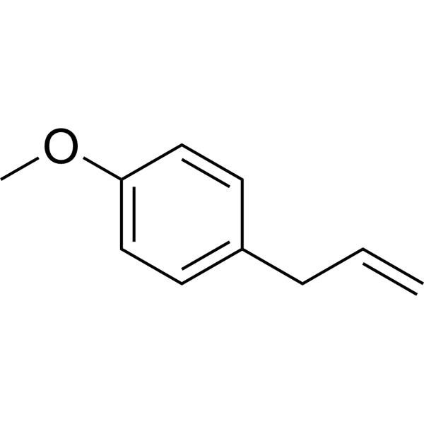
Estragole (4-Allylanisole) is a relatively nontoxic volatile terpenoid ether and major component of the essential oil from many plants. Estragole significantly triggers Apoptosis, suppresses LPS-induced intracellular ROS production. Estragole activats Nrf-2 and regulates NF-κB. Estragole has anti-toxoplasma, anti-inflammatory, anti-edema, antioxidant and immunomodulatory properties. Estragole blocks DRG neuron excitability. Estragole has improves gastric ulcer activity.
For research use only. We do not sell to patients.
- Purity : 99.21%
- CAS No.: 140-67-0
- Formula: C10H12O
- Molecular Weight:148.20
-
Storage:Pure form -20°C, 3 years , 4°C, 2 years ; In solvent -80°C, 6 months , -20°C, 1 month

To place orders, for customer services and technical support, please contact: MedChemExpress USA
Tel: 609-228-6898 E-mail: [email protected] [email protected]


-
 Biological Activity
-
Chemical Information
-
Solvent & Solubility
- Protocols
- Purity & Documentation
- References
-
 Help & FAQs



All Parasite Isoforms
More

Biological Activity
Description
IC50 & Target
|
Toxoplasma |
In Vitro
Estragole (0-200 μg/mL, 24 h) exhibits a dose-dependent cytotoxic activity in the monolayer culture of MCF7 cell line with IC50 value of 74 μg/mL[3].
Estragole (2000 μM, 24 h) induces apoptosis in AA8 and EM9 cells[4].
Estragole (84.5-674 μM, 2 h) exhibits anti-inflammatory activity with the regulation of NF-κB and activation of Nrf-2 signaling pathways in LPS-induced RAW 264.7 cells[5].
Estragole (1-300 μM) increases micronuclei counts (3.2-7.1 fold) in HepG2-CYP1A2 cells[6].
Estragole (31.25-500 μg/mL, 24 h) does not exhibit significant cytotoxic effects in the A. Salina and hemolysis tests[7].
Estragole (3-60 μg/mL, 30 min) inhibits neutrophil migration toward fMLP[8].
Estragole (0.6-14 mM) blocks DRG neuronal excitability by direct inhibition of Na+ channels[9].
MedChemExpress (MCE) has not independently confirmed the accuracy of these methods. They are for reference only.
-
Cell Line:MCF7
-
Concentration:25, 37, 50, 74, 75, 100 μg/mL
-
Incubation Time:4 h
-
Result:Showed apparent apoptotic morphological changes such as nuclear fragmentation, chromatin condensation, and destruction of cell membrane integrity.
Triggered apoptosis.
Showed the early apoptotic cells population of 43% after treatment with IC50/2 (IC50: 74 μg/mL) concentration for 4 h.
Increased and reached 55% after treatment with IC50 (74 μg/mL) concentration for 4 h.
Increased the activity of caspase-3.
In Vivo
Estragole (30-60 mg/kg, p.o.) shows anti-inflammatory and antiedematogenic activity, with reducing paw edema in mice[10].
Estragole (31.2-250 mg/kg, p.o., pre-treated 30 min- 1 h) prevents gastric ulcers via cytoprotective, antioxidant and immunoregulatory mechanisms in Swiss mice[11].
Estragole (100 mg/kg, p.o., daily, 6 consecutive days) shows anti-toxoplasma activity in murine models of congenital and noncongenital toxoplasmosis[12].
Estragole (100 g, i.p., once a week for 17 weeks) decreases the HNF4 and FOXA DNA-binding activities and inhibits the induction of tyrosine aminotransferase and tryptophan oxygenase only in susceptible female mice[13].
MedChemExpress (MCE) has not independently confirmed the accuracy of these methods. They are for reference only.
-
Animal Model:Carrageenan-induced peritonitis model[8]
-
Dosage:250, 500, 750 mg/kg
-
Administration:Oral gavage (p.o.), 30 min before the intraperitoneal injection carrageenan solution
-
Result:Inhibited the leukocyte migration at doses of 500 (51%) and 750 mg/kg (52%).
Had a similar effect on anethole.


Chemical Information
-
CAS No. 140-67-0
-
Appearance Liquid (Density: 0.965 g/cm3 )
-
Molecular Weight 148.20
-
Formula C10H12O
-
Color Colorless to light yellow
-
SMILES
C=CCC1=CC=C(OC)C=C1
-
Synonyms
4-Allylanisole
-
Structure Classification
-
Initial Source
-
Shipping
Room temperature in continental US; may vary elsewhere.
-
Storage
Pure form -20°C 3 years 4°C 2 years In solvent -80°C 6 months -20°C 1 month


Solvent & Solubility
In Vitro:
DMSO : 100 mg/mL (674.76 mM; Need ultrasonic; Hygroscopic DMSO has a significant impact on the solubility of product, please use newly opened DMSO)
Please refer to the solubility information to select the appropriate solvent. Once prepared, please aliquot and store the solution to prevent product inactivation from repeated freeze-thaw cycles.
Storage method and period of stock solution: -80°C, 6 months; -20°C, 1 month. When stored at -80°C, please use it within 6 months. When stored at -20°C, please use it within 1 month.
Please refer to the solubility information to select the appropriate solvent. Once prepared, please aliquot and store the solution to prevent product inactivation from repeated freeze-thaw cycles.
Storage method and period of stock solution: -80°C, 6 months; -20°C, 1 month. When stored at -80°C, please use it within 6 months. When stored at -20°C, please use it within 1 month.
Concentration (start) × Volume (start) = Concentration (final) × Volume (final)
In Vivo:
Select the appropriate dissolution method based on your experimental animal and administration route.
- For the following dissolution methods, please ensure to first prepare a clear stock solution using an In Vitro approach and then sequentially add co-solvents:
- To ensure reliable experimental results, the clarified stock solution can be appropriately stored based on storage conditions. As for the working solution for In Vivo experiments, it is recommended to prepare freshly and use it on the same day.
- The percentages shown for the solvents indicate their volumetric ratio in the final prepared solution. If precipitation or phase separation occurs during preparation, heat and/or sonication can be used to aid dissolution.
Add each solvent one by one: 10% DMSO 40% PEG300 5% Tween-80 45% Saline
Solubility: ≥ 2.5 mg/mL (16.87 mM); Clear solution
This protocol yields a clear solution of ≥ 2.5 mg/mL (saturation unknown).
Taking 1 mL working solution as an example, add 100 μL DMSO stock solution (25.0 mg/mL) to 400 μL PEG300, and mix evenly; then add 50 μL Tween-80 and mix evenly; then add 450 μL Saline to adjust the volume to 1 mL.
Preparation of Saline: Dissolve 0.9 g sodium chloride in ddH₂O and dilute to 100 mL to obtain a clear Saline solution.
Add each solvent one by one: 10% DMSO 90% (20% SBE-β-CD in Saline)
Solubility: ≥ 2.5 mg/mL (16.87 mM); Clear solution
This protocol yields a clear solution of ≥ 2.5 mg/mL (saturation unknown).
Taking 1 mL working solution as an example, add 100 μL DMSO stock solution (25.0 mg/mL) to 900 μL 20% SBE-β-CD in Saline, and mix evenly.
Preparation of 20% SBE-β-CD in Saline (4°C, storage for one week): 2 g SBE-β-CD powder is dissolved in 10 mL Saline, completely dissolve until clear.
In Vivo Dissolution Calculator
Please enter the basic information of animal experiments:
-
-
-
-
Recommended: Prepare an additional quantity of animals to account for potential losses during experiments.
Please enter your animal formula composition:
-
%DMSO +
Recommended: Keep the proportion of DMSO in working solution below 2% if your animal is weak.
-
%+
-
+%Tween-80 + +
-
%Saline +
The co-solvents required include: DMSO, . All of co-solvents are available by MedChemExpress (MCE). , Tween 80. All of co-solvents are available by MedChemExpress (MCE).
Working solution concentration: 0.22 mg/mL
Method for preparing stock solution: mg drug dissolved in μL DMSO. Stock solution concentration: mg/mL.
1. Take μL DMSO stock solution;
2. Add μL .
μL , mix evenly;
3. Then add μL Tween 80, mix evenly;
4. Then add μL
Please ensure that the stock solution in the first step is dissolved to a clear state, and add co-solvents in sequence. You can use ultrasonic heating (ultrasonic cleaner, recommended frequency 20-40 kHz), vortexing, etc. to assist dissolution.


Purity & Documentation
-
Data Sheet (278 KB)
-
SDS (762 KB)
- English - EN (762 KB)
- Français - FR (762 KB)
- Deutsch - DE (762 KB)
- Norwegian - NO (762 KB)
- Español - ES (762 KB)
- Swedish - SV (762 KB)
- Italian - IT (762 KB)
- Korean - KR (762 KB)
- Portuguese - PT (762 KB)
-
Handling Instructions (2659 KB)
References
[1]. Leal-Cardoso JH, et al. Effects of estragole on the compound action potential of the rat sciatic nerve. Braz J Med Biol Res. 2004 Aug;37(8):1193-8. [Content Brief]
[2]. Oliveira CB, et al. Anti-Toxoplasma Activity of Estragole and Thymol in Murine Models of Congenital and Noncongenital Toxoplasmosis. J Parasitol. 2016 Jun;102(3):369-76. [Content Brief]
[4]. Martins C, et al. Estragole: a weak direct-acting food-borne genotoxin and potential carcinogen. Mutat Res. 2012 Aug 30;747(1):86-92. [Content Brief]
[6]. Schulte-Hubbert R, et al. Estragole: DNA adduct formation in primary rat hepatocytes and genotoxic potential in HepG2-CYP1A2 cells. Toxicology. 2020 Nov;444:152566. [Content Brief]
[7]. Coêlho ML, et al. Cytotoxic and Antioxidant Properties of Natural Bioactive Monoterpenes Nerol, Estragole, and 3,7-Dimethyl-1-Octanol. Adv Pharmacol Pharm Sci. 2022 Nov 23;2022:8002766. [Content Brief]
[8]. Silva-Comar FM, et al. Effect of estragole on leukocyte behavior and phagocytic activity of macrophages. Evid Based Complement Alternat Med. 2014;2014:784689. [Content Brief]
[9]. Silva-Alves KS, et al. Estragole blocks neuronal excitability by direct inhibition of Na+ channels. Braz J Med Biol Res. 2013 Dec;46(12):1056-1063. [Content Brief]
[10]. Rodrigues LB, et al. Anti-inflammatory and antiedematogenic activity of the Ocimum basilicum essential oil and its main compound estragole: In vivo mouse models. Chem Biol Interact. 2016 Sep 25;257:14-25. [Content Brief]
[11]. Alves Júnior EB, et al. Estragole prevents gastric ulcers via cytoprotective, antioxidant and immunoregulatory mechanisms in animal models. Biomed Pharmacother. 2020 Oct;130:110578. [Content Brief]
[12]. Oliveira CB, et al. Anti-Toxoplasma Activity of Estragole and Thymol in Murine Models of Congenital and Noncongenital Toxoplasmosis. J Parasitol. 2016 Jun;102(3):369-76. [Content Brief]
[13]. Kaledin VI, et al. Effect of hepatocarcinogenicity of estragole on the glucocorticoid-mediated induction of liver-specific enzymes and the activity of the transcription factors FOXA and HNF4 in the liver of mouse and rat. Biofizika. 2010 Mar-Apr;55(2):326-35. Russian. [Content Brief]


Complete Stock Solution Preparation Table
Please refer to the solubility information to select the appropriate solvent. Once prepared, please aliquot and store the solution to prevent product inactivation from repeated freeze-thaw cycles.
Storage method and period of stock solution: -80°C, 6 months; -20°C, 1 month. When stored at -80°C, please use it within 6 months. When stored at -20°C, please use it within 1 month.
| Optional Solvent | Concentration Solvent Mass | 1 mg | 5 mg | 10 mg | 25 mg |
|---|---|---|---|---|---|
| DMSO | 1 mM | 6.7476 mL | 33.7382 mL | 67.4764 mL | 168.6910 mL |
| 5 mM | 1.3495 mL | 6.7476 mL | 13.4953 mL | 33.7382 mL | |
| 10 mM | 0.6748 mL | 3.3738 mL | 6.7476 mL | 16.8691 mL | |
| 15 mM | 0.4498 mL | 2.2492 mL | 4.4984 mL | 11.2461 mL | |
| 20 mM | 0.3374 mL | 1.6869 mL | 3.3738 mL | 8.4345 mL | |
| 25 mM | 0.2699 mL | 1.3495 mL | 2.6991 mL | 6.7476 mL | |
| 30 mM | 0.2249 mL | 1.1246 mL | 2.2492 mL | 5.6230 mL | |
| 40 mM | 0.1687 mL | 0.8435 mL | 1.6869 mL | 4.2173 mL | |
| 50 mM | 0.1350 mL | 0.6748 mL | 1.3495 mL | 3.3738 mL | |
| 60 mM | 0.1125 mL | 0.5623 mL | 1.1246 mL | 2.8115 mL | |
| 80 mM | 0.0843 mL | 0.4217 mL | 0.8435 mL | 2.1086 mL | |
| 100 mM | 0.0675 mL | 0.3374 mL | 0.6748 mL | 1.6869 mL |


Estragole Related Classifications
HY-N5060 Related Classifications
 Powered by Bioz