



Serplulimab

Based on 1 Customer Validation
Serplulimab (HLX 10) is a humanized anti-PD-1 monoclonal antibody. Serplulimab can inhibit tumor growth, regulate the tumor microenvironment, and has anti-tumor activity. Serplulimab can be used in the research of cancer such as lung cancer and colon cancer.
For research use only. We do not sell to patients.
- Purity : 99.30%
- CAS No.: 2231029-82-4
- Molecular Weight:144.18 kDa
-
Storage:
Please store the product under the recommended conditions in the Certificate of Analysis.


To place orders, for customer services and technical support, please contact: MedChemExpress USA
Tel: 609-228-6898 E-mail: [email protected] [email protected]


-
 Biological Activity
-
Chemical Information
- Protocols
- Purity & Documentation
- References
-
 Help & FAQs


-

HY-P9902
PembrolizumabPD-1 Inhibitor -

HY-P9904
AtezolizumabAnti-PD-L1 Antibody -
HY-P9903
NivolumabPD-1 Inhibitor -

HY-P99052
TislelizumabPD-1 Inhibitor -

HY-P9978
ToripalimabPD-1 Inhibitor -

HY-P99048
SintilimabPD-1/PD-L1 Inhibitor -

HY-P9971
CamrelizumabPD-1 Neutralizing Antibody -

HY-P99115
EnvafolimabAnti PD-L1 Antibody -

HY-P9919
DurvalumabPD-L1 Blocking Antibody -
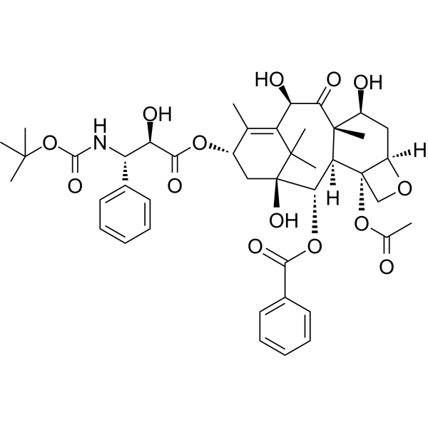
HY-B0011
DocetaxelTubulin Stabilizer -
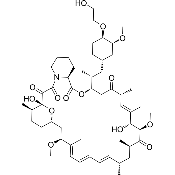
HY-10218
EverolimusmTORC Inhibitor -
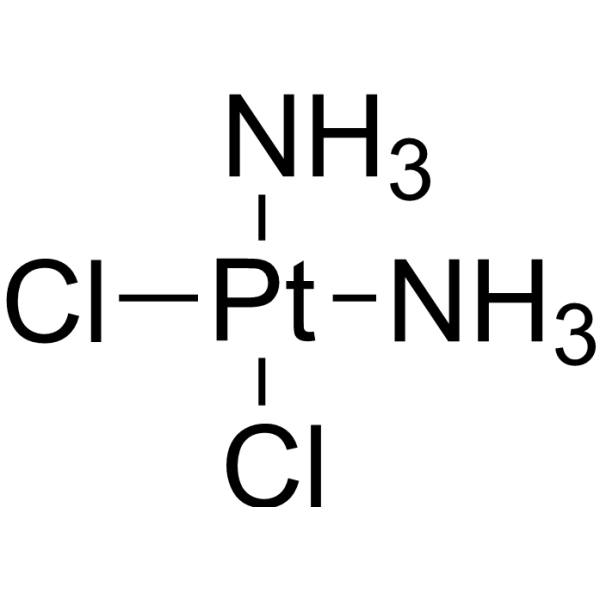
HY-17394
CisplatinDNA Alkylator


Biological Activity
Description
Isotype
Human IgG4 kappa
Recommend Isotype Controls
Species Reactivity
Human
IC50 & Target
[1]|
p-STAT3 |
Akt1 |
Akt2 |
Akt3 |
In Vitro
Serplulimab (10 μg/mL; 0-16 h) can increase the phosphorylation levels of AKT1/2/3 (T308), AKT1/2/3 (S473), and STAT3 (Y705) in Jurkat-PD-1 cells in response to PD-1 signaling blockade[1].
Serplulimab (0-1000 ng/mL; 2 days) can enhance the production of IFNγ in T cells, increase the cytotoxicity against SGC7901, and reverse the inhibitory effect of SGC7901 on T-cell effector functions in the co-culture system of CD8+ T cells and SGC7901 gastric cancer cells. The effect is even better when combined with HLX53[1].
MedChemExpress (MCE) has not independently confirmed the accuracy of these methods. They are for reference only.
In Vivo
MedChemExpress (MCE) has not independently confirmed the accuracy of these methods. They are for reference only.
-
Animal Model:BALB/c-hPD-1/hTIGIT dKI HuGEMM mice engrafted with CT26
-
Dosage:/
-
Administration:Intraperitoneal injection; twice a week; until the end of the experiment
-
Result:Showed moderate anti-tumor efficacy. When combined with HLX53, it exerted a notably enhanced inhibition on tumor growth, with a tumor growth inhibition (TGI) of 66.09%.
Enhanced survival of mice.
Clinical Trial
| NCT Number | Sponsor | Condition | Start Date |
Phase

|
|---|---|---|---|---|
| NCT01329991 | Plexxikon| | 2011-05 | PHASE1 |
Gene ID
Accession
Conjugated
Unconjugated
Reconsititution
The product can be reconstituted/diluted with sterile PBS or saline.
Format
-
Human IgG4 kappa
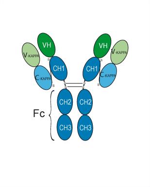
Application
ELISA, FACS, Functional assay
Verified Bioactivity
-
 Flow cytometric analysis of 1X10⁶ 10 ng/mL PMA and 1 μg/mL Ionomycin co-stimulated Jurkat cells (24h) with Serplulimab (HY-P99118, red). Cells were fixed with 4% paraformaldehyde. Then stained with the primary antibody at 1/200 dilution for an hour at 4℃. Alexa Fluor 488-conjugated AffiniPure Goat Anti-Human IgG H&L (HY-P83776) was used as the secondary antibody at 1/1,000 dilution for 30 minutes at 4℃. Human IgG4 kappa (HY-P99003, blue) was used as the isotype control, cells without incubation with primary antibody were used as the unlabeled control (black).


Chemical Information
-
CAS No. 2231029-82-4
-
Appearance Liquid
-
Molecular Weight 144.18 kDa
-
Color Colorless to light yellow
-
SMILES
[Serplulimab]
-
Synonyms
HLX10
-
Shipping
Shipping with dry ice.
-
Formulation
Please refer to the lot-specific COA for specific buffer information.
-
Storage
Please store the product under the recommended conditions in the Certificate of Analysis.


Purity & Documentation
-
Data Sheet (278 KB)
-
SDS (251 KB)
- English - EN (251 KB)
- Français - FR (251 KB)
- Deutsch - DE (251 KB)
- Norwegian - NO (251 KB)
- Español - ES (251 KB)
- Swedish - SV (251 KB)
- Italian - IT (251 KB)
- Korean - KR (251 KB)
- Portuguese - PT (251 KB)
-
Inhibitory Antibodies User Guide (603 KB)
References
[1]. Zhang Y, et al. Insights into the mechanisms of serplulimab: a distinctive anti-PD-1 monoclonal antibody, in combination with a TIGIT or LAG3 inhibitor in preclinical tumor immunotherapy studies. MAbs. 2024 Jan-Dec;16(1):2419838. [Content Brief]
[2]. Lee A. Serplulimab: First Approval. Drugs. 2022 Jul;82(10):1137-1141. [Content Brief]

Calculators
Concentration (start) × Volume (start) = Concentration (final) × Volume (final)


Serplulimab Related Classifications
HY-P99118 Related Classifications
 Powered by Bioz