



Nesfrotamig

Based on 1 Customer Validation
Nesfrotamig (YH32367; ABL105) is a bispecific activator targeting HER2 and 4-1BB. The Kd values of Nesfrotamig for human HER2 and human 4-1BB are 0.48 nM and 3.36 nM, respectively. By blocking tumor cell growth signals, activating HER2-dependent local 4-1BB in tumors to maintain T cell survival, and inducing NK cell-mediated antibody-dependent cellular cytotoxicity, Nesfrotamig enhances the cytotoxicity and tumor infiltration ability of immune cells. Nesfrotamig promotes the generation of tumor-specific memory T cells, drives T cell-mediated tumor lysis, exhibits significant anti-tumor efficacy against both HER2-positive and HER2-low-expressing tumors, and shows synergistic activity when combined with anti-PD-1 antibodies. In cynomolgus monkey studies, Nesfrotamig demonstrates good safety and is suitable for research related to HER2-positive and HER2-low-expressing tumors.
Para uso exclusivo en investigación. No vendemos a pacientes.
- Pureza : 99.22%
- No. CAS: 2966936-23-0
-
Almacenamiento:
Please store the product under the recommended conditions in the Certificate of Analysis.


To place orders, for customer services and technical support, please contact: MedChemExpress USA
Tel: 609-228-6898 E-mail: [email protected] [email protected]


-
 Actividad biológica
-
Chemical Information
- Protocolo
- Pureza y Documentación
- Referencias
-
 Help & FAQs


Ver todos los productos específicos de isoformas TNF Receptor
More

Actividad biológica
Descripciòn
Isotype
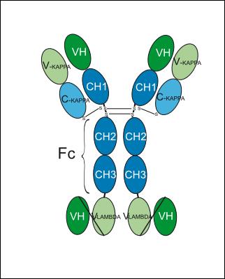
IgG1-kappa-[scFv]2-lambda-heavy
Recommend Isotype Controls
Species Reactivity
Human
IC50 & Target
ERBB2 & TNFRSF9
In Vitro
Nesfrotamig binds to 4-1BB-expressing Jurkat cells and HER2-expressing HCC1954 cells in a dose-dependent manner[1].
Nesfrotamig induces 4-1BB activation in a HER2 expression-dependent manner across multiple human cancer cell lines, with higher induction fold observed in cell lines with higher normalized HER2 expression levels[1].
Nesfrotamig (0.01 μg/mL-10 μg/mL) inhibits the proliferation of BT-474 cells in a dose-dependent manner, and its potency is comparable to that of Trastuzumab (HY-P9907)[1].
ABL105 potently activates the 4-1BB signaling pathway in three HER2-expressing tumor cell lines, NCI-N87, JIMT-1 and HCC1954[2].
ABL105 induces IFN-γ secretion and subsequent tumor cell lysis in a co-culture system of hPBMC and HER2-expressing tumor cells[2].
YH32367 induces IFN-γ secretion and subsequent tumor cell death in a co-culture system of hPBMC and HER2-expressing tumor cells[3].
MedChemExpress (MCE) has not independently confirmed the accuracy of these methods. They are for reference only. Further protocols information, click here.
In Vivo
Nesfrotamig (ABL105) (10 mg/kg; i.p.; twice weekly; 4 weeks) induces complete tumor regression in h4-1BB KI mice bearing hHER2/MC38 colorectal tumors and establishes durable, tumor-specific memory T cell immunity that prevents tumor re-challenge[1].
Nesfrotamig exhibits superior tumor-eradicating efficacy and induces long-term anti-tumor immunity in MC38/hHER2-bearing h4-1BB KI mice compared to benchmark HER2-targeted and 4-1BB/HER2-bispecific agents[3].
Nesfrotamig demonstrates greater monotherapy efficacy than a benchmark 4-1BB/HER2-bispecific agent in HER2-low tumor-bearing h4-1BB KI mice, and exhibits synergistic anti-tumor activity when combined with anti-PD-1 antibody[3].
MedChemExpress (MCE) has not independently confirmed the accuracy of these methods. They are for reference only.
-
Animal Model:humanized PBMC-engrafted mice with Breast cancer[1]
-
Dosage:10 mg/kg
-
Administration:i.v.; twice weekly; 4 weeks
-
Result:Reduced mean tumor volume to ~50 mm3 by 29 days post-tumor inoculation.
Increased tumor infiltration of CD4+, CD8+, CD4+ effector memory, and CD8+ effector memory T cells significantly relative to control groups.
Showed lower fold induction of hCD45-positive cells in peripheral blood than anti-4-1BB antibody control.
Maintained F4/80+ cell counts in the liver comparable to control groups.
-
Animal Model:human 4-1BB knock-in (h4-1BB KI) mice with Colorectal cancer[1]
-
Dosage:10 mg/kg
-
Administration:i.p.; twice weekly; 4 weeks
-
Result:Reduced mean tumor volume to near 0 mm3 by 40 days post-tumor inoculation and maintained suppression through 90 days.
Enabled complete protection against re-challenge with hHER2/MC38 cells in mice cured of initial tumors, while B16F10 tumors grew normally.
Gene ID
Accession
Conjugated
Unconjugated
Reconsititution
The product can be reconstituted/diluted with sterile PBS or saline.
Format
-
IgG1-kappa-[scFv]2-lambda-heavy
Aplicación
ELISA, FACS, Functional assay


Chemical Information
-
No. CAS 2966936-23-0
-
Appearance Liquid
-
Color Colorless to light yellow
-
SMILES
N/A
-
Synonyms
YH32367; ABL105
-
Envío
Shipping with dry ice.
-
Formulation
Please refer to the lot-specific COA for specific buffer information.
-
Almacenamiento
Please store the product under the recommended conditions in the Certificate of Analysis.

Protocolo
-
Cell Cytotoxicity Assay
Cytotoxicity assays are usually based on the assessment of cell membrane damage, which can also be indirectly detected by measuring cell viability. Detection methods include MTT assay, CKK-8 assay, LDH assay and ATP assay, etc.
-
Mammalian live/dead viability and cytotoxicity staining
Live/dead viability and cytotoxicity staining assays are based on the simultaneous detection of intracellular esterase activity in metabolically active (viable) cells and membrane integrity loss in non-viable cells. In commonly used dual-staining approaches, membrane-permeant fluorogenic substrates are converted by intracellular esterases into fluorescent products in live cells, while impermeant DNA-binding dyes selectively enter cells with compromised plasma membranes and label nucleic acids in dead or dying cells, enabling discrimination between viable and non-viable populations by fluorescence microscopy or flow cytometry.
-
Protocol for Cell Counting and Cell Density Analysis
Cell counting and cell-density analysis estimate the number of cells in a known volume or field area. Manual hemocytometer counting uses a chamber of defined geometry to convert counted cells into cells/mL, while automated counters and image-analysis workflows detect cell objects from optical, brightfield, fluorescence, impedance, or digital-image features. Trypan blue viability counting is based on dye exclusion: viable cells with intact membranes exclude dye, while non-viable cells with compromised membranes stain blue. The readout is total cell density, viable-cell density, dead-cell density, and percent viability. Cell density can also be estimated from microscopy images by counting objects per image area, from flow cytometry using calibrated volume or reference particles, or from in situ microscopy in bioreactors after calibration against reference methods such as hemocytometer or flow cytometry.
-
Somatic Cell Culture
A method of simulating the in vivo environment in vitro to maintain the cell growth, differentation and main functions.
-
CCK-8/WST-8 Cell Proliferation Assay
The CCK-8/WST-8 assay is based on the reduction of the water-soluble tetrazolium salt WST-8 to a water-soluble formazan product by cellular dehydrogenases in metabolically active cells, where the generated formazan amount is proportional to the number of living cells and is quantified by measuring absorbance in the visible range, providing a colorimetric readout for cell viability and proliferation assessment. This class of tetrazolium-based assays improves upon earlier MTT-based systems by producing a water-soluble formazan, eliminating the need for organic solubilization steps and enabling direct spectrophotometric measurement in culture medium.
-
Cell Counting-Based Growth Curve Assay
Cell counting-based growth curve assays quantify cell proliferation by directly measuring changes in viable cell number over time using manual or automated counting methods such as hemocytometer-based counting or instrument-assisted cell enumeration, enabling construction of growth curves that reflect population expansion dynamics in response to culture conditions. A widely used approach is trypan blue exclusion with hemocytometer counting, where membrane-compromised (non-viable) cells take up the dye, allowing discrimination between viable and non-viable cells while simultaneously enabling total cell number quantification. Repeated sampling across time points allows estimation of proliferation rate, growth phases, and comparative growth kinetics between experimental conditions.
-
MTT Cell Proliferation Assay
The MTT assay is a colorimetric endpoint assay for estimating viable cell number, cell growth, cytotoxicity, or cell activation in cultured mammalian cells. Living cells reduce the yellow tetrazolium salt MTT into purple/blue formazan, while dead cells do not generate the same signal; the resulting color can be quantified with a multiwell spectrophotometer. MTT reduction is commonly interpreted as a readout of metabolic activity that often correlates with viable cell number, but it should not be treated as a direct cell-counting method unless the assay is optimized for the cell type and experimental condition. Studies show that MTT reduction can involve mitochondrial and non-mitochondrial reducing systems, and formazan may accumulate in intracellular lipid droplets rather than simply marking mitochondria.
-
Research Protocol for Cancer Immunology
Cancer immunology studies how the immune system recognizes, suppresses, edits, or fails to eliminate malignant cells through tumor antigen release, antigen presentation, T-cell priming, immune trafficking, tumor-cell killing, and feedback inhibition in the tumor microenvironment. The cancer-immunity cycle links tumor antigenicity, dendritic-cell priming, CD8+ T-cell infiltration, cytotoxic function, and immune-checkpoint regulation to tumor rejection or immune escape. Immune-checkpoint pathways such as PD-1/PD-L1 and CTLA-4 suppress antitumor T-cell activity and can be therapeutically blocked, but many tumors remain resistant because of poor antigen presentation, weak T-cell infiltration, suppressive myeloid cells, regulatory T cells, and tumor-intrinsic immune-exclusion programs. Unresolved questions include which immune-cell states predict response, how tumor-intrinsic pathways exclude immune cells, how myeloid suppression limits checkpoint blockade, and which combination strategies
-
Cell Viability Determination by MTT Colorimetric Assay
The following protocol uses the MTT colorimetric assay as a classic literature-established method for assessing cell viability/metabolic activity in cultured mammalian cells. MTT[3-(4,5-dimethylthiazol-2-yl)-2,5-diphenyltetrazolium bromide] is reduced by metabolically active cells to a colored formazan product; the amount of formazan is quantified spectrophotometrically and provides an indirect measure of metabolically active viable cells. Importantly, MTT reduction reflects cellular oxidoreductase/metabolic activity rather than an absolute direct count of living cells, so changes in cellular metabolism can alter the signal independently of cell number.


Pureza y Documentación
-
Ficha de datos (263 KB)
-
SDS (251 KB)
- English - EN (251 KB)
- Français - FR (251 KB)
- Deutsch - DE (251 KB)
- Norwegian - NO (251 KB)
- Español - ES (251 KB)
- Swedish - SV (251 KB)
- Italian - IT (251 KB)
- Korean - KR (251 KB)
- Portuguese - PT (251 KB)
-
Inhibitory Antibodies User Guide (603 KB)
Referencias

Calculators
Concentration (start) × Volume (start) = Concentration (final) × Volume (final)


Nesfrotamig Related Classifications
HY-P991149 Related Classifications
 Powered by Bioz