



SR 16832

Based on 1 Customer Validation
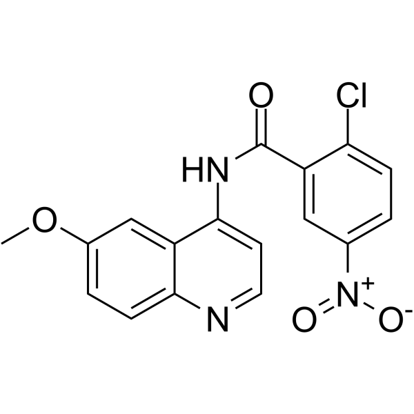
SR 16832 is a dual-site covalent, orthosteric and allosteric PPARγ antagonist. SR 16832 activates the TGF-β signaling pathway and upregulates the expression of Vimentin and Fibronectin (Fibronectin). SR 16832 is toxic to bronchial epithelium. SR 16832 can be used in research related to type 2 diabetes and pulmonary fibrosis.
Para uso exclusivo en investigación. No vendemos a pacientes.
- Pureza : 99.58%
- No. CAS: 2088135-12-8
- Fòrmula: C17H12ClN3O4
- Peso molecular:357.75
-
Almacenamiento:Powder -20°C, 3 years , 4°C, 2 years ; In solvent -80°C, 6 months , -20°C, 1 month

To place orders, for customer services and technical support, please contact: MedChemExpress USA
Tel: 609-228-6898 E-mail: [email protected] [email protected]


-
 Actividad biológica
-
Chemical Information
-
Solvente y solubilidad
- Protocolo
- Pureza y Documentación
- Referencias
-
 Help & FAQs





Actividad biológica
Descripciòn
IC50 & Target
[1]|
PPARγ |
In Vitro
SR 16832 (5 μM MRL20 post-treatment) completely inhibits allosteric cellular activation of Gal4-PPARγ LBD by MRL20 in HEK293T cells, while having minimal impact on basal Gal4-PPARγ LBD activity[1].
SR 16832 lowers basal transactivation of full-length PPARγ and completely inhibits allosteric cellular activation of full-length PPARγ by MRL20 in HEK293T cells[1].
SR 16832 weakens the allosteric potency of MRL20 to recruit TRAP220 coactivator peptide to purified PPARγ LBD, including in the presence of RXRα LBD[1].
SR 16832 completely blocks allosteric binding of rosiglitazone to purified PPARγ LBD, preventing TRAP220 coactivator peptide recruitment[1].
SR 16832 weakens the allosteric potency of MRL20 to modulate NCoR corepressor peptide binding to purified PPARγ LBD[1].
SR 16832 completely inhibits allosteric cellular activation of Gal4-PPARγ LBD by Rosiglitazone (HY-17386) in HEK293T cells[1].
SR 16832 (1, 2, ..., 200 μM) exhibits cytotoxicity in human bronchial epithelial BEAS-2B cells with an EC20 of 5 μM[2].
SR 16832 (5 μM; 4, 24, and 72 h) activates TGF-β signaling, increases inflammation, and upregulates vimentin and fibronectin expression in human bronchial epithelial BEAS-2B cells[2].
SR 16832 (10 μM; 4, 24, and 72 h) activates TGF-β signaling, increases inflammation, and upregulates vimentin and fibronectin expression in primary normal human bronchial epithelial (NHBE) cells[2].
MedChemExpress (MCE) has not independently confirmed the accuracy of these methods. They are for reference only. Further protocols information, click here.
-
Cell Line:human bronchial epithelial BEAS-2B cells
-
Concentration:5 μM
-
Incubation Time:4 h, 24 h, 72 h
-
Result:Increased TGF-β expression 2-fold relative to control at 4 h (p < 0.01).
Increased TNF-α expression 1.5-fold relative to control at 24 h (p < 0.01).
Increased vimentin expression 3-fold relative to control at 24 h (p < 0.001).
Increased fibronectin expression 3.5-fold relative to control at 72 h (p < 0.001).
-
Cell Line:primary normal human bronchial epithelial (NHBE) cells
-
Concentration:10 μM
-
Incubation Time:4 h, 24 h, 72 h
-
Result:Increased TGF-β expression 4-fold relative to control at 4 h (p < 0.001).
Increased TNF-α expression 1.5-fold relative to control at 24 h (p < 0.01).
Increased vimentin expression 3.5-fold relative to control at 24 h (p < 0.01).
Increased fibronectin expression 4.5-fold relative to control at 72 h (p < 0.001).


Chemical Information
-
No. CAS 2088135-12-8
-
Appearance Solid
-
Peso molecular 357.75
-
Fòrmula C17H12ClN3O4
-
Color White to off-white
-
SMILES
O=C(NC1=CC=NC2=CC=C(OC)C=C12)C3=CC([N+]([O-])=O)=CC=C3Cl
-
Envío
Room temperature in continental US; may vary elsewhere.
-
Almacenamiento
Powder -20°C 3 years 4°C 2 years In solvent -80°C 6 months -20°C 1 month


Solvente y solubilidad
In Vitro:
DMSO : 250 mg/mL (698.81 mM; Need ultrasonic; Hygroscopic DMSO has a significant impact on the solubility of product, please use newly opened DMSO)
Please refer to the solubility information to select the appropriate solvent. Once prepared, please aliquot and store the solution to prevent product inactivation from repeated freeze-thaw cycles.
Storage method and period of stock solution: -80°C, 6 months; -20°C, 1 month. When stored at -80°C, please use it within 6 months. When stored at -20°C, please use it within 1 month.
Please refer to the solubility information to select the appropriate solvent. Once prepared, please aliquot and store the solution to prevent product inactivation from repeated freeze-thaw cycles.
Storage method and period of stock solution: -80°C, 6 months; -20°C, 1 month. When stored at -80°C, please use it within 6 months. When stored at -20°C, please use it within 1 month.
Concentration (start) × Volume (start) = Concentration (final) × Volume (final)

Protocolo
-
Fibrosis/Collagen Morphometry
Fibrosis and collagen morphometry is based on the quantitative visualization of fibrillar collagen deposition in tissue sections using histochemical stains such as Sirius Red (Picrosirius Red) or Masson's trichrome, followed by image-based or polarization-enhanced analysis to estimate collagen proportional area as a surrogate of extracellular matrix accumulation during fibrotic remodeling. Sirius Red combined with polarized light microscopy enhances detection of collagen fibers due to birefringence properties, enabling more specific visualization of collagen type I and III fibrils compared to conventional bright-field histology, while whole-section or region-restricted digital morphometry reduces field-selection bias in fibrosis assessment. Alternative quantitative approaches include second harmonic generation (SHG) and two-photon excited fluorescence microscopy, which enable label-free detection of fibrillar collagen and have been validated against histological staining and biochemica
-
RNA extraction experimental
By lysing cells, releasing RNA, and removing impurities such as proteins and DNA, high-purity RNA products are finally obtained. The commonly used traditional method is the guanidine isothiocyanate/phenol/chloroform method (Trizol), which is suitable for a variety of animal materials including animal tissues, microorganisms, cultured cells, etc., and most plant materials.
-
Collagen: Sirius Red Staining
Sirius Red or picrosirius red staining is a histochemical method for visualizing collagen-rich extracellular matrix in tissue sections, and collagen fibers are detected as red-stained structures under bright-field microscopy with enhanced birefringence under polarized light. Picrosirius red is useful for assessing total collagen organization, distribution, and fibrosis burden, but polarized color should not be interpreted as a definitive collagen type I versus type III readout because color is affected by fiber orientation, thickness, and packing.
-
Connective Tissue: Masson's Trichrome/Collagen Trichrome Staining
Masson’s Trichrome (collagen/trichrome staining) is a histological technique that differentially stains tissue compartments using sequential acidic dyes to distinguish collagen from muscle and cytoplasmic components based on dye affinity and tissue permeability differences, enabling visualization of fibrosis and connective tissue architecture in histological sections. The classical formulation typically uses Weigert's iron hematoxylin for nuclear staining, Biebrich scarlet-acid fuchsin for cytoplasm and muscle, and aniline blue (or light green variants) for collagen, producing a characteristic blue/green collagen signal contrasted against red cytoplasm and dark nuclei. The staining principle relies on selective displacement of smaller dye molecules by larger anionic dyes in collagen-rich regions under controlled acidified conditions, which enhances collagen-specific dye retention. This property makes the method widely used for fibrosis assessment in organs such as heart, liver, lung, a
-
Research Protocol for Metabolic Diseases
AMP-activated protein kinase, AMPK, is a conserved cellular energy sensor that responds to reduced cellular energy status and coordinates metabolism by increasing ATP-generating catabolic pathways while suppressing ATP-consuming anabolic processes. In metabolic disease research, the AMPK pathway is experimentally relevant because it regulates hepatic lipid synthesis, fatty acid oxidation, glucose production, skeletal-muscle glucose disposal, mTORC1-linked biosynthesis, autophagy, mitochondrial homeostasis, and whole-body energy balance. The central pathway logic is that energy stress, metformin, exercise-like stimulation, or direct AMPK activators increase AMPKα Thr172 phosphorylation and downstream substrate phosphorylation, including ACC and RAPTOR. Phosphorylation of ACC suppresses lipogenesis and supports fatty acid oxidation, whereas phosphorylation of RAPTOR suppresses mTORC1 signaling and links cellular energy status to growth and protein synthesis control. The pathway is linked


Pureza y Documentación
-
Ficha de datos (278 KB)
-
SDS (254 KB)
- English - EN (254 KB)
- Français - FR (254 KB)
- Deutsch - DE (254 KB)
- Norwegian - NO (254 KB)
- Español - ES (254 KB)
- Swedish - SV (254 KB)
- Italian - IT (254 KB)
- Korean - KR (254 KB)
- Portuguese - PT (254 KB)
-
Instrucciones de manejo (2659 KB)
Referencias
[1]. Brust R, et al. Modification of the Orthosteric PPARγ Covalent Antagonist Scaffold Yields an Improved Dual-Site Allosteric Inhibitor. ACS Chem Biol. 2017;12(4):969-978. [Content Brief]
[2]. Jeong J, et al. Advancing the Adverse Outcome Pathway for PPARγ Inactivation Leading to Pulmonary Fibrosis Using Bradford-Hill Consideration and the Comparative Toxicogenomics Database. Chem Res Toxicol. 2022 Feb 21;35(2):233-243. [Content Brief]


Complete Stock Solution Preparation Table
Please refer to the solubility information to select the appropriate solvent. Once prepared, please aliquot and store the solution to prevent product inactivation from repeated freeze-thaw cycles.
Storage method and period of stock solution: -80°C, 6 months; -20°C, 1 month. When stored at -80°C, please use it within 6 months. When stored at -20°C, please use it within 1 month.
| Optional Solvent | Concentration Solvent Mass | 1 mg | 5 mg | 10 mg | 25 mg |
|---|---|---|---|---|---|
| DMSO | 1 mM | 2.7952 mL | 13.9762 mL | 27.9525 mL | 69.8812 mL |
| 5 mM | 0.5590 mL | 2.7952 mL | 5.5905 mL | 13.9762 mL | |
| 10 mM | 0.2795 mL | 1.3976 mL | 2.7952 mL | 6.9881 mL | |
| 15 mM | 0.1863 mL | 0.9317 mL | 1.8635 mL | 4.6587 mL | |
| 20 mM | 0.1398 mL | 0.6988 mL | 1.3976 mL | 3.4941 mL | |
| 25 mM | 0.1118 mL | 0.5590 mL | 1.1181 mL | 2.7952 mL | |
| 30 mM | 0.0932 mL | 0.4659 mL | 0.9317 mL | 2.3294 mL | |
| 40 mM | 0.0699 mL | 0.3494 mL | 0.6988 mL | 1.7470 mL | |
| 50 mM | 0.0559 mL | 0.2795 mL | 0.5590 mL | 1.3976 mL | |
| 60 mM | 0.0466 mL | 0.2329 mL | 0.4659 mL | 1.1647 mL | |
| 80 mM | 0.0349 mL | 0.1747 mL | 0.3494 mL | 0.8735 mL | |
| 100 mM | 0.0280 mL | 0.1398 mL | 0.2795 mL | 0.6988 mL |


SR 16832 Related Classifications
HY-112247 Related Classifications
 Powered by Bioz