



GSK-3484862
Based on 26 publication(s) in Google Scholar
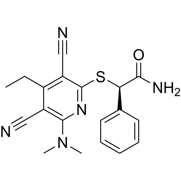
GSK-3484862 is a highly potent non-covalent inhibitor and demethylating agent of DNMT1. GSK-3484862 induces genome-wide DNA demethylation, including the regulatory elements of DNMT3B and the promoter region of TERT, and significantly inhibits cell viability, growth, proliferation and self-renewal. GSK-3484862 blocks the transformation of young AT2 cells, induces apoptosis, and generates transcriptomic features similar to those of senescent cells. GSK-3484862 is widely used in studies related to lung cancer, oral squamous cell carcinoma and lung adenocarcinoma.
For research use only. We do not sell to patients.
- Purity : 99.95%
- CAS No.: 2170136-65-7
- Formula: C19H19N5OS
- Molecular Weight:365.45
-
Storage:Powder -20°C, 3 years , 4°C, 2 years ; In solvent -80°C, 1 year , -20°C, 6 months

To place orders, for customer services and technical support, please contact: MedChemExpress USA
Tel: 609-228-6898 E-mail: [email protected] [email protected]


-
 Biological Activity
-
Chemical Information
-
Solvent & Solubility
- Protocols
- Purity & Documentation
- References
-
 Help & FAQs


-
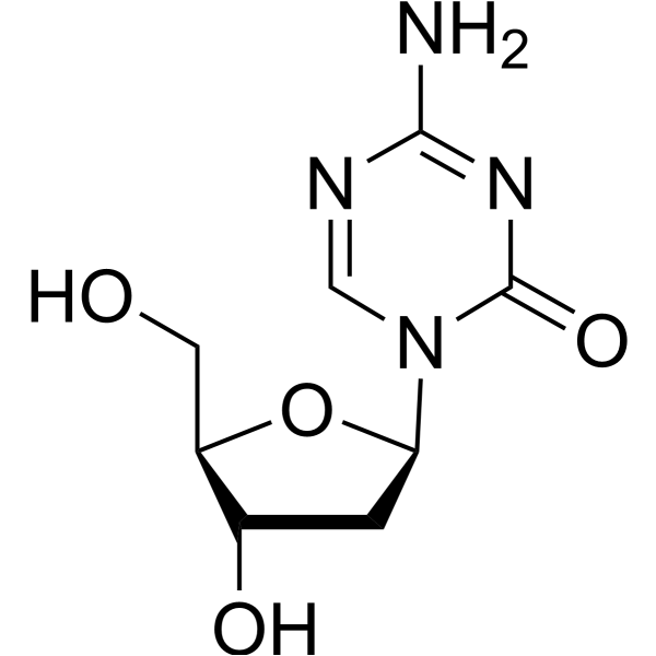
HY-A0004
DecitabineDNMT Inhibitor -
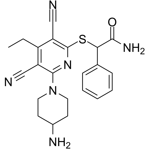
HY-139664
GSK-3685032DNMT1 Inhibitor -
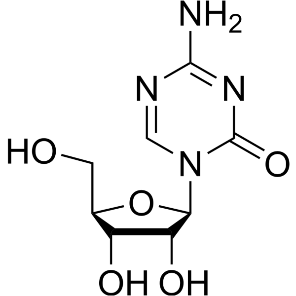
HY-10586
5-AzacytidineDNMT Inhibitor -
HY-13642
RG108DNMT Inhibitor -
HY-13803
TazemetostatEZH2 Inhibitor -
HY-70002
EnzalutamideAndrogen Receptor Antagonist -
HY-13470
GSK126EZH2 Inhibitor -
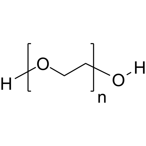
HY-Y0873
PEG300Neutral Polymer -
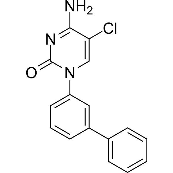
HY-111558
Bobcat339TET Inhibitor -
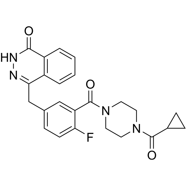
HY-10162
OlaparibPARP Inhibitor -

HY-15141
StaurosporinePKC/PKA Inhibitor -

HY-100579
Ferrostatin-1Ferroptosis Inhibitor
Publications Citing Use of MedChemExpress (MCE) GSK-3484862
More- Nat Genet. 2022 Nov;54(11):1702-1710. [Abstract]
- Nat Commun. 2025 Feb 5;16(1):1377. [Abstract]
- Nat Commun. 2025 Jan 22;16(1):929. [Abstract]
- Nat Commun. 2023 Apr 14;14(1):2122. [Abstract]
- Nat Ecol Evol. 2026 Jun 2. [Abstract]
- Nucleic Acids Res. 2026 Feb 5;54(4):gkag089. [Abstract]
- Nucleic Acids Res. 2024 Sep 23;52(17):10194-10219. [Abstract]
- Nucleic Acids Res. 2024 Mar 21;52(5):e24. [Abstract]
- J Clin Invest. 2023 Apr 17;133(8):e167953. [Abstract]
- Adv Sci (Weinh). 2025 Jan;12(4):e2410360. [Abstract]
- Cell Death Dis. 2025 Apr 4;16(1):251. [Abstract]
- Environ Sci Technol. 2024 Aug 20;58(33):14629-14640. [Abstract]
- Arch Toxicol. 2025 May;99(5):2179-2196. [Abstract]
- Mol Cancer Res. 2022 Nov 3;20(11):1598-1610. [Abstract]
- Drug Dev Res. 2026 May;87(3):e70299. [Abstract]
- J Biochem. 2026 Feb 13:mvag011. [Abstract]
- J Oral Biosci. 2024 Sep;66(3):530-538. [Abstract]
- bioRxiv. 2026 Jun 15.
- bioRxiv. 2026 Feb 8:2026.02.06.704516. [Abstract]
- bioRxiv. 2025 Nov 14.
- bioRxiv. 2025 Oct 20:2025.10.19.683051. [Abstract]
- bioRxiv. 2025 April 08.
- Patent. US20240252638A1.
- biorxiv. 2024 Jun 07.
- bioRxiv. 2024 Apr 3:2024.04.03.587980. [Abstract]
- bioRxiv. September 13, 2021.
 Customer Validation & Images

-
Bio/Physico-chemical Assay
-
IF
-
Bio/Physico-chemical Assay
-
RT-PCR
-
Flow Cytometry
All DNA Methyltransferase Isoforms
More

Biological Activity
Description
IC50 & Target
|
DNMT1 |
In Vitro
GSK-3484862 (2 μM, 10 μM; 6 days) induces global DNA demethylation in mouse embryonic stem cells, reducing the global CpG methylation level of the cells from nearly 70% to less than 18%, and the methylation pattern after induction is similar to that of Dnmt1-/- deficient mouse embryonic stem cells[1].
GSK-3484862 (1-10 μM; 1 week) inhibits proliferation, promotes apoptosis, and reduces the self-renewal capacity of Cal27 and FaDu oral squamous cell carcinoma (OSCC) cells by downregulating the expression of DNMT1[2].
GSK-3484862 inhibits the in vitro transformation of primary alveolar type 2 cells isolated from young KP-Cas9 mice, but has no effect on alveolar type 2 cells from aged KP-Cas9 mice, and induces the expression of Nupr1 and Lcn2 in this system[3].
GSK-3484862 induces hypomethylation at specific CpG sites in Nupr1 enhancers (E1, E7, E8), which correlates with upregulated Nupr1 gene expression[3].
MedChemExpress (MCE) has not independently confirmed the accuracy of these methods. They are for reference only. Further protocols information, click here.
-
Cell Line:Murine embryonic stem cells (mESC, wild-type (WT) or Dnmt1/3a/3b triple knockout (TKO))
-
Concentration:2 µM and 10 µM
-
Incubation Time:6 or 14 days
-
Result:Induced dramatic DNA methylation loss, with global CpG methylation levels falling from near 70% in WT mESC to less than 18% after 6 days.
-
Cell Line:Murine embryonic stem cells (mESC, wild-type (WT) or Dnmt1/3a/3b triple knockout (TKO))
-
Concentration:2 µM and 10 µM
-
Incubation Time:4 days
-
Result:Resulted in a modest reduction in DNMT1 protein level.
In Vivo
Systemic administration of GSK-3484862 inhibits the transformation of AT2 cells in young mice, but has no such effect on AT2 cells in aged mice, while it induces the expression of Nupr1 and Lcn2 in cells from both age groups[3].
GSK-3484862 induces pyroptosis of tumor cells in the mouse 4T1 breast cancer model[4].
MedChemExpress (MCE) has not independently confirmed the accuracy of these methods. They are for reference only.


Chemical Information
-
CAS No. 2170136-65-7
-
Appearance Solid
-
Molecular Weight 365.45
-
Formula C19H19N5OS
-
Color White to light yellow
-
SMILES
O=C(N)[C@H](SC1=NC(N(C)C)=C(C#N)C(CC)=C1C#N)C2=CC=CC=C2
-
Synonyms
(R)-GSK3482364
-
Shipping
Room temperature in continental US; may vary elsewhere.
-
Storage
Powder -20°C 3 years 4°C 2 years In solvent -80°C 1 year -20°C 6 months


Publications (26)
-
Journal Impact Factor
-
Most Recent
-
Nat Genet
DNA sequence and chromatin modifiers cooperate to confer epigenetic bistability at imprinting control regions. [Abstract]2022 Nov;54(11):1702-1710. PMID: 36333500
GSK-3484862 purchased from MedChemExpress. Usage Cited in: Nat Genet. 2022 Nov;54(11):1702-1710. [Abstract]
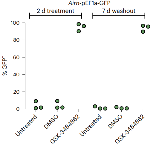
Flow cytometric analysis of three independent clones with the methylated Airn-CAG reporter after 2 days treatment with the DNA methylation inhibitor GSK-3484862 (10 µM) and untreated and DMSO controls. Measurements were repeated 7 days after washout of the drug to test for reversion of the reporter silencing.
-
Nat Commun
A biallelically active embryonic enhancer dictates GNAS imprinting through allele-specific conformations. [Abstract]2025 Feb 5;16(1):1377. PMID: 39910084
GSK-3484862 purchased from MedChemExpress. Usage Cited in: Nat Commun. 2025 Feb 5;16(1):1377. [Abstract]
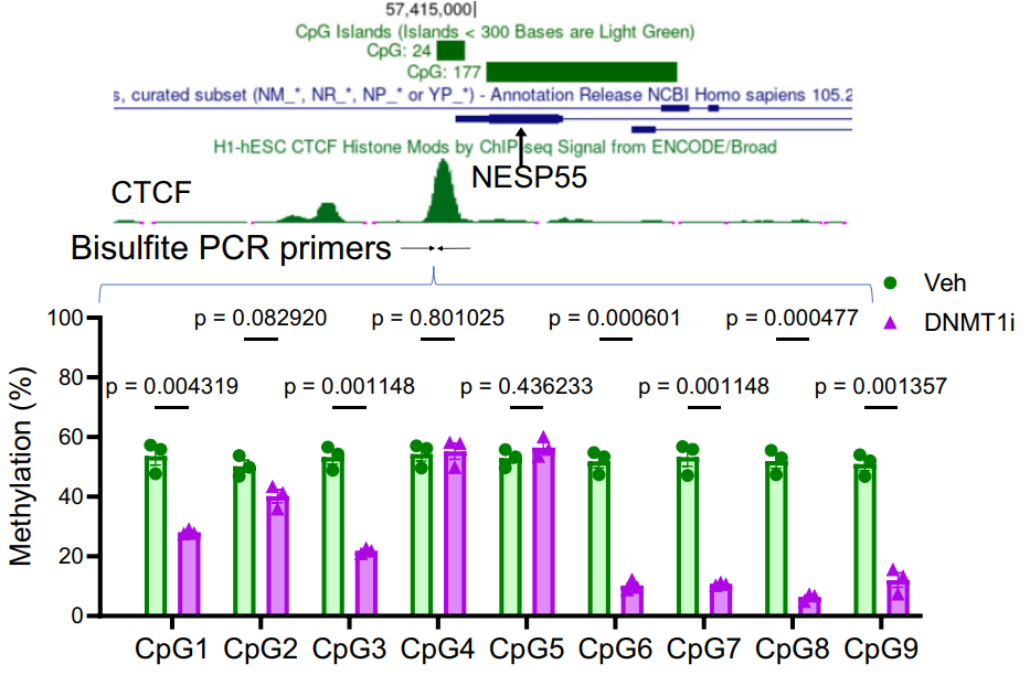
WT hESCs were treated with a DNTM1 inhibitor (DNMT1i), GSK-3484862, 10 µM for 17 days.
-
Nat Commun
Motif distribution and DNA methylation underlie distinct Cdx2 binding during development and homeostasis. [Abstract]2025 Jan 22;16(1):929. PMID: 39843425
GSK-3484862 purchased from MedChemExpress. Usage Cited in: Nat Commun. 2025 Jan 22;16(1):929. [Abstract]
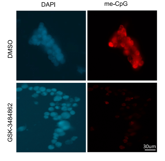
Fluorescence micrographs showing loss of DNA methylation in cells upon treatment with inhibitor GSK-3484862 (10 μM) for 6 days, (4 independent experiments).
GSK-3484862 purchased from MedChemExpress. Usage Cited in: Nat Commun. 2025 Jan 22;16(1):929. [Abstract]
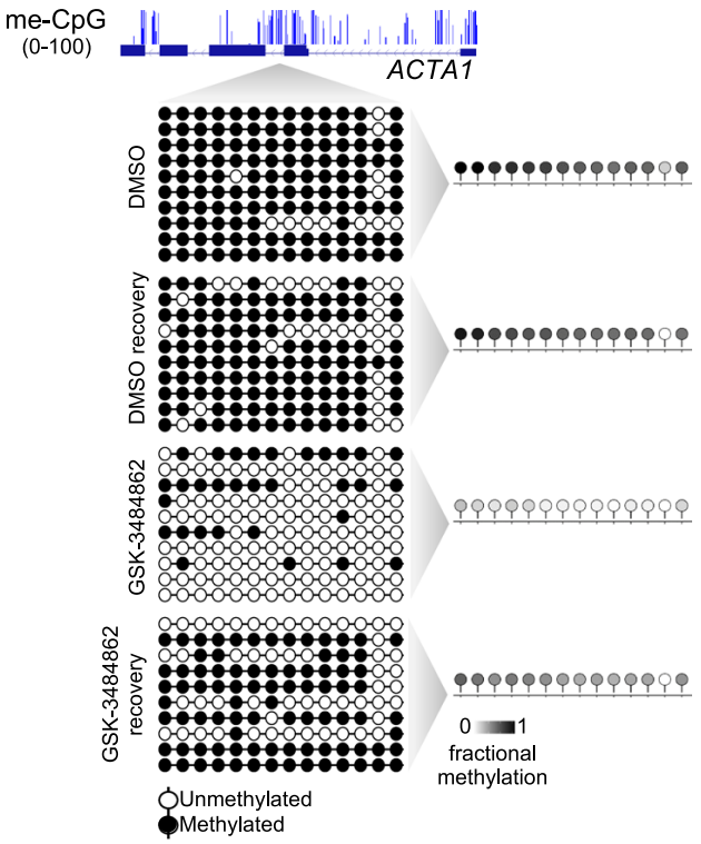
Representative genomic track showing fractional methylation of CpGs within ACTA1 gene body in HCT116 cells. Lollipop plots show 14 consecutive CpGs within 10 random representative amplicon sequences (from > 100× coverage on nanopore amplicon bisulfite sequencing) and their methylation status upon DMSO or GSK-3484862 (10 µM) treatment and 3 days of recovery; average methylation of all sequenced amplicons (>100× coverage) is represented for all treatment conditions in the right panel.
-
Nat Commun
2023 Apr 14;14(1):2122. PMID: 37055433 -
Nat Ecol Evol
Gene body methylation suppresses intragenic transcription and permits epigenetic inheritance in a cnidarian. [Abstract]2026 Jun 2. PMID: 42231006 -
Nucleic Acids Res
Single-molecule tracking of DNMT1 in living cells reveals its cell cycle dynamics and its redistribution upon drug treatment. [Abstract]2026 Feb 5;54(4):gkag089. PMID: 41641704 -
Nucleic Acids Res
The C-terminal 4CXXC-type zinc finger domain of CDCA7 recognizes hemimethylated DNA and modulates activities of chromatin remodeling enzyme HELLS. [Abstract]2024 Sep 23;52(17):10194-10219. PMID: 39142653 -
Nucleic Acids Res
Uncovering the roles of DNA hemi-methylation in transcriptional regulation using MspJI-assisted hemi-methylation sequencing. [Abstract]2024 Mar 21;52(5):e24. PMID: 38261991 -
J Clin Invest
The long-range interaction between two GNAS imprinting control regions delineates pseudohypoparathyroidism type 1B pathogenesis. [Abstract]2023 Apr 17;133(8):e167953. PMID: 36853809
GSK-3484862 purchased from MedChemExpress. Usage Cited in: J Clin Invest. 2023 Apr 17;133(8):e167953. [Abstract]
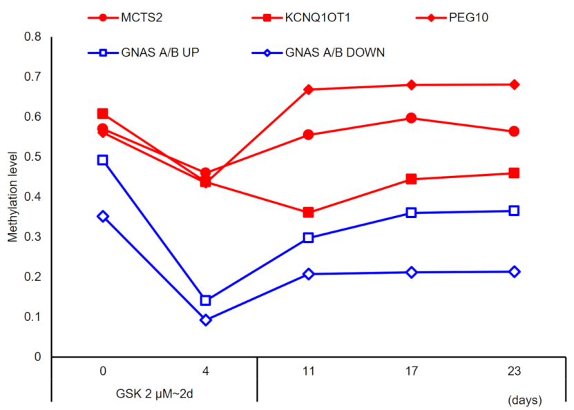
WT hESCs were treated with 2 μM GSK-3484862 for 2 days. Following the removal of GSK3484862, genomic DNA was purified at the indicated time points, and methylation levels at upstream (UP) and downstream (DOWN) of A/B DMR, MCTS2, PEG10, and KCNQ1OT1 were calculated by MSRE-qPCR. The results showed that in the hESCs, methylation levels at the A/B DMR decreased substantially by day 4 following a 2-day treatment with GSK-3484862 (14.1% and 9.2% methylation for the UP and DOWN regions, respectively).
-
Adv Sci (Weinh)
Targeted SPP1 Inhibition of Tumor-Associated Myeloid Cells Effectively Decreases Tumor Sizes. [Abstract]2025 Jan;12(4):e2410360. PMID: 39639496 -
Cell Death Dis
Growth factor receptor plasticity drives therapeutic persistence of metastatic breast cancer. [Abstract]2025 Apr 4;16(1):251. PMID: 40185706 -
Environ Sci Technol
Retinal Degeneration Response to Graphene Quantum Dots: Disruption of the Blood-Retina Barrier Modulated by Surface Modification-Dependent DNA Methylation. [Abstract]2024 Aug 20;58(33):14629-14640. PMID: 39102579 -
Arch Toxicol
Integrative genome-wide aberrant DNA methylation and transcriptome analysis identifies diagnostic markers for colorectal cancer. [Abstract]2025 May;99(5):2179-2196. PMID: 40059124 -
Mol Cancer Res
2022 Nov 3;20(11):1598-1610. PMID: 35925047 -
Drug Dev Res
Sustained DNA Hypomethylation Induced by a DNA Methyltransferase 1 Inhibitor Triggers Apoptosis in Thyroid Cancer Cells. [Abstract]2026 May;87(3):e70299. PMID: 42048599 -
J Biochem
2026 Feb 13:mvag011. PMID: 41679964 -
J Oral Biosci
Exploring the Role of DNMT1 in Dental Papilla Cell Fate Specification during Mouse Tooth Germ Development through Integrated Single-Cell Transcriptomics and Bulk RNA Sequencing. [Abstract]2024 Sep;66(3):530-538. PMID: 38942194 -
-
bioRxiv
Regulation of BCL11A DNA binding and expression in human erythrocyte precursor HUDEP-2 cells. [Abstract]2026 Feb 8:2026.02.06.704516. PMID: 41822826 -
-
bioRxiv
Divergent roles of DNA methylation, TRIM28, and p53 surveillance in human embryonic and trophoblast stem cells. [Abstract]2025 Oct 20:2025.10.19.683051. PMID: 41279747 -
-
-
-
bioRxiv
2024 Apr 3:2024.04.03.587980. PMID: 38617249 -


Solvent & Solubility
In Vitro:
DMSO : 20 mg/mL (54.73 mM; ultrasonic and warming and heat to 60°C; Hygroscopic DMSO has a significant impact on the solubility of product, please use newly opened DMSO)
Please refer to the solubility information to select the appropriate solvent. Once prepared, please aliquot and store the solution to prevent product inactivation from repeated freeze-thaw cycles.
Storage method and period of stock solution: -80°C, 1 year; -20°C, 6 months. When stored at -80°C, please use it within 1 year. When stored at -20°C, please use it within 6 months.
Please refer to the solubility information to select the appropriate solvent. Once prepared, please aliquot and store the solution to prevent product inactivation from repeated freeze-thaw cycles.
Storage method and period of stock solution: -80°C, 1 year; -20°C, 6 months. When stored at -80°C, please use it within 1 year. When stored at -20°C, please use it within 6 months.
Concentration (start) × Volume (start) = Concentration (final) × Volume (final)
In Vivo:
Select the appropriate dissolution method based on your experimental animal and administration route.
- For the following dissolution methods, please ensure to first prepare a clear stock solution using an In Vitro approach and then sequentially add co-solvents:
- To ensure reliable experimental results, the clarified stock solution can be appropriately stored based on storage conditions. As for the working solution for In Vivo experiments, it is recommended to prepare freshly and use it on the same day.
- The percentages shown for the solvents indicate their volumetric ratio in the final prepared solution. If precipitation or phase separation occurs during preparation, heat and/or sonication can be used to aid dissolution.
Add each solvent one by one: 10% DMSO 40% PEG300 5% Tween-80 45% Saline
Solubility: 2.08 mg/mL (5.69 mM); Suspended solution; Need ultrasonic
This protocol yields a suspended solution of 2.08 mg/mL. Suspended solution can be used for oral and intraperitoneal injection.
Taking 1 mL working solution as an example, add 100 μL DMSO stock solution (20.8 mg/mL) to 400 μL PEG300, and mix evenly; then add 50 μL Tween-80 and mix evenly; then add 450 μL Saline to adjust the volume to 1 mL.
Preparation of Saline: Dissolve 0.9 g sodium chloride in ddH₂O and dilute to 100 mL to obtain a clear Saline solution.
Add each solvent one by one: 10% DMSO 90% (20% SBE-β-CD in Saline)
Solubility: 2.08 mg/mL (5.69 mM); Suspended solution; Need ultrasonic
This protocol yields a suspended solution of 2.08 mg/mL. Suspended solution can be used for oral and intraperitoneal injection.
Taking 1 mL working solution as an example, add 100 μL DMSO stock solution (20.8 mg/mL) to 900 μL 20% SBE-β-CD in Saline, and mix evenly.
Preparation of 20% SBE-β-CD in Saline (4°C, storage for one week): 2 g SBE-β-CD powder is dissolved in 10 mL Saline, completely dissolve until clear.
In Vivo Dissolution Calculator
Please enter the basic information of animal experiments:
-
-
-
-
Recommended: Prepare an additional quantity of animals to account for potential losses during experiments.
Please enter your animal formula composition:
-
%DMSO +
Recommended: Keep the proportion of DMSO in working solution below 2% if your animal is weak.
-
%+
-
+%Tween-80 + +
-
%Saline +
The co-solvents required include: DMSO, . All of co-solvents are available by MedChemExpress (MCE). , Tween 80. All of co-solvents are available by MedChemExpress (MCE).
Working solution concentration: 0.22 mg/mL
Method for preparing stock solution: mg drug dissolved in μL DMSO. Stock solution concentration: mg/mL.
1. Take μL DMSO stock solution;
2. Add μL .
μL , mix evenly;
3. Then add μL Tween 80, mix evenly;
4. Then add μL
Please ensure that the stock solution in the first step is dissolved to a clear state, and add co-solvents in sequence. You can use ultrasonic heating (ultrasonic cleaner, recommended frequency 20-40 kHz), vortexing, etc. to assist dissolution.

Protocols
-
Apoptosis
Apoptosis, also called programmed cell death, is generally characterized by distinct morphological characteristics.
-
TUNEL staining for apoptotic DNA fragmentation
TUNEL staining detects DNA strand breaks by using terminal deoxynucleotidyl transferase to add labeled nucleotides to exposed 3′-OH DNA termini, generating either microscopic staining in fixed cells or tissue sections, or fluorescence/cytometric signal in cell suspensions. TUNEL positivity reflects DNA fragmentation but should not be interpreted alone as definitive apoptosis, because TUNEL can also label necrotic, autolytic, mechanically damaged, or DNA-repair-associated DNA breaks.
-
Cell Cytotoxicity Assay
Cytotoxicity assays are usually based on the assessment of cell membrane damage, which can also be indirectly detected by measuring cell viability. Detection methods include MTT assay, CKK-8 assay, LDH assay and ATP assay, etc.
-
Mammalian live/dead viability and cytotoxicity staining
Live/dead viability and cytotoxicity staining assays are based on the simultaneous detection of intracellular esterase activity in metabolically active (viable) cells and membrane integrity loss in non-viable cells. In commonly used dual-staining approaches, membrane-permeant fluorogenic substrates are converted by intracellular esterases into fluorescent products in live cells, while impermeant DNA-binding dyes selectively enter cells with compromised plasma membranes and label nucleic acids in dead or dying cells, enabling discrimination between viable and non-viable populations by fluorescence microscopy or flow cytometry.
-
Annexin V plus membrane-impermeant dye apoptosis staining
Annexin V-based apoptosis assays rely on the detection of phosphatidylserine (PS) externalization from the inner leaflet of the plasma membrane to the outer leaflet, an early biochemical hallmark of apoptosis. Fluorescently labeled Annexin V binds PS in a calcium-dependent manner, enabling identification of early apoptotic cells by flow cytometry or fluorescence microscopy. When combined with a membrane-impermeant DNA-binding dye (e. g. , propidium iodide), this approach allows discrimination between viable (Annexin V−/dye−), early apoptotic (Annexin V+/dye−), and late apoptotic or necrotic (Annexin V+/dye+) cell populations by assessing membrane integrity and PS exposure.
-
Apoptosis Solutions
Apoptosis is a regulated, generally non-lytic cell-death pathway that removes unwanted, damaged, infected, or abnormal cells through coordinated morphological changes, caspase activation, DNA fragmentation, and membrane remodeling. The intrinsic apoptosis pathway is controlled mainly by mitochondrial outer membrane permeabilization, BCL-2 family proteins, cytochrome c release, apoptosome formation, caspase-9 activation, and downstream executioner caspase-3/7 activation. The extrinsic apoptosis pathway is initiated by death receptors such as Fas, TNFR, and TRAIL receptors, which recruit adaptor proteins and activate caspase-8 before engaging executioner caspases or mitochondrial amplification through BID cleavage. Apoptosis is linked to many phenotypes, including cancer cell killing, tissue homeostasis, immune regulation, neurodegeneration, infection response, and treatment-induced cytotoxicity; unresolved questions include how apoptosis interacts with necroptosis, pyroptosis, ferroptos
-
CCK-8/WST-8 Cell Proliferation Assay
The CCK-8/WST-8 assay is based on the reduction of the water-soluble tetrazolium salt WST-8 to a water-soluble formazan product by cellular dehydrogenases in metabolically active cells, where the generated formazan amount is proportional to the number of living cells and is quantified by measuring absorbance in the visible range, providing a colorimetric readout for cell viability and proliferation assessment. This class of tetrazolium-based assays improves upon earlier MTT-based systems by producing a water-soluble formazan, eliminating the need for organic solubilization steps and enabling direct spectrophotometric measurement in culture medium.
-
MTT Cell Proliferation Assay
The MTT assay is a colorimetric endpoint assay for estimating viable cell number, cell growth, cytotoxicity, or cell activation in cultured mammalian cells. Living cells reduce the yellow tetrazolium salt MTT into purple/blue formazan, while dead cells do not generate the same signal; the resulting color can be quantified with a multiwell spectrophotometer. MTT reduction is commonly interpreted as a readout of metabolic activity that often correlates with viable cell number, but it should not be treated as a direct cell-counting method unless the assay is optimized for the cell type and experimental condition. Studies show that MTT reduction can involve mitochondrial and non-mitochondrial reducing systems, and formazan may accumulate in intracellular lipid droplets rather than simply marking mitochondria.
-
hPSC maintenance and expansion
This protocol maintains and expands human pluripotent stem cells under feeder-free, chemically defined conditions using E8 medium and vitronectin-coated culture surfaces; the readout is sustained adherent colony growth with undifferentiated morphology and retained pluripotency-marker expression during serial passaging. E8-based hPSC culture relies on defined soluble factors and matrix-dependent adhesion rather than feeder cells; vitronectin supports hPSC attachment through integrin-mediated interactions, and EDTA passaging dissociates colonies as small aggregates without enzymatic digestion, centrifugation, or routine ROCK-inhibitor treatment.
-
Senescence-associated β-galactosidase staining
Senescence-associated β-galactosidase staining detects β-galactosidase activity that is histochemically visible at pH 6. 0 in senescent cells, where X-gal cleavage produces an insoluble blue precipitate observable by bright-field microscopy. This activity reflects increased lysosomal β-galactosidase/lysosomal mass rather than a senescence-essential enzyme, because GLB1 depletion or genetic lysosomal β-galactosidase deficiency can abolish SA-β-gal staining while cells still undergo senescence. SA-β-gal was originally reported in senescent but not presenescent fibroblasts and keratinocytes, absent from quiescent fibroblasts and terminally differentiated keratinocytes, and increased with donor age in human skin samples. Because SA-β-gal can also appear in some non-senescent or tissue-specific contexts, interpretation should be paired with experimental controls and, when possible, independent senescence markers.
-
Cell Viability Determination by MTT Colorimetric Assay
The following protocol uses the MTT colorimetric assay as a classic literature-established method for assessing cell viability/metabolic activity in cultured mammalian cells. MTT[3-(4,5-dimethylthiazol-2-yl)-2,5-diphenyltetrazolium bromide] is reduced by metabolically active cells to a colored formazan product; the amount of formazan is quantified spectrophotometrically and provides an indirect measure of metabolically active viable cells. Importantly, MTT reduction reflects cellular oxidoreductase/metabolic activity rather than an absolute direct count of living cells, so changes in cellular metabolism can alter the signal independently of cell number.


Purity & Documentation
-
Data Sheet (282 KB)
-
SDS (393 KB)
- English - EN (393 KB)
- Français - FR (393 KB)
- Deutsch - DE (393 KB)
- Norwegian - NO (393 KB)
- Español - ES (393 KB)
- Swedish - SV (393 KB)
- Italian - IT (393 KB)
- Korean - KR (393 KB)
- Portuguese - PT (393 KB)
-
Handling Instructions (2659 KB)
References
[1]. Azevedo Portilho N, et al. The DNMT1 inhibitor GSK-3484862 mediates global demethylation in murine embryonic stem cells. Epigenetics Chromatin. 2021;14(1):56. Published 2021 Dec 15. [Content Brief]
[2]. Liu Y, et al. DNMT1-targeting remodeling global DNA hypomethylation for enhanced tumor suppression and circumvented toxicity in oral squamous cell carcinoma. Mol Cancer. 2024;23(1):104. Published 2024 May 16. [Content Brief]
[4]. Chen Q, et al. GSK-3484862, a DNMT1 degrader, promotes DNMT3B expression in lung cancer cells. NAR Cancer. 2025;7(2):zcaf018. Published 2025 May 27. [Content Brief]


Complete Stock Solution Preparation Table
Please refer to the solubility information to select the appropriate solvent. Once prepared, please aliquot and store the solution to prevent product inactivation from repeated freeze-thaw cycles.
Storage method and period of stock solution: -80°C, 1 year; -20°C, 6 months. When stored at -80°C, please use it within 1 year. When stored at -20°C, please use it within 6 months.
| Optional Solvent | Concentration Solvent Mass | 1 mg | 5 mg | 10 mg | 25 mg |
|---|---|---|---|---|---|
| DMSO | 1 mM | 2.7364 mL | 13.6818 mL | 27.3635 mL | 68.4088 mL |
| 5 mM | 0.5473 mL | 2.7364 mL | 5.4727 mL | 13.6818 mL | |
| 10 mM | 0.2736 mL | 1.3682 mL | 2.7364 mL | 6.8409 mL | |
| 15 mM | 0.1824 mL | 0.9121 mL | 1.8242 mL | 4.5606 mL | |
| 20 mM | 0.1368 mL | 0.6841 mL | 1.3682 mL | 3.4204 mL | |
| 25 mM | 0.1095 mL | 0.5473 mL | 1.0945 mL | 2.7364 mL | |
| 30 mM | 0.0912 mL | 0.4561 mL | 0.9121 mL | 2.2803 mL | |
| 40 mM | 0.0684 mL | 0.3420 mL | 0.6841 mL | 1.7102 mL | |
| 50 mM | 0.0547 mL | 0.2736 mL | 0.5473 mL | 1.3682 mL |


GSK-3484862 Related Classifications
HY-135146 Related Classifications
 Powered by Bioz