



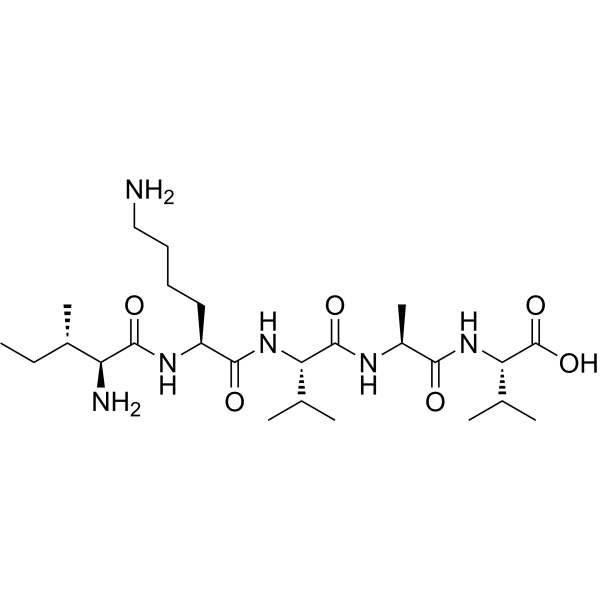
H-Ile-Lys-Val-Ala-Val-OH
Based on 2 publication(s) in Google Scholar
H-Ile-Lys-Val-Ala-Val-OH is one of the most potent active sites of laminin-1. H-Ile-Lys-Val-Ala-Val-OH promotes cell adhesion, neurite outgrowth, and tumor growth. H-Ile-Lys-Val-Ala-Val-OH stimulates BMMSC population growth and proliferation by activating MAPK/ERK1/2 and PI3K/Akt signalling pathways.
For research use only. We do not sell to patients.
- Purity : 99.35%
- CAS No.: 131167-89-0
- Formula: C25H48N6O6
- Molecular Weight:528.69
-
Storage:
Sealed storage, away from moisture.
Powder -80°C, 2 years , -20°C, 1 year* In solvent : -80°C, 6 months; -20°C, 1 month (sealed storage, away from moisture)

To place orders, for customer services and technical support, please contact: MedChemExpress USA
Tel: 609-228-6898 E-mail: [email protected] [email protected]


-
 Biological Activity
-
Chemical Information
-
Solvent & Solubility
- Protocols
- Purity & Documentation
- References
-
 Help & FAQs


-
HY-P3513
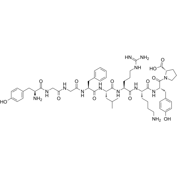
β-Neo-EndorphinErk1/2 Activator -
HY-129566
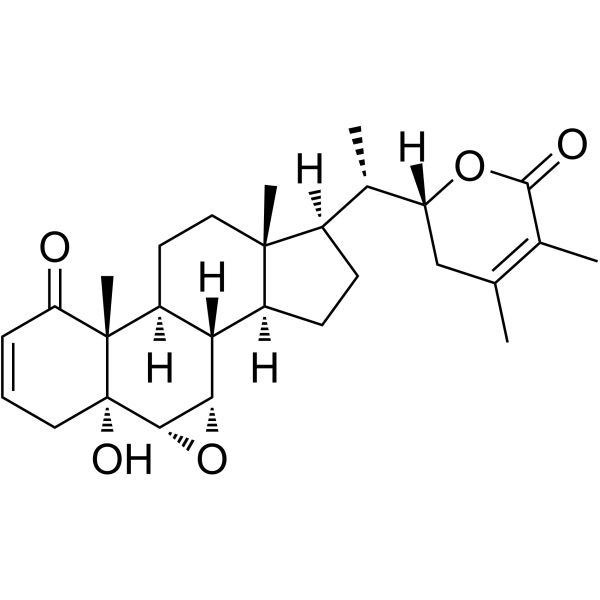
Withanolide BNeuroprotective Agent -
HY-17587
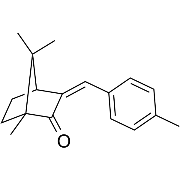
4-Methylbenzylidene camphorEstrogen-Like Drug -
HY-W010907
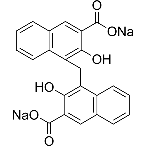
Pamoic acid disodiumGPR35 Agonist -
HY-137977
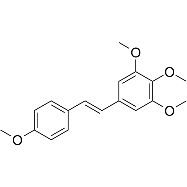
DMU-212Anticancer Compound -

HY-10071
Y-27632ROCK Inhibitor -
HY-N8303
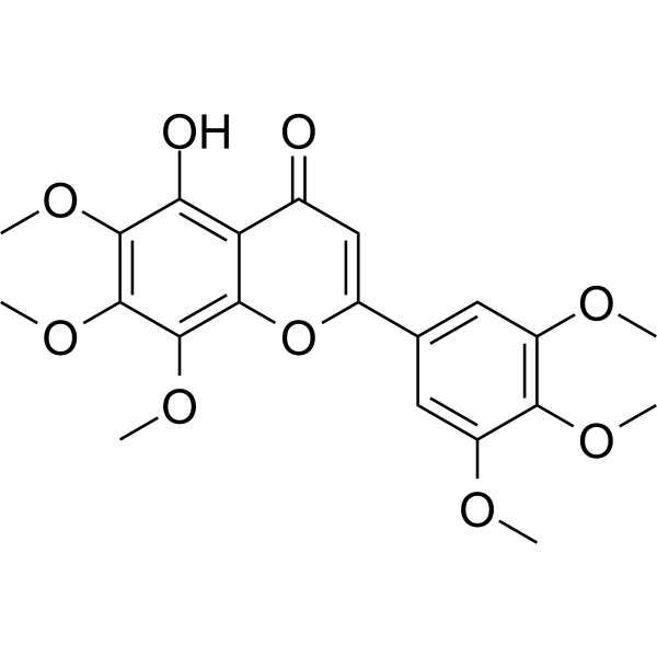
Gardenin AMAPK/ERK/PKC/PKA Activator -
HY-108635
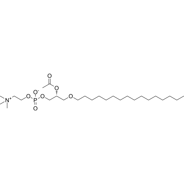
C16-PAFPAFR Activator -
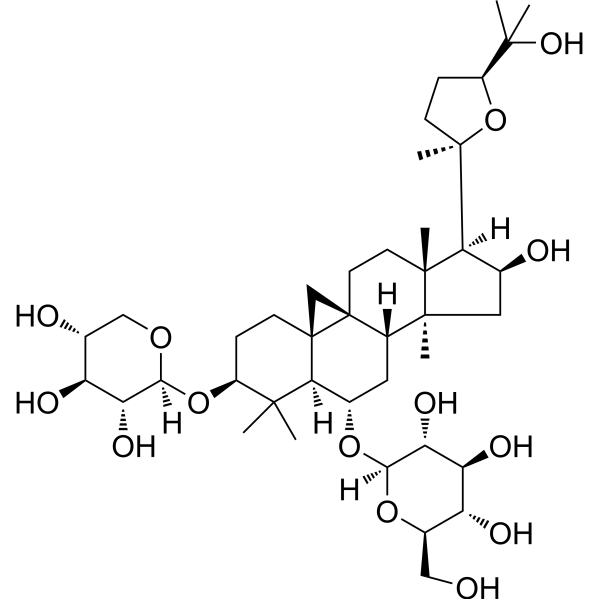
HY-N0431
Astragaloside IVMMP Inhibitor -
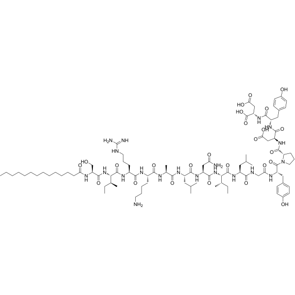
HY-P5985
mSIRKERK1/2 Activator -
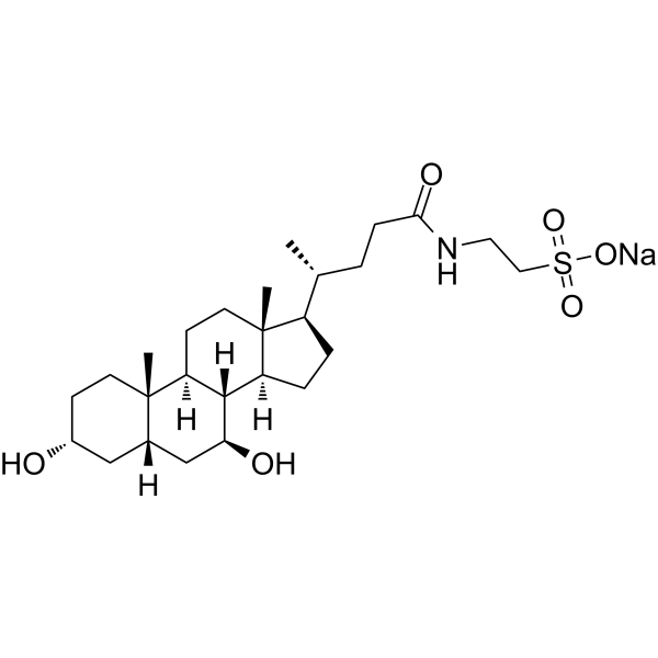
HY-19696A
Tauroursodeoxycholate sodiumEndoplasmic Reticulum Stress Inhibitor -

HY-P3418
CKLF1-C27CKLF1 C-terminal Peptide
Publications Citing Use of MedChemExpress (MCE) H-Ile-Lys-Val-Ala-Val-OH
More

Biological Activity
Description
IC50 & Target
|
ERK1 |
ERK2 |
Akt |
In Vitro
H-Ile-Lys-Val-Ala-Val-OH (0-2.5 mM, 0-48 h) induces proliferation and proliferating cell nuclear antigen (PCNA) synthesis of bone marrow mesenchymal stem cell (BMMSC)[2].
H-Ile-Lys-Val-Ala-Val-OH (5 mM, 24 h) induces the BMMSC cell cycle for cells to enter S phase from G0/G1 and arrests them from entering G2/M phase[2].
H-Ile-Lys-Val-Ala-Val-OH (0-2.5 mM, 0-48 h) activats MAPK/ERK and PI3K/Akt signalling pathways by enhancing phosphorylation levels of ERK1/2 and Akt in the BMMSCs[2].
MedChemExpress (MCE) has not independently confirmed the accuracy of these methods. They are for reference only. Further protocols information, click here.
-
Cell Line:BMMSC
-
Concentration:0, 0.004, 0.02, 0.1, 0.5 and 2.5 mM
-
Incubation Time:0, 12, 24, 36, 48 h
-
Result:Stimulated proliferating cell nuclear antigen (PCNA) synthesis and induced proliferation of bone marrow mesenchymal stem cell (BMMSC) in a dose- and time-dependent manner.
-
Cell Line:BMMSC
-
Concentration:5 mM
-
Incubation Time:24 h
-
Result:Induced the BMMSC cell cycle for cells to enter S phase from G0/G1 and arrested them from entering G2/M phase; the increased proportion of S phase in cell cycle was considered to be a sign of BMMSC proliferation.
-
Cell Line:BMMSC
-
Concentration:0, 0.004, 0.02, 0.1, 0.5 and 2.5 mM
-
Incubation Time:0, 12, 24, 36, 48 h
-
Result:Increased significantly the levels of p-ERK1/2 and p-Akt in a dose- and time-dependent manner.


Chemical Information
-
CAS No. 131167-89-0
-
Appearance Solid
-
Molecular Weight 528.69
-
Formula C25H48N6O6
-
Color White to off-white
-
Sequence
H-Ile-Lys-Val-Ala-Val-OH
-
Sequence Shortening
IKVAV
-
Shipping
Room temperature in continental US; may vary elsewhere.
-
Storage
Sealed storage, away from moisture
Powder -80°C 2 years -20°C 1 year * In solvent : -80°C, 6 months; -20°C, 1 month (sealed storage, away from moisture)


Publications (2)
-
Journal Impact Factor
-
Most Recent
-
Int J Mol Sci
miR-107 Targets NSG1 to Regulate Hypopharyngeal Squamous Cell Carcinoma Progression through ERK Pathway. [Abstract]2024 May 29;25(11):5961. PMID: 38892156 -


Solvent & Solubility
In Vitro:
H2O : 100 mg/mL (189.15 mM; Need ultrasonic)
Please refer to the solubility information to select the appropriate solvent. Once prepared, please aliquot and store the solution to prevent product inactivation from repeated freeze-thaw cycles.
Storage method and period of stock solution: -80°C, 6 months; -20°C, 1 month (sealed storage, away from moisture). When stored at -80°C, please use it within 6 months. When stored at -20°C, please use it within 1 month.
* Note: If you choose water as the stock solution, please dilute it to the working solution, then filter and sterilize it with a 0.22 μm filter before use.
Please refer to the solubility information to select the appropriate solvent. Once prepared, please aliquot and store the solution to prevent product inactivation from repeated freeze-thaw cycles.
Storage method and period of stock solution: -80°C, 6 months; -20°C, 1 month (sealed storage, away from moisture). When stored at -80°C, please use it within 6 months. When stored at -20°C, please use it within 1 month.
* Note: If you choose water as the stock solution, please dilute it to the working solution, then filter and sterilize it with a 0.22 μm filter before use.
Concentration (start) × Volume (start) = Concentration (final) × Volume (final)

Protocols
-
PC12 NGF-Induced Neuronal Differentiation Culture
PC12 cells are a rat adrenal pheochromocytoma clonal line that responds to NGF by stopping proliferation and extending branching neurite-like processes; after longer NGF exposure, cells develop long processes and neuronal-like ultrastructural and functional features. NGF-induced differentiation is read out mainly by neurite outgrowth, reduced proliferation, microtubule assembly, and neuronal differentiation-associated proteins such as MAPs, tau, GAP-43, and synapsin-1.
-
SH-SY5Y Neuronal Differentiation Culture
SH-SY5Y neuronal differentiation culture uses sequential exposure to retinoic acid and neurotrophic factors to reduce proliferative neuroblastoma-like behavior and induce neuron-like morphology, including neurite extension, neuronal marker expression, and, in RA/BDNF protocols, greater synaptic-marker expression than undifferentiated culture. Retinoic acid is commonly used as the initiating differentiation cue, while BDNF in serum-reduced or serum-free medium supports later maturation and neurotrophic-factor-dependent neuron-like survival.
-
SH-SY5Y neuronal-like differentiation
SH-SY5Y neuronal-like differentiation uses defined culture conditions to shift proliferative human neuroblastoma cells toward a neuron-like state, mainly assessed by reduced proliferation, neurite extension, neuronal-marker expression, and, in some protocols, increased dependence on neurotrophic support. Retinoic acid (RA) is commonly used for the first differentiation phase, and sequential RA followed by brain-derived neurotrophic factor (BDNF) in serum-free medium is a well-characterized approach for generating neuron-like SH-SY5Y cultures with extensive neurite outgrowth. The primary readouts are morphology-based neurite outgrowth and marker-based confirmation using proteins such as βIII-tubulin, MAP2, GAP43, synaptophysin, NeuN, NSE, TH, or related neuronal/synaptic markers, depending on the study endpoint.
-
Patient-Derived Xenograft (PDX)
Patient-derived xenograft (PDX) models are generated by engrafting primary human tumor tissue directly into immunodeficient mice, allowing in vivo propagation of patient tumor biology without initial in vitro adaptation. These models are used to preserve key histopathological and molecular characteristics of the original tumor and enable assessment of tumor growth dynamics and therapeutic response in a living organism. The biological readout is tumor engraftment and subsequent growth in the murine host, which reflects the ability of human tumor cells to survive, vascularize, and expand in an immunocompromised microenvironment.
-
Patient-Derived Orthotopic Xenograft (PDOX)
Patient-derived orthotopic xenograft (PDOX) modeling implants fresh patient tumor tissue or patient-derived tumor cells into the anatomically corresponding organ or tissue site of immunodeficient mice, usually by surgical orthotopic implantation, to preserve patient tumor histology, local microenvironmental context, invasion, metastatic behavior, and treatment-response features better than subcutaneous implantation. PDOX readouts include tumor engraftment, orthotopic tumor growth, local invasion, metastasis, recurrence after resection, histologic similarity to the donor tumor, biomarker retention, molecular concordance, survival, and response or resistance to therapy. PDOX models are used for preclinical drug testing and individualized therapy evaluation, but engraftment success varies by tumor type and specimen quality.
-
Cell differentiation
Cell differentiation refers to the process in which cells of the same origin gradually produce cell groups with different morphological structure and functional characteristics.
-
PC12 NGF-induced neuronal-like differentiation
PC12 cells are a rat adrenal pheochromocytoma-derived clonal cell line that responds to nerve growth factor by stopping proliferation and extending neurites, producing a sympathetic neuron-like phenotype used to study neuronal differentiation and neurite outgrowth. NGF acts through TrkA-dependent signaling, and neurite outgrowth is associated with ERK/Akt signaling, microtubule organization, neuronal-marker expression, and increased electrophysiological neuronal features such as sodium-channel density. The main assay readout is morphological differentiation, usually measured as the percentage of neurite-bearing cells, neurite length, neurite number, or total neurite length per cell. Additional readouts include GAP-43, tyrosine hydroxylase, βIII-tubulin, neurofilament, synapsin I, synaptophysin, ERK phosphorylation, Akt phosphorylation, and sodium-channel current density.
-
Subcutaneous Cell-Line-Derived Xenograft
Subcutaneous cell-line-derived xenograft (CDX) models are established by implanting cultured human cancer cell lines into immunodeficient mice, where the injected cells form localized tumors that can be monitored in vivo as a measure of tumorigenic potential, growth kinetics, and treatment response. These models are widely used in oncology research because they allow reproducible tumor formation and enable comparative assessment of tumor growth between different cell lines or genetic manipulations in a controlled in vivo microenvironment. Subcutaneous implantation of cancer cells in immunodeficient mice is a standard approach for evaluating tumor growth behavior and therapeutic response across multiple cancer types, including prostate, esophageal, pancreatic, and colon cancer models.
-
Orthotopic Cell-Line Xenograft
Orthotopic cell-line xenograft models involve implantation of human cancer cell lines into the anatomically corresponding organ of immunodeficient mice to reproduce tumor growth within a native microenvironment, enabling more clinically relevant tumor behavior compared with subcutaneous models. These models are widely used because orthotopic placement better recapitulates tumor progression, including invasion and metastatic spread, which are often underrepresented in heterotopic implantation systems. Compared with conventional xenografts, orthotopic implantation is described as more technically complex but provides improved simulation of tumor-microenvironment interactions and metastatic behavior, making it particularly valuable for translational oncology research. Surgical orthotopic implantation approaches have been emphasized as enabling faithful reproduction of clinical cancer features, including metastasis and disease progression patterns that align with the tumor’s organ of origi
-
Cell Viability Determination by MTT Colorimetric Assay
The following protocol uses the MTT colorimetric assay as a classic literature-established method for assessing cell viability/metabolic activity in cultured mammalian cells. MTT[3-(4,5-dimethylthiazol-2-yl)-2,5-diphenyltetrazolium bromide] is reduced by metabolically active cells to a colored formazan product; the amount of formazan is quantified spectrophotometrically and provides an indirect measure of metabolically active viable cells. Importantly, MTT reduction reflects cellular oxidoreductase/metabolic activity rather than an absolute direct count of living cells, so changes in cellular metabolism can alter the signal independently of cell number.


Purity & Documentation
-
Data Sheet (274 KB)
-
SDS (252 KB)
- English - EN (252 KB)
- Français - FR (252 KB)
- Deutsch - DE (252 KB)
- Norwegian - NO (252 KB)
- Español - ES (252 KB)
- Swedish - SV (252 KB)
- Italian - IT (252 KB)
- Korean - KR (252 KB)
- Portuguese - PT (252 KB)
-
Handling Instructions (2659 KB)
References
[1]. Nomizu M, et al. The all-D-configuration segment containing the IKVAV sequence of laminin A chain has similar activities to the all-L-peptide in vitro and in vivo. J Biol Chem. 1992 Jul 15;267(20):14118-21. [Content Brief]
[2]. Li B, et al. IKVAV regulates ERK1/2 and Akt signalling pathways in BMMSC population growth and proliferation. Cell Prolif. 2014 Apr;47(2):133-45. [Content Brief]


Complete Stock Solution Preparation Table
Please refer to the solubility information to select the appropriate solvent. Once prepared, please aliquot and store the solution to prevent product inactivation from repeated freeze-thaw cycles.
Storage method and period of stock solution: -80°C, 6 months; -20°C, 1 month (sealed storage, away from moisture). When stored at -80°C, please use it within 6 months. When stored at -20°C, please use it within 1 month.
| Optional Solvent | Concentration Solvent Mass | 1 mg | 5 mg | 10 mg | 25 mg |
|---|---|---|---|---|---|
| H2O | 1 mM | 1.8915 mL | 9.4573 mL | 18.9147 mL | 47.2867 mL |
| 5 mM | 0.3783 mL | 1.8915 mL | 3.7829 mL | 9.4573 mL | |
| 10 mM | 0.1891 mL | 0.9457 mL | 1.8915 mL | 4.7287 mL | |
| 15 mM | 0.1261 mL | 0.6305 mL | 1.2610 mL | 3.1524 mL | |
| 20 mM | 0.0946 mL | 0.4729 mL | 0.9457 mL | 2.3643 mL | |
| 25 mM | 0.0757 mL | 0.3783 mL | 0.7566 mL | 1.8915 mL | |
| 30 mM | 0.0630 mL | 0.3152 mL | 0.6305 mL | 1.5762 mL | |
| 40 mM | 0.0473 mL | 0.2364 mL | 0.4729 mL | 1.1822 mL | |
| 50 mM | 0.0378 mL | 0.1891 mL | 0.3783 mL | 0.9457 mL | |
| 60 mM | 0.0315 mL | 0.1576 mL | 0.3152 mL | 0.7881 mL | |
| 80 mM | 0.0236 mL | 0.1182 mL | 0.2364 mL | 0.5911 mL | |
| 100 mM | 0.0189 mL | 0.0946 mL | 0.1891 mL | 0.4729 mL |
* Note: If you choose water as the stock solution, please dilute it to the working solution, then filter and sterilize it with a 0.22 μm filter before use.


H-Ile-Lys-Val-Ala-Val-OH Related Classifications
HY-P4322 Related Classifications
 Powered by Bioz