



XQ2B

Based on 1 Customer Validation
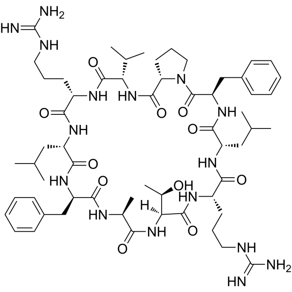
XQ2B is a specific cGAS inhibitor targeting protein-DNA interaction and phase separation. XQ2B markedly reduces ISD-induced Autophagy. XQ2B inhibits herpes simplex virus 1 (HSV-1)-induced antiviral immune responses and enhances HSV-1 infection.
For research use only. We do not sell to patients.
- Purity : 99.45%
- CAS No.: 2104728-28-9
- Formula: C59H92N16O11
- Molecular Weight:1201.46
-
Storage:
Sealed storage, away from moisture.
Powder -80°C, 2 years , -20°C, 1 year* In solvent : -80°C, 6 months; -20°C, 1 month (sealed storage, away from moisture)

To place orders, for customer services and technical support, please contact: MedChemExpress USA
Tel: 609-228-6898 E-mail: [email protected] [email protected]


-
 Biological Activity
-
Chemical Information
-
Solvent & Solubility
- Protocols
- Purity & Documentation
- References
-
 Help & FAQs




Biological Activity
Description
In Vitro
XQ2B (10 μM; 24 h) inhibits the expression of Ifnb1, Cxcl10 and Il6 in Trex1-/- BMDMs[1].
MedChemExpress (MCE) has not independently confirmed the accuracy of these methods. They are for reference only.
In Vivo
XQ2B (10 mg/kg; i.v.; every other day; 7 days) suppresses systemic inflammation in Trex1-/- mice[1].
MedChemExpress (MCE) has not independently confirmed the accuracy of these methods. They are for reference only.
-
Animal Model:C57BL/6 Trex1-/- male mice (8-week-old) with autoimmune disease model[1]
-
Dosage:10 mg/kg
-
Administration:Intravenous injection, every other day, 7-day cycle for evaluating anti-inflammatory effect and 11 - day cycle for survival analysis
-
Result:Attenuated the inflammation in heart, stomach, tongue, kidney, and skeletal muscle of Trex1-/- mice compared to untreated Trex1-/- mice (the 7-day cycle experiment).
Suppressed the mRNA level of Ifnb1, Cxcl10 and Il6 caused by Trex1 deficiency.
Showed no effect on mouse survival during the test period (the 11-day cycle experiment).


Chemical Information
-
CAS No. 2104728-28-9
-
Appearance Solid
-
Molecular Weight 1201.46
-
Formula C59H92N16O11
-
Color White to off-white
-
Sequence
Cyclo({d-Phe}-Ala-Thr-Arg-Leu-{d-phe}-Pro-Val-Arg-Leu)
-
Sequence Shortening
Cyclo({d-Phe}-ATRL-{d-phe}-PVRL)
-
Shipping
Room temperature in continental US; may vary elsewhere.
-
Storage
Sealed storage, away from moisture
Powder -80°C 2 years -20°C 1 year * In solvent : -80°C, 6 months; -20°C, 1 month (sealed storage, away from moisture)


Solvent & Solubility
In Vitro:
H2O : 100 mg/mL (83.23 mM; Need ultrasonic)
Please refer to the solubility information to select the appropriate solvent. Once prepared, please aliquot and store the solution to prevent product inactivation from repeated freeze-thaw cycles.
Storage method and period of stock solution: -80°C, 6 months; -20°C, 1 month (sealed storage, away from moisture). When stored at -80°C, please use it within 6 months. When stored at -20°C, please use it within 1 month.
* Note: If you choose water as the stock solution, please dilute it to the working solution, then filter and sterilize it with a 0.22 μm filter before use.
Please refer to the solubility information to select the appropriate solvent. Once prepared, please aliquot and store the solution to prevent product inactivation from repeated freeze-thaw cycles.
Storage method and period of stock solution: -80°C, 6 months; -20°C, 1 month (sealed storage, away from moisture). When stored at -80°C, please use it within 6 months. When stored at -20°C, please use it within 1 month.
* Note: If you choose water as the stock solution, please dilute it to the working solution, then filter and sterilize it with a 0.22 μm filter before use.
Concentration (start) × Volume (start) = Concentration (final) × Volume (final)


Purity & Documentation
-
Data Sheet (284 KB)
-
SDS (252 KB)
- English - EN (252 KB)
- Français - FR (252 KB)
- Deutsch - DE (252 KB)
- Norwegian - NO (252 KB)
- Español - ES (252 KB)
- Swedish - SV (252 KB)
- Italian - IT (252 KB)
- Korean - KR (252 KB)
- Portuguese - PT (252 KB)
-
Handling Instructions (2659 KB)
References


Complete Stock Solution Preparation Table
Please refer to the solubility information to select the appropriate solvent. Once prepared, please aliquot and store the solution to prevent product inactivation from repeated freeze-thaw cycles.
Storage method and period of stock solution: -80°C, 6 months; -20°C, 1 month (sealed storage, away from moisture). When stored at -80°C, please use it within 6 months. When stored at -20°C, please use it within 1 month.
| Optional Solvent | Concentration Solvent Mass | 1 mg | 5 mg | 10 mg | 25 mg |
|---|---|---|---|---|---|
| H2O | 1 mM | 0.8323 mL | 4.1616 mL | 8.3232 mL | 20.8080 mL |
| 5 mM | 0.1665 mL | 0.8323 mL | 1.6646 mL | 4.1616 mL | |
| 10 mM | 0.0832 mL | 0.4162 mL | 0.8323 mL | 2.0808 mL | |
| 15 mM | 0.0555 mL | 0.2774 mL | 0.5549 mL | 1.3872 mL | |
| 20 mM | 0.0416 mL | 0.2081 mL | 0.4162 mL | 1.0404 mL | |
| 25 mM | 0.0333 mL | 0.1665 mL | 0.3329 mL | 0.8323 mL | |
| 30 mM | 0.0277 mL | 0.1387 mL | 0.2774 mL | 0.6936 mL | |
| 40 mM | 0.0208 mL | 0.1040 mL | 0.2081 mL | 0.5202 mL | |
| 50 mM | 0.0166 mL | 0.0832 mL | 0.1665 mL | 0.4162 mL | |
| 60 mM | 0.0139 mL | 0.0694 mL | 0.1387 mL | 0.3468 mL | |
| 80 mM | 0.0104 mL | 0.0520 mL | 0.1040 mL | 0.2601 mL |
* Note: If you choose water as the stock solution, please dilute it to the working solution, then filter and sterilize it with a 0.22 μm filter before use.


XQ2B Related Classifications
HY-P5997 Related Classifications
 Powered by Bioz