



IN-1130
Based on 2 publication(s) in Google Scholar
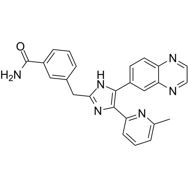
IN-1130 is a highly selective transforming growth factor-β type I receptor kinase (ALK5) inhibitor with an IC50 of 5.3 nM for ALK5-mediated Smad3 phosphorylation. IN-1130 inhibits ALK5 phosphorylation of casein (IC50=36 nM) and p38α mitogen-activated protein kinase (IC50=4.3 μM). IN-1130 suppresses renal fibrosis in obstructive nephropathy and blocks breast cancer lung metastasis.
For research use only. We do not sell to patients.
- Purity : 99.93%
- CAS No.: 868612-83-3
- Formula: C25H20N6O
- Molecular Weight:420.47
-
Storage:Powder -20°C, 3 years , 4°C, 2 years ; In solvent -80°C, 6 months , -20°C, 1 month

To place orders, for customer services and technical support, please contact: MedChemExpress USA
Tel: 609-228-6898 E-mail: [email protected] [email protected]


-
 Biological Activity
-
Chemical Information
-
Solvent & Solubility
- Protocols
- Purity & Documentation
- References
-
 Help & FAQs


-
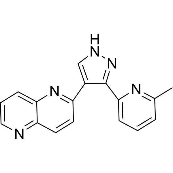
HY-13012
RepSoxTGFβR-1/ALK5 Inhibitor -
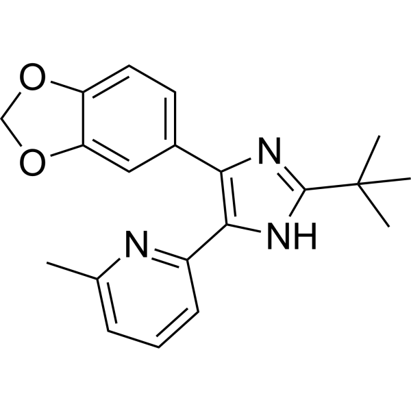
HY-13521
SB-505124ALK4/5/7 Inhibitor -
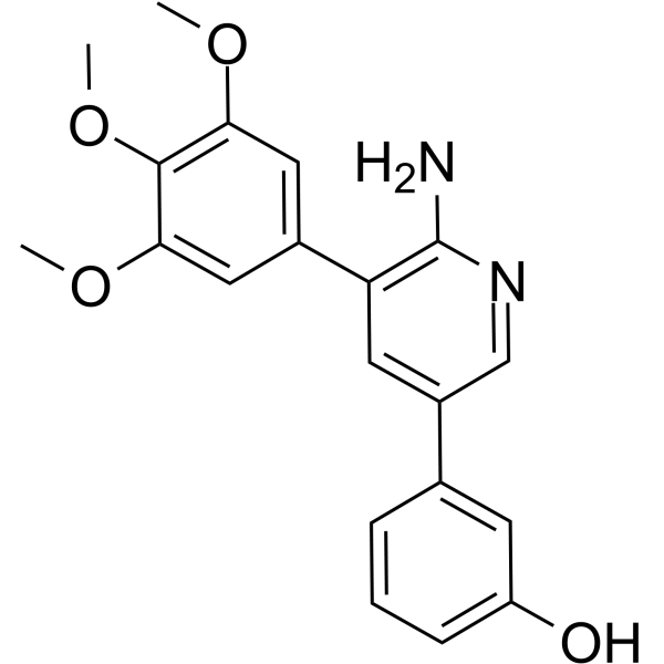
HY-12278
K02288BMPI Inhibitor -
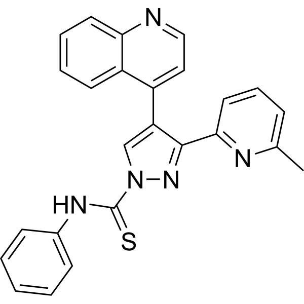
HY-10432
A 83-01ALK4/5/7 Inhibitor -
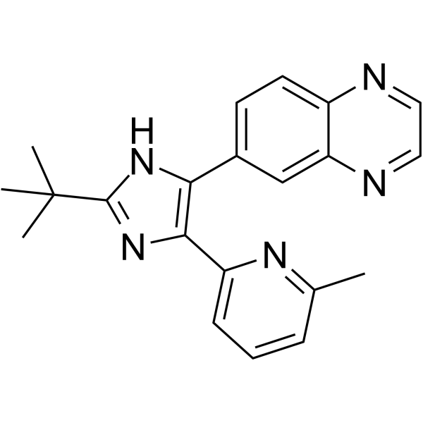
HY-12043
SB 525334ALK5 Inhibitor -
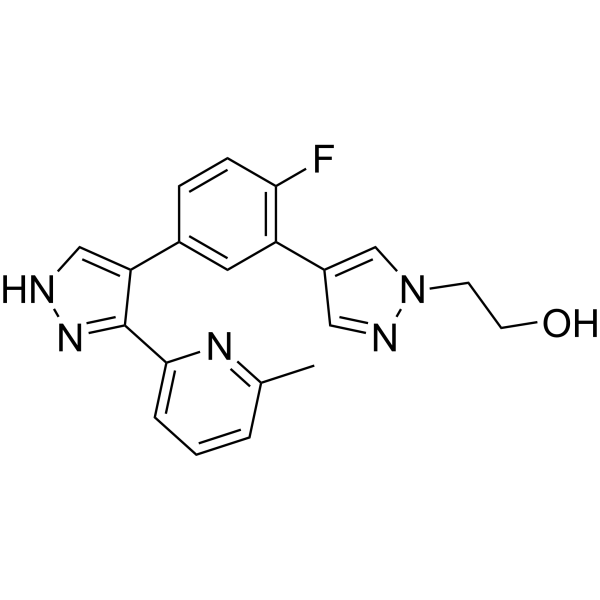
HY-12953
R-268712ALK-5 Inhibitor -
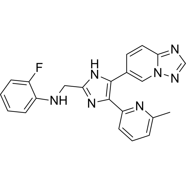
HY-19928
VactosertibALK5 Inhibitor -

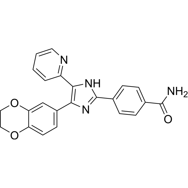
HY-10431
SB-431542TGF-β Receptor Kinase Inhibitor -
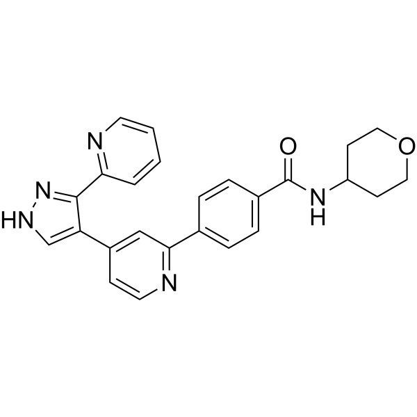
HY-10324
D4476CK1 Inhibitor -
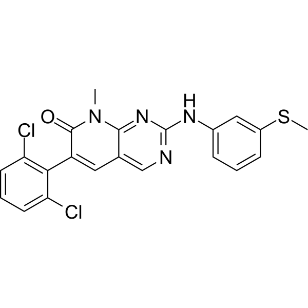
HY-10326
GW788388ALK5 Inhibitor -
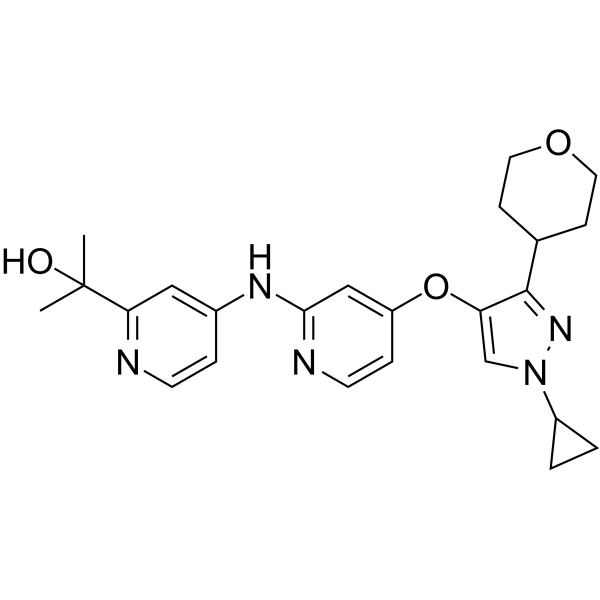
HY-10395
PD173955Src Inhibitor -
HY-103021
LY3200882ALK5 Inhibitor
Publications Citing Use of MedChemExpress (MCE) IN-1130
More

Biological Activity
Description
IC50 & Target
[1]|
ALK5 |
Cellular Effect
|
Cell Line

|
Type | Value | Description | References |
|---|---|---|---|---|
| 4T1 | IC50 |
0.0442 μM
Compound: 6, IN-1130
|
Inhibition of ALK5 in mouse 4T1 cells assessed as inhibition of TGFbeta1-induced luciferase activity after 24 hrs by luciferase reporter gene assay
Inhibition of ALK5 in mouse 4T1 cells assessed as inhibition of TGFbeta1-induced luciferase activity after 24 hrs by luciferase reporter gene assay
|
[PMID: 24786585] |
| HaCaT | IC50 |
0.0196 μM
Compound: 6, IN-1130
|
Inhibition of ALK5 in human HaCaT cells assessed as inhibition of TGFbeta1-induced luciferase activity after 24 hrs by luciferase reporter gene assay
Inhibition of ALK5 in human HaCaT cells assessed as inhibition of TGFbeta1-induced luciferase activity after 24 hrs by luciferase reporter gene assay
|
[PMID: 24786585] |
| Sf9 | IC50 |
0.017 μM
Compound: 5, IN-1130
|
Inhibition of GST-fused recombinant human ALK5 expressed in baculovirus-infected insect Sf9 cells using casein as substrate by radioisotope-based assay
Inhibition of GST-fused recombinant human ALK5 expressed in baculovirus-infected insect Sf9 cells using casein as substrate by radioisotope-based assay
|
[PMID: 24704197] |
| Sf9 | IC50 |
0.017 μM
Compound: 6, IN-1130
|
Inhibition of human recombinant GST-fused ALK5 expressed in insect Sf9 cells using casein as substrate by radiometric kinase assay
Inhibition of human recombinant GST-fused ALK5 expressed in insect Sf9 cells using casein as substrate by radiometric kinase assay
|
[PMID: 21696866] |
| Sf9 | IC50 |
0.017 μM
Compound: 6, IN-1130
|
Inhibition of human recombinant ALK5 expressed in insect Sf9 cells using casein as substrate by radioisotopic assay
Inhibition of human recombinant ALK5 expressed in insect Sf9 cells using casein as substrate by radioisotopic assay
|
[PMID: 24786585] |
| Sf9 | IC50 |
0.017 μM
Compound: 6, IN-1130
|
Inhibition of human recombinant GST-fused ALK5 expressed in Sf9 cells
Inhibition of human recombinant GST-fused ALK5 expressed in Sf9 cells
|
[PMID: 21435890] |
| Sf9 | IC50 |
0.036 μM
Compound: 8, IN-1130
|
Inhibition of human recombinant GST-fused ALK5 expressed in Sf9 cells
Inhibition of human recombinant GST-fused ALK5 expressed in Sf9 cells
|
[PMID: 20472445] |
| Sf9 | IC50 |
17.2 nM
Compound: 5, IN-1130
|
Inhibition of human recombinant GST-fused ALK5 expressed in Sf9 cells using casein as a substrate by radioisotopic protein kinase assay
Inhibition of human recombinant GST-fused ALK5 expressed in Sf9 cells using casein as a substrate by radioisotopic protein kinase assay
|
[PMID: 23047226] |
| Sf9 | IC50 |
17.2 nM
Compound: 6, IN-1130
|
Inhibition of human recombinant GST-fused ALK5 expressed in Sf9 cells using casein as a substrate by radioisotopic protein kinase assay
Inhibition of human recombinant GST-fused ALK5 expressed in Sf9 cells using casein as a substrate by radioisotopic protein kinase assay
|
[PMID: 21911290] |
| Sf9 | IC50 |
36 nM
Compound: IN-1130
|
Inhibition of GST fused human recombinant ALK5 expressed in Sf9 cells by radioisotopic protein kinase assay
Inhibition of GST fused human recombinant ALK5 expressed in Sf9 cells by radioisotopic protein kinase assay
|
[PMID: 18571921] |
In Vitro
IN-1130 (0.5, 1 μM; for 2 hours) inhibits TGF-β-stimulated Smad2 phosphorylation and subsequent nuclear translocation in HepG2 and 4T1 cells[2].
?
IN-1130 (1 μM; for 72 hours) restores the TGF-β-mediated decrease in E-cadherin protein expression. IN-1130 (1 μM; for 72 hours) inhibits TGF-β-induced MMPs mRNA expression and the gelatinolytic activity of secreted MMPs in MCF10A cells[2].
?
IN-1130 (1 μM; pretreated for 30 min) inhibits TGF-β-induced MDA-MB-231 cells, NMuMG, and MCF10A cells mobility and invasion[2].
MedChemExpress (MCE) has not independently confirmed the accuracy of these methods. They are for reference only. Further protocols information, click here.
-
Cell Line:HepG2 and 4T1 cells
-
Concentration:0.5, 1 μM
-
Incubation Time:For 2 hours
-
Result:Inhibited TGF-β-stimulated Smad2 phosphorylation.
-
Cell Line:MCF10A cells
-
Concentration:1 μM
-
Incubation Time:For 72 hours
-
Result:Inhibited TGF-β-induced MMPs mRNA expression and the gelatinolytic activity of secreted MMPs.
In Vivo
? IN-1130 (10, 20 mg/kg/day; for 14 days) dose-dependently decreases levels of TGF-β1 mRNA and suppresses phosphorylation of Smad2, α-SMA, myofibroblasts in rat UUO kidneys[1].
? IN-1130 (40 mg/kg; IP; 3 times per week for 3 weeks) inhibits in vivo breast cancer metastasis to the lungs in MMTV/c-Neu mice (Eight-week-old female BALB/c mice)[2].
MedChemExpress (MCE) has not independently confirmed the accuracy of these methods. They are for reference only.
-
Animal Model:Six-week-old male Sprague–Dawley rats weighing 180-200 g[1]
-
Dosage:10 and 20 mg/kg
-
Administration:IP; daily; for 7 and 14 days
-
Result:Reduced the extent of interstitial nephritis and fibrosis (arrowheads) with 10 mg/kg.


Chemical Information
-
CAS No. 868612-83-3
-
Appearance Solid
-
Molecular Weight 420.47
-
Formula C25H20N6O
-
Color White to light yellow
-
SMILES
O=C(N)C1=CC=CC(CC2=NC(C3=NC(C)=CC=C3)=C(C4=CC=C5N=CC=NC5=C4)N2)=C1
-
Shipping
Room temperature in continental US; may vary elsewhere.
-
Storage
Powder -20°C 3 years 4°C 2 years In solvent -80°C 6 months -20°C 1 month


Publications (2)
-
Journal Impact Factor
-
Most Recent
-
Oncogene
Olfactomedin 4 orchestrates TGF-β/CEACAM6 axis, promoting cell-autonomous epithelial to mesenchymal transition during gallbladder epithelium carcinogenesis. [Abstract]2026 Jul;45(24):2359-2374. PMID: 42050078 -
iScience
Organic phosphate but not inorganic phosphate regulates Fgf23 expression through MAPK and TGF-ꞵ signaling. [Abstract]2024 Mar 29;27(6):109625. PMID: 38883842


Solvent & Solubility
In Vitro:
DMSO : 100 mg/mL (237.83 mM; Need ultrasonic; Hygroscopic DMSO has a significant impact on the solubility of product, please use newly opened DMSO)
Please refer to the solubility information to select the appropriate solvent. Once prepared, please aliquot and store the solution to prevent product inactivation from repeated freeze-thaw cycles.
Storage method and period of stock solution: -80°C, 6 months; -20°C, 1 month. When stored at -80°C, please use it within 6 months. When stored at -20°C, please use it within 1 month.
Please refer to the solubility information to select the appropriate solvent. Once prepared, please aliquot and store the solution to prevent product inactivation from repeated freeze-thaw cycles.
Storage method and period of stock solution: -80°C, 6 months; -20°C, 1 month. When stored at -80°C, please use it within 6 months. When stored at -20°C, please use it within 1 month.
Concentration (start) × Volume (start) = Concentration (final) × Volume (final)
In Vivo:
Select the appropriate dissolution method based on your experimental animal and administration route.
- For the following dissolution methods, please ensure to first prepare a clear stock solution using an In Vitro approach and then sequentially add co-solvents:
- To ensure reliable experimental results, the clarified stock solution can be appropriately stored based on storage conditions. As for the working solution for In Vivo experiments, it is recommended to prepare freshly and use it on the same day.
- The percentages shown for the solvents indicate their volumetric ratio in the final prepared solution. If precipitation or phase separation occurs during preparation, heat and/or sonication can be used to aid dissolution.
Add each solvent one by one: 10% DMSO 90% (20% SBE-β-CD in Saline)
Solubility: ≥ 1.43 mg/mL (3.40 mM); Clear solution
This protocol yields a clear solution of ≥ 1.43 mg/mL (saturation unknown).
Taking 1 mL working solution as an example, add 100 μL DMSO stock solution (14.3 mg/mL) to 900 μL 20% SBE-β-CD in Saline, and mix evenly.
Preparation of 20% SBE-β-CD in Saline (4°C, storage for one week): 2 g SBE-β-CD powder is dissolved in 10 mL Saline, completely dissolve until clear.
In Vivo Dissolution Calculator
Please enter the basic information of animal experiments:
-
-
-
-
Recommended: Prepare an additional quantity of animals to account for potential losses during experiments.
Please enter your animal formula composition:
-
%DMSO +
Recommended: Keep the proportion of DMSO in working solution below 2% if your animal is weak.
-
%+
-
+%Tween-80 + +
-
%Saline +
The co-solvents required include: DMSO, . All of co-solvents are available by MedChemExpress (MCE). , Tween 80. All of co-solvents are available by MedChemExpress (MCE).
Working solution concentration: 0.22 mg/mL
Method for preparing stock solution: mg drug dissolved in μL DMSO. Stock solution concentration: mg/mL.
1. Take μL DMSO stock solution;
2. Add μL .
μL , mix evenly;
3. Then add μL Tween 80, mix evenly;
4. Then add μL
Please ensure that the stock solution in the first step is dissolved to a clear state, and add co-solvents in sequence. You can use ultrasonic heating (ultrasonic cleaner, recommended frequency 20-40 kHz), vortexing, etc. to assist dissolution.

Protocols
-
Kinase activity and phosphorylation assays
Kinase activity assays measure the ability of kinases to transfer phosphate groups from ATP to specific substrates, while phosphorylation assays detect the presence and levels of phosphorylated proteins. Common methods include radiolabeled ATP incorporation (e. g. ,), ADP release detection via bioluminescence (e. g. ,[3]), enzyme-linked immunosorbent assays (ELISA) for phospho-specific epitopes (e. g. ,[6]), and microtiter-based formats for high-throughput screening (e. g. ,[8]). The ADP-Glo assay quantifies kinase activity by measuring ADP produced during phosphorylation using a luciferase-based system. Radiometric assays involve autoradiography or scintillation counting after incorporation of 32P-labeled ATP into substrate proteins. ELISA-based approaches rely on phospho-specific antibodies to detect activated kinases in cell lysates or purified samples.
-
Fibrosis/Collagen Morphometry
Fibrosis and collagen morphometry is based on the quantitative visualization of fibrillar collagen deposition in tissue sections using histochemical stains such as Sirius Red (Picrosirius Red) or Masson's trichrome, followed by image-based or polarization-enhanced analysis to estimate collagen proportional area as a surrogate of extracellular matrix accumulation during fibrotic remodeling. Sirius Red combined with polarized light microscopy enhances detection of collagen fibers due to birefringence properties, enabling more specific visualization of collagen type I and III fibrils compared to conventional bright-field histology, while whole-section or region-restricted digital morphometry reduces field-selection bias in fibrosis assessment. Alternative quantitative approaches include second harmonic generation (SHG) and two-photon excited fluorescence microscopy, which enable label-free detection of fibrillar collagen and have been validated against histological staining and biochemica
-
Western Blot
Western blotting (WB) is a commonly used experimental method in molecular biology, biochemistry, and immunogenetics for identifying and quantifying target proteins. It combines gel electrophoresis with immunoassay, enabling researchers to analyze protein expression, post-translational modifications, and molecular weight.
-
Collagen: Sirius Red Staining
Sirius Red or picrosirius red staining is a histochemical method for visualizing collagen-rich extracellular matrix in tissue sections, and collagen fibers are detected as red-stained structures under bright-field microscopy with enhanced birefringence under polarized light. Picrosirius red is useful for assessing total collagen organization, distribution, and fibrosis burden, but polarized color should not be interpreted as a definitive collagen type I versus type III readout because color is affected by fiber orientation, thickness, and packing.
-
Cell Cytotoxicity Assay
Cytotoxicity assays are usually based on the assessment of cell membrane damage, which can also be indirectly detected by measuring cell viability. Detection methods include MTT assay, CKK-8 assay, LDH assay and ATP assay, etc.
-
Connective Tissue: Masson's Trichrome/Collagen Trichrome Staining
Masson’s Trichrome (collagen/trichrome staining) is a histological technique that differentially stains tissue compartments using sequential acidic dyes to distinguish collagen from muscle and cytoplasmic components based on dye affinity and tissue permeability differences, enabling visualization of fibrosis and connective tissue architecture in histological sections. The classical formulation typically uses Weigert's iron hematoxylin for nuclear staining, Biebrich scarlet-acid fuchsin for cytoplasm and muscle, and aniline blue (or light green variants) for collagen, producing a characteristic blue/green collagen signal contrasted against red cytoplasm and dark nuclei. The staining principle relies on selective displacement of smaller dye molecules by larger anionic dyes in collagen-rich regions under controlled acidified conditions, which enhances collagen-specific dye retention. This property makes the method widely used for fibrosis assessment in organs such as heart, liver, lung, a
-
Tail-Vein Experimental Metastasis Xenograft
Tail-vein experimental metastasis xenograft models assess the ability of injected tumor cells to survive circulation, arrest in vascular beds, extravasate, and colonize distant organs, most commonly lung after lateral tail-vein injection; this model bypasses primary-tumor formation, local invasion, and intravasation, so the readout reflects late metastatic colonization rather than the full metastatic cascade. The main readouts are metastatic burden measured by bioluminescence imaging, gross metastatic nodules, histology, organ weight, survival, or ex vivo tumor-cell quantification; luciferase-labeled tumor cells permit longitudinal noninvasive monitoring, while histology confirms organ colonization and tissue localization.
-
Protocol for Kinase activity and phosphorylation assays
Kinase activity assays measure transfer of phosphate from ATP to a protein or peptide substrate, generating phosphorylated substrate, ADP, or incorporated radiolabeled phosphate as the readout; phosphorylation assays measure site-specific phosphorylation in cells or tissues as a proxy for kinase-pathway activation, inhibition, or substrate regulation. Phosphorylation can be detected by phospho-specific Western blot, immunoprecipitation kinase assay, phospho-immunofluorescence, phospho-flow cytometry, luminescent ADP detection, radiolabeled ATP incorporation, or reporter-based pathway assays, and these readouts can be applied to cancer cells, primary neurons, mouse tumors, organoids, inflammatory macrophages, ferroptosis studies, and mitophagy studies when the kinase target is biologically relevant.
-
Breast Cancer Modeling
Breast cancer is a heterogeneous cancer, and it has been distinguished into four subtypes: luminal A, luminal B, HER2-positive and basal-like. Molecular mutations, epigenetic alterations, hormone exposure and immune microenvironment are related to the progression of breast cancer.
-
Orthotopic Cell-Line Xenograft
Orthotopic cell-line xenograft models involve implantation of human cancer cell lines into the anatomically corresponding organ of immunodeficient mice to reproduce tumor growth within a native microenvironment, enabling more clinically relevant tumor behavior compared with subcutaneous models. These models are widely used because orthotopic placement better recapitulates tumor progression, including invasion and metastatic spread, which are often underrepresented in heterotopic implantation systems. Compared with conventional xenografts, orthotopic implantation is described as more technically complex but provides improved simulation of tumor-microenvironment interactions and metastatic behavior, making it particularly valuable for translational oncology research. Surgical orthotopic implantation approaches have been emphasized as enabling faithful reproduction of clinical cancer features, including metastasis and disease progression patterns that align with the tumor’s organ of origi


Purity & Documentation
-
Data Sheet (281 KB)
-
SDS (393 KB)
- English - EN (393 KB)
- Français - FR (393 KB)
- Deutsch - DE (393 KB)
- Norwegian - NO (393 KB)
- Español - ES (393 KB)
- Swedish - SV (393 KB)
- Italian - IT (393 KB)
- Korean - KR (393 KB)
- Portuguese - PT (393 KB)
-
Handling Instructions (2659 KB)
References
[1]. Moon JA, et al. IN-1130, a novel transforming growth factor-beta type I receptor kinase (ALK5) inhibitor, suppresses renal fibrosis in obstructive nephropathy. Kidney Int. 2006 Oct;70(7):1234-43. [Content Brief]
[2]. Park CY, et al. An novel inhibitor of TGF-β type I receptor, IN-1130, blocks breast cancer lung metastasis through inhibition of epithelial-mesenchymal transition. Cancer Lett. 2014 Aug 28;351(1):72-80. [Content Brief]


Complete Stock Solution Preparation Table
Please refer to the solubility information to select the appropriate solvent. Once prepared, please aliquot and store the solution to prevent product inactivation from repeated freeze-thaw cycles.
Storage method and period of stock solution: -80°C, 6 months; -20°C, 1 month. When stored at -80°C, please use it within 6 months. When stored at -20°C, please use it within 1 month.
| Optional Solvent | Concentration Solvent Mass | 1 mg | 5 mg | 10 mg | 25 mg |
|---|---|---|---|---|---|
| DMSO | 1 mM | 2.3783 mL | 11.8915 mL | 23.7829 mL | 59.4573 mL |
| 5 mM | 0.4757 mL | 2.3783 mL | 4.7566 mL | 11.8915 mL | |
| 10 mM | 0.2378 mL | 1.1891 mL | 2.3783 mL | 5.9457 mL | |
| 15 mM | 0.1586 mL | 0.7928 mL | 1.5855 mL | 3.9638 mL | |
| 20 mM | 0.1189 mL | 0.5946 mL | 1.1891 mL | 2.9729 mL | |
| 25 mM | 0.0951 mL | 0.4757 mL | 0.9513 mL | 2.3783 mL | |
| 30 mM | 0.0793 mL | 0.3964 mL | 0.7928 mL | 1.9819 mL | |
| 40 mM | 0.0595 mL | 0.2973 mL | 0.5946 mL | 1.4864 mL | |
| 50 mM | 0.0476 mL | 0.2378 mL | 0.4757 mL | 1.1891 mL | |
| 60 mM | 0.0396 mL | 0.1982 mL | 0.3964 mL | 0.9910 mL | |
| 80 mM | 0.0297 mL | 0.1486 mL | 0.2973 mL | 0.7432 mL | |
| 100 mM | 0.0238 mL | 0.1189 mL | 0.2378 mL | 0.5946 mL |


IN-1130 Related Classifications
HY-18758 Related Classifications
 Powered by Bioz