



D-Isofloridoside

Based on 1 Customer Validation
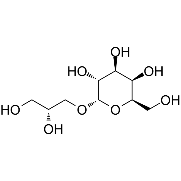
D-Isofloridoside, one of the polysaccharide precursors, has the activity of scavenging free radicals, inhibiting ROS expression, and inhibiting MMP-2 and MMP-9.
商品は「研究用試薬」です。人や動物の医療用・臨床診断用・食品用の製品ではありません。
研究用途以外に使用した場合、当社は一切の責任を負いかねます。
- 純度 : 98.59%
- CAS 番号: 23202-76-8
- 分子式: C9H18O8
- 分子量:254.23
-
保管条件:
-20°C, sealed storage, away from moisture
* In solvent : -80°C, 6 months; -20°C, 1 month (sealed storage, away from moisture)

To place orders, for customer services and technical support, please contact: MedChemExpress USA
Tel: 609-228-6898 E-mail: [email protected] [email protected]


-
 生物活性
生物活性
-
化学情報
- プロトコル
- 純度とドキュメンテーション
- 参考文献
-
 Help & FAQs


-
HY-12169
MarimastatMMP Inhibitor -
HY-13564
BatimastatMMP Inhibitor -
HY-118694
TAPI-0TACE/MMP Inhibitor -
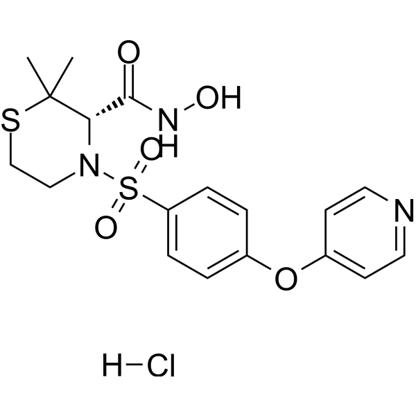
HY-12170A
Prinomastat hydrochlorideMMP Inhibitor -
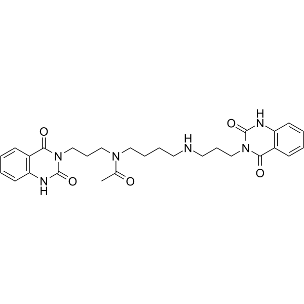
HY-109097
OtaplimastatMMP Inhibitor -
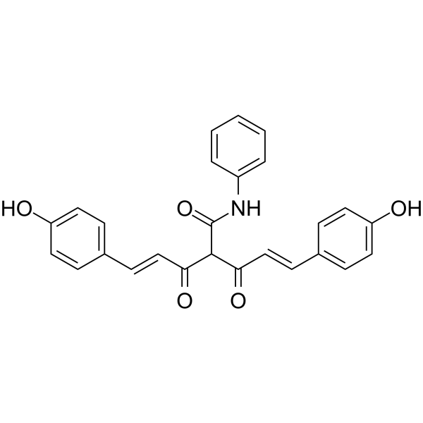
HY-120793
CMC2.24Ras/MMPs Inhibitor -

HY-13564R
Batimastat (Standard)MMP Inhibitor -

HY-12169R
Marimastat (Standard)MMP Inhibitor -

HY-109097R
Otaplimastat (Standard)Reference Standard


生物活性
製品説明
体外実験
D-Isofloridoside can reduce the activity of MMP-2/9, and can inhibit the expression of hypoxia-inducible factor-1α (HIF-1α) by regulating the downstream PI3K/AKT and MAPK pathways, thereby down-regulating the production of vascular endothelial growth factor (VEGF) in CoCl2-induced HT1080 cell. D-Isofloridoside can inhibit the activation of VEGF receptor (VEGFR-2), regulate downstream PI3K/AKT, MAPK, NF-κB signal pathways, activate apoptosis, and thus down-regulate the production of platelet-derived growth factor (PDGF) in VEGF-induced HUVEC[2].
MedChemExpress (MCE) has not independently confirmed the accuracy of these methods. They are for reference only. Further protocols information, click here.


化学情報
-
CAS 番号 23202-76-8
-
性状 Oil
-
分子量 254.23
-
分子式 C9H18O8
-
Color Light yellow to yellow
-
SMILES
OC[C@@H](O)CO[C@H]1O[C@@H]([C@@H]([C@@H]([C@H]1O)O)O)CO
-
Structure Classification
-
Initial Source
-
輸送条件
Room temperature in continental US; may vary elsewhere.
-
保管条件
-20°C, sealed storage, away from moisture
* In solvent : -80°C, 6 months; -20°C, 1 month (sealed storage, away from moisture)

プロトコル
-
RNA extraction experimental
By lysing cells, releasing RNA, and removing impurities such as proteins and DNA, high-purity RNA products are finally obtained. The commonly used traditional method is the guanidine isothiocyanate/phenol/chloroform method (Trizol), which is suitable for a variety of animal materials including animal tissues, microorganisms, cultured cells, etc., and most plant materials.
-
Mitochondrial membrane-potential fluorescent assay
Mitochondrial membrane potential fluorescent assays estimate ΔΨm in living cells using lipophilic cationic dyes such as TMRM, TMRE, rhodamine 123, and JC-1, which accumulate in mitochondria according to membrane polarization; loss of signal after FCCP or CCCP treatment is interpreted as mitochondrial depolarization. TMRM/TMRE and rhodamine 123 are commonly used for semi-quantitative live-cell microscopy or flow cytometry, while JC-1 can report a shift from red aggregate fluorescence to green monomer fluorescence during depolarization; interpretation requires controls because dye concentration, quenching mode, cell type, dye efflux, and mitochondrial mass can affect fluorescence independently of ΔΨm.
-
ROS/oxidative-stress fluorescent staining
ROS/oxidative-stress fluorescent staining uses cell-permeant fluorogenic probes that become fluorescent after oxidation inside cells or tissues; commonly used examples include DCFH-DA/DCFDA for broad cellular oxidant detection, DHE for superoxide-related signal detection, MitoSOX for mitochondrial superoxide-related signal detection, and CellROX probes for oxidative-stress-associated fluorescence readouts. The assay detects probe oxidation rather than a single ROS species unless the probe and analysis method have been chemically validated for that species. DCFH-DA enters cells, is deacetylated by intracellular esterases to DCFH, and produces fluorescent DCF after oxidation, so the readout is used as an operational measure of total cellular oxidative stress rather than a species-specific ROS measurement. DHE and MitoSOX can report superoxide-related oxidation, but red fluorescence alone can include non-specific ethidium-like oxidation products; HPLC or optimized spectral approaches are
-
Mitochondrial membrane-potential and mitochondrial mass staining
Mitochondrial membrane potential staining measures the electrochemical polarization across the mitochondrial inner membrane in live cells using lipophilic cationic fluorescent probes; early rhodamine-based work showed that selective mitochondrial dye accumulation is lost when the mitochondrial transmembrane potential is dissipated. JC-1 reports mitochondrial polarization by shifting from green monomer fluorescence to red J-aggregate fluorescence as dye concentration increases within energized mitochondria; therefore, the red/green fluorescence ratio is used as a relative readout of mitochondrial membrane potential. TMRE or TMRM staining provides a single-channel relative readout because these cationic rhodamine esters accumulate in polarized mitochondria, and lower fluorescence indicates reduced mitochondrial polarization when acquisition and dye-loading conditions are controlled. Mitochondrial mass staining is commonly performed with MitoTracker Green FM or related MitoTracker dyes as


純度とドキュメンテーション
-
データシート (267 KB)
-
SDS (252 KB)
- English - EN (252 KB)
- Français - FR (252 KB)
- Deutsch - DE (252 KB)
- Norwegian - NO (252 KB)
- Español - ES (252 KB)
- Swedish - SV (252 KB)
- Italian - IT (252 KB)
- Korean - KR (252 KB)
- Portuguese - PT (252 KB)
-
取扱説明書 (2659 KB)
参考文献
[1]. Yang S, et al. The Protective Effect of the Polysaccharide Precursor, D-Isofloridoside, from Laurencia undulata on Alcohol-Induced Hepatotoxicity in HepG2 Cells. Molecules. 2020;25(5):1024. Published 2020 Feb 25. [Content Brief]
[2]. Yang S, et al. Mechanism Analysis of Antiangiogenic d-Isofloridoside from Marine Edible Red algae Laurencia undulata in HUVEC and HT1080 cell. J Agric Food Chem. 2021 Nov 12. [Content Brief]

Calculators
濃度 (開始) × 体積 (開始) = 濃度 (終了) × 体積 (終了)


D-Isofloridoside 関連分類
HY-N10176 関連分類
 Powered by Bioz