



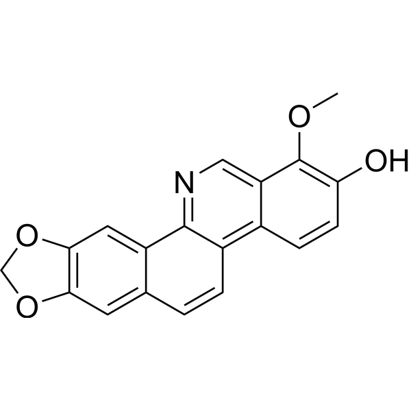
Decarine
Decarine (Rutaceline) is a benzophenanthridine alkaloid found in Zanthoxylum species. Decarinewith shows anti-inflammatory, antimycobacterial, and anti-HIV activity. Decarine inhibits NO, TNF-α, IL-1β, IL-6, and IL-8 production in inflammatory cell models. Decarine inhibits growth of Mycobacterium tuberculosis strains, reduces intracellular Mycobacterium tuberculosis survival, and shows low cytotoxicity toward human macrophages. Decarine inhibits HIV replication in acutely infected lymphocytes. Decarine can be used for the researches of inflammation, tuberculosis, and HIV infection.
商品は「研究用試薬」です。人や動物の医療用・臨床診断用・食品用の製品ではありません。
研究用途以外に使用した場合、当社は一切の責任を負いかねます。
- CAS 番号: 54354-62-0
- 分子式: C19H13NO4
- 分子量:319.31
-
保管条件:
Please store the product under the recommended conditions in the Certificate of Analysis.

To place orders, for customer services and technical support, please contact: MedChemExpress USA
Tel: 609-228-6898 E-mail: [email protected] [email protected]


-
 生物活性
生物活性
-
化学情報
- プロトコル
- 純度とドキュメンテーション
- 参考文献
-
 Help & FAQs




生物活性
製品説明
IC50 & Target
[1]|
IL-1β |
IL-6 |
IL-8 |
Cellular Effect
|
Cell Line

|
Type | Value | Description | References |
|---|---|---|---|---|
| Neutrophil | IC50 |
1.29 μg/mL
Compound: Decarine
|
Antiinflammatory activity against human neutrophils assessed as inhibition of fMLP/CB-induced superoxide anion generation
Antiinflammatory activity against human neutrophils assessed as inhibition of fMLP/CB-induced superoxide anion generation
|
[PMID: 19128011] |
| Neutrophil | IC50 |
1.94 μg/mL
Compound: Decarine
|
Antiinflammatory activity against human neutrophils assessed as inhibition of fMLP/CB-induced elastase release
Antiinflammatory activity against human neutrophils assessed as inhibition of fMLP/CB-induced elastase release
|
[PMID: 19128011] |
| RAW264.7 | IC50 |
48.4 μM
Compound: 127
|
Inhibition of NO production in LPS-stimulated mouse RAW264.7 cells preincubated with compound for 30 mins followed by LPS stimulation and measured after 24 hrs by Griess reagent-based assay
Inhibition of NO production in LPS-stimulated mouse RAW264.7 cells preincubated with compound for 30 mins followed by LPS stimulation and measured after 24 hrs by Griess reagent-based assay
|
[PMID: 33454546] |
体外実験
Decarine (24 h) potently inhibits NO production in LPS (HY-D1056)-induced RAW 264.7 macrophages with an IC50 of 48.43 μM, displaying anti-inflammatory activity without cytotoxicity[1].
Decarine (10-20 μM; 24 h) significantly inhibits TNF-α and IL-1β production in LPS-induced THP-1 macrophages[1].
Decarine (20 μM; 24 h) significantly inhibits IL-6 and IL-8 production in TNF-α + IL-1β-induced Caco-2 cells[1].
Decarine (10-80 μM; 24 h) is non-toxic to THP-1 macrophages at concentrations up to 20 μM, but exhibits cytotoxicity at 40 μM and 80 μM[1].
Decarine (5-40 μM; 24 h) is non-toxic to Caco-2 cells at concentrations up to 40 μM[1].
Decarine (7-8 day) potently inhibits the in vitro growth of virulent Mycobacterium tuberculosis H37Rv (MIC = 1.6 μg/mL) and avirulent Mycobacterium tuberculosis H37Ra (MIC = 3.1 μg/mL)[2].
Decarine (0.8-50 μg/mL; 7 day) exhibits low cytotoxicity against human THP-1 macrophages (IC50 = 66.0 μg/mL) and has high selectivity indices (21.3 for Mycobacterium tuberculosis H37Ra, 41.2 for Mycobacterium tuberculosis H37Rv) for antimycobacterial activity[2].
Decarine (1.6-25 μg/mL; 5 days) reduces intracellular Mycobacterium tuberculosis H37Rv survival in human THP-1 macrophages by almost two log units at 6.2 μg/mL after 5 days of exposure, with dose-dependent bactericidal activity[2].
Decarine (4 days) potently inhibits HIV replication in acutely infected H9 lymphocyte cells with an EC50 of <0.1 μg/mL and has a therapeutic index of >226, as it inhibits uninfected H9 cell growth with an IC50 of 22.6 μg/mL[3].
MedChemExpress (MCE) has not independently confirmed the accuracy of these methods. They are for reference only.
-
Cell Line:human THP-1 macrophages
-
Concentration:10 μM, 20 μM
-
Incubation Time:24 h
-
Result:Inhibited TNF-α production significantly in a dose-dependent manner at 10 μM and 20 μM.
Inhibited IL-1β production significantly in a dose-dependent manner at 10 μM and 20 μM.
-
Cell Line:human Caco-2 colon cancer cells
-
Concentration:5 μM, 10 μM, 20 μM
-
Incubation Time:24 h
-
Result:Reduced IL-6 production significantly at 20 μM.
Reduced IL-8 production significantly at 20 μM.
-
Cell Line:human THP-1 macrophages
-
Concentration:10 μM, 20 μM, 40 μM, 80 μM
-
Incubation Time:24 h
-
Result:Did not reduce cell viability at 10 μM and 20 μM.
Reduced cell viability at 40 μM.
Reduced cell viability at 80 μM.


化学情報
-
CAS 番号 54354-62-0
-
分子量 319.31
-
分子式 C19H13NO4
-
SMILES
OC1=C(OC)C2=CN=C3C(C(C=CC3=C2C=C1)=C4)=CC5=C4OCO5
-
別名
Rutaceline
-
Structure Classification
-
Initial Source
-
輸送条件
Room temperature in continental US; may vary elsewhere.
-
保管条件
Please store the product under the recommended conditions in the Certificate of Analysis.


純度とドキュメンテーション
参考文献
[1]. Zhang HL, et al. Chemical constituents and anti-inflammatory activities of Maqian (Zanthoxylum myriacanthum var. pubescens) bark extracts. Sci Rep. 2017;7:45805. Published 2017 Apr 6. [Content Brief]
[2]. Luo X, et al. Zanthoxylum capense constituents with antimycobacterial activity against Mycobacterium tuberculosis in vitro and ex vivo within human macrophages. J Ethnopharmacol. 2013;146(1):417-422. [Content Brief]
[3]. Cheng MJ, et al. Two new sesquiterpenoids and anti-HIV principles from the root bark of Zanthoxylum ailanthoides. Bioorg Med Chem. 2005;13(21):5915-5920. [Content Brief]

Calculators
濃度 (開始) × 体積 (開始) = 濃度 (終了) × 体積 (終了)


Decarine 関連分類
HY-N3700 関連分類
 Powered by Bioz