



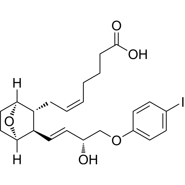
I-BOP
I-BOP is an agonist for thromboxane A2 receptor (TP) with a KD of 0.61 nM. I-BOP promotes proliferation through activation of PI3K pathway in vascular smooth muscle. I-BOP dose-dependently biphasicly affects the excitatory postsynaptic potential (e.p.s.p.) in hippocampal neurons.
商品は「研究用試薬」です。人や動物の医療用・臨床診断用・食品用の製品ではありません。
研究用途以外に使用した場合、当社は一切の責任を負いかねます。
- CAS 番号: 128719-90-4
- 分子式: C23H29IO5
- 分子量:512.38
-
保管条件:
Please store the product under the recommended conditions in the Certificate of Analysis.

To place orders, for customer services and technical support, please contact: MedChemExpress USA
Tel: 609-228-6898 E-mail: [email protected] [email protected]


-
 生物活性
生物活性
-
化学情報
- プロトコル
- 純度とドキュメンテーション
- 参考文献
-
 Help & FAQs


-
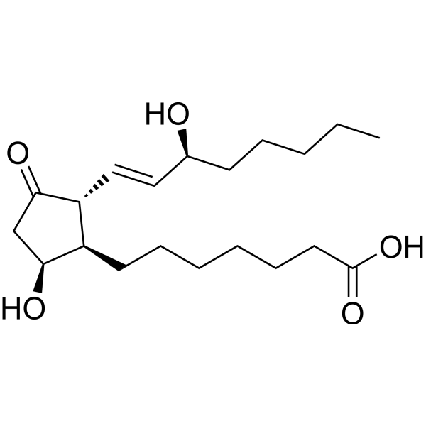
HY-113096
Prostaglandin D1CatSper Activator -
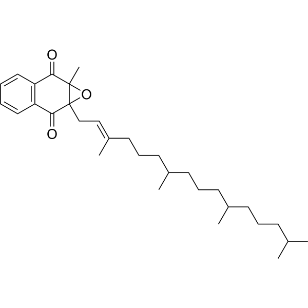
HY-113164
Vitamin K1 2,3-epoxideEndogenous Metabolite -
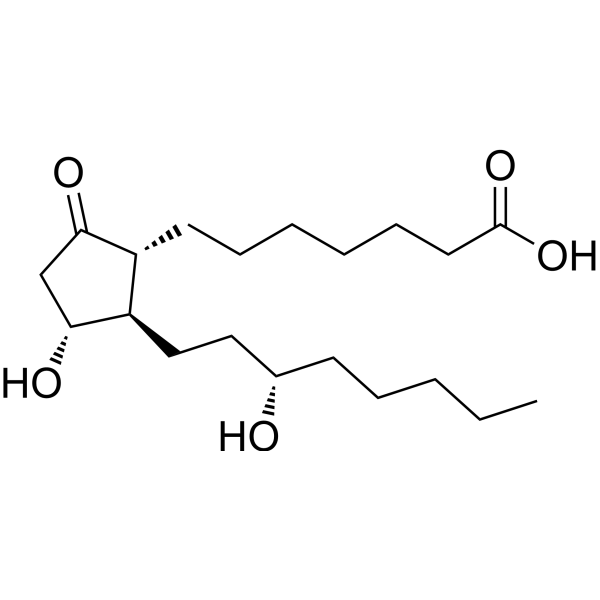
HY-113113
13,14-Dihydro PGE1PGE1 Metabolite -
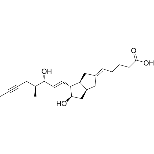
HY-118670
16(S)-IloprostStereoisomer -
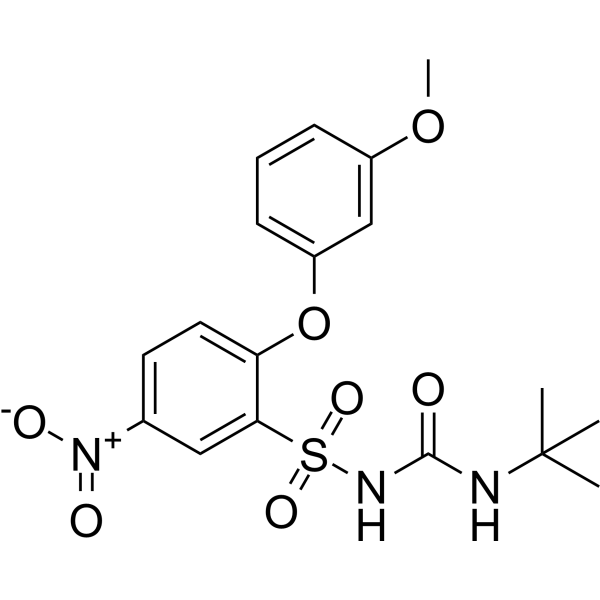
HY-118014
CAY10535FP/TP Receptor Inhibitor -
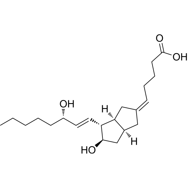
HY-124363
5-cis CarbaprostacyclinPGI2 Analog -
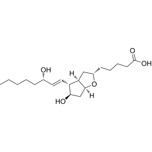
HY-130522
6β-Prostaglandin I1Prostacycline Analogue -
HY-130594
6α-Prostaglandin I1PGI2 Analog -

HY-120975
2,3-Dinor thromboxane b1Thromboxane B2 Metabolite -

HY-114660
9,11-Methane-epoxy prostaglandin F1αDerivative of Prostaglandin H1 -
HY-118670A
16(R)-IloprostStereoisomer


生物活性
製品説明
体外実験
I-BOP (1-100 nM) activates the MAP kinase, stimulates the tyrosine phosphorylation, and thus causes the activation of PI3K signaling pathway[1].
I-BOP (0.1-3 μM) enhances the synaptic transmission in hippocampal CA1 neuron at low concentration by increasing the neurotransmitter release, inhibits the excitatory synaptic transmission in hippocampal CA1 neuron through reduction of membrane input resistance at high concentration[2].
MedChemExpress (MCE) has not independently confirmed the accuracy of these methods. They are for reference only.
-
Cell Line:A7r5.HEL.TP cell
-
Concentration:1-100 nM
-
Incubation Time:24 h
-
Result:Increased phosphorylated tyrosine.


化学情報
-
CAS 番号 128719-90-4
-
分子量 512.38
-
分子式 C23H29IO5
-
SMILES
O=C(O)CCC/C=C\C[C@H]1[C@](O2)([H])CC[C@]2([H])[C@@H]1/C=C/[C@@H](O)COC3=CC=C(I)C=C3
-
輸送条件
Room temperature in continental US; may vary elsewhere.
-
保管条件
Please store the product under the recommended conditions in the Certificate of Analysis.


純度とドキュメンテーション
参考文献
[1]. Morinelli TA, et al., Tyrosine phosphorylation of phosphatidylinositol 3-kinase and of the thromboxane A2 (TXA2) receptor by the TXA2 mimetic I-BOP in A7r5 cells. Biochem Pharmacol. 1997 Jun 15;53(12):1823-32. [Content Brief]
[2]. Hsu KS, et al., Thromboxane A2 agonist modulation of excitatory synaptic transmission in the rat hippocampal slice. Br J Pharmacol. 1996 Aug;118(8):2220-7. [Content Brief]

Calculators
濃度 (開始) × 体積 (開始) = 濃度 (終了) × 体積 (終了)


I-BOP 関連分類
HY-116431 関連分類
 Powered by Bioz