



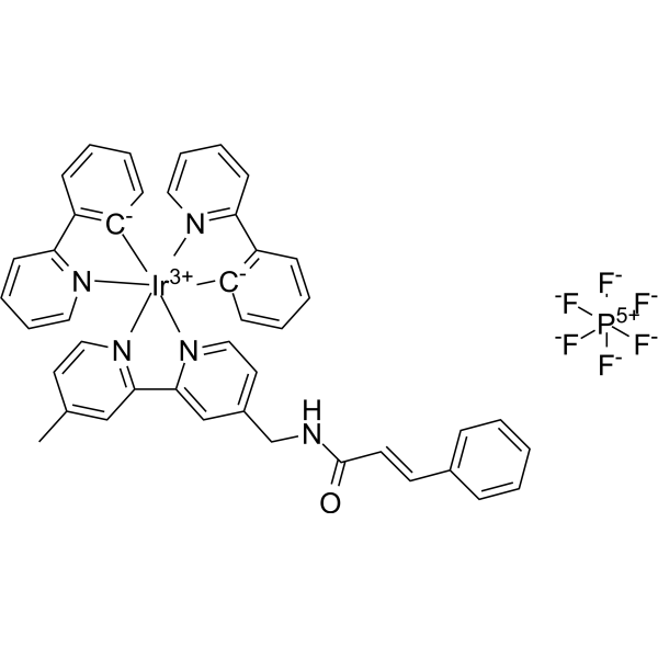
Ir-CA
Ir-CA is an antitumor agent. Ir-CA can accumulate in mitochondria and induces mitochondria dysfunction. Ir-CA induces apoptosis and autophagy. Ir-CA initiates mitophagy and cell cycle arrest to kill Cisplatin (HY-17394)-resistant A549R cells. Ir-CA can effectively inhibit metastasis by inhibiting MMP-2/MMP-9.
商品は「研究用試薬」です。人や動物の医療用・臨床診断用・食品用の製品ではありません。
研究用途以外に使用した場合、当社は一切の責任を負いかねます。
- CAS 番号: 2870682-93-0
- 分子式: C43H35F6IrN5OP
- 分子量:974.95
-
保管条件:
Please store the product under the recommended conditions in the Certificate of Analysis.

To place orders, for customer services and technical support, please contact: MedChemExpress USA
Tel: 609-228-6898 E-mail: [email protected] [email protected]


-
 生物活性
-
化学情報
- プロトコル
- 純度とドキュメンテーション
- 参考文献
-
 Help & FAQs




生物活性
製品説明
体外実験
Ir-CA shows cytotoxicity against A549, A549R, HLF, BEAS-2B, MCF-7, 4T1 cells with IC50s of 4.4, 4.5, 6.6, 2.3, 2.6, 5.7 μM respectively[1].
Ir-CA is quickly absorbed by the A549R cells (indicated by the red fluorescence after 1 h of incubation)[1].
Ir-CA (10 μM, 2 h) can accumulate in the mitochondria of living A549 and A549R cells (Co-staining experiments of Ir-CA with Mito-Tracker)[1].
Ir-CA (10 μM, 24 h) damages mitochondrial morphology and the mitochondrial function in A549R cells, and induces ROS production[1].
Ir-CA (10 μM, 24 h) induces cell apoptosis in A549R cells via a nonapoptotic/necrotic pathway, and also increases conversion from LC3-I to LC3-II, suggesting the autophagy[1].
Ir-CA (2.5 μM, 24 h) resensitizes the A549R cells to Cisplatin (HY-17394)[1].
Ir-CA (5 μM, 24 h) inhibitS the metastasis of A549R cells via down-regulation of MMPs[1].
MedChemExpress (MCE) has not independently confirmed the accuracy of these methods. They are for reference only.
体内実験
Ir-CA (5 mg/kg, i.p., once every other day for 14 d) inhibits tumor growth in A549 tumor-bearing Balb/c nude mice[1].
MedChemExpress (MCE) has not independently confirmed the accuracy of these methods. They are for reference only.
- Mitochondrial membrane-potential fluorescent assay
- Annexin V plus membrane-impermeant dye apoptosis staining
- BrdU Incorporation Assay
- Protocol for Cell Cycle
- Detection of Apoptosis
- TUNEL staining for apoptotic DNA fragmentation
- Flow cytometric DNA-content cell-cycle staining
- Autophagy
- Mitochondrial membrane-potential and mitochondrial mass staining
- Apoptosis Solutions
- Lysosome and acidic-vesicle live-cell staining
- Mitophagy Solutions
- Macroautophagy Solutions
- Orthotopic Cell-Line Xenograft


化学情報
-
CAS 番号 2870682-93-0
-
分子量 974.95
-
分子式 C43H35F6IrN5OP
-
SMILES
CC1=CC2=[N](C=C1)[Ir+3]34([C-]5=C(C=CC=C5)C6=[N]3C=CC=C6)([C-]7=C(C=CC=C7)C8=[N]4C=CC=C8)[N]9=CC=C(CNC(/C=C/C%10=CC=CC=C%10)=O)C=C29.[F-][P+5]([F-])([F-])([F-])([F-])[F-]
-
輸送条件
Room temperature in continental US; may vary elsewhere.
-
保管条件
Please store the product under the recommended conditions in the Certificate of Analysis.


純度とドキュメンテーション
-
データシート (275 KB)
-
SDS (252 KB)
- Français - FR (252 KB)
- Deutsch - DE (252 KB)
- Norwegian - NO (252 KB)
- Español - ES (252 KB)
- Swedish - SV (252 KB)
- Italian - IT (252 KB)
- Korean - KR (252 KB)
- Portuguese - PT (252 KB)
-
取扱説明書 (2659 KB)
参考文献
ホットトピック
-
All
-
Topics
-
Most Recent
-
Oldest
-
Topics Mitophagy: A Central Target in Neurodegenerative Diseases
Mitophagy is a selective form of autophagy that eliminates damaged or dysfunctional mitochondria and thereby preserves mitochondrial quality control, cellular homeostasis, and neuronal survival.
2026-06-18
-
Topics When Mitophagy Becomes An Innate Immune Checkpoint
Mitophagy becomes an innate immune checkpoint when selective autophagic removal of damaged mitochondria regulates the availability of mitochondrial danger signals that activate innate immune pathways.
2026-06-18

Calculators
濃度 (開始) × 体積 (開始) = 濃度 (終了) × 体積 (終了)


Ir-CA 関連分類
HY-162344 関連分類
 Powered by Bioz