



Resistomycin

Based on 1 Customer Validation
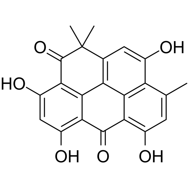
Resistomycin, a pentacyclic polyketide antibiotic, possesses anticancer activity and induces apoptosis.
商品は「研究用試薬」です。人や動物の医療用・臨床診断用・食品用の製品ではありません。
研究用途以外に使用した場合、当社は一切の責任を負いかねます。
- 純度 : 99.9%
- CAS 番号: 20004-62-0
- 分子式: C22H16O6
- 分子量:376.36
-
保管条件:Powder -20°C, 3 years ; In solvent -80°C, 6 months , -20°C, 1 month

To place orders, for customer services and technical support, please contact: MedChemExpress USA
Tel: 609-228-6898 E-mail: [email protected] [email protected]


-
 生物活性
生物活性
-
化学情報
- プロトコル
- 純度とドキュメンテーション
-
 Help & FAQs


Antibiotic アイソフォーム固有の製品をすべて表示
More

生物活性
製品説明
Cellular Effect
|
Cell Line

|
Type | Value | Description | References |
|---|---|---|---|---|
| CCRF-CEM | IC50 |
0.5 μM
Compound: 1
|
Antiproliferative activity against human CEM cells after 72 hrs by MTT assay
Antiproliferative activity against human CEM cells after 72 hrs by MTT assay
|
[PMID: 29137865] |
| CCRF-CEM | IC50 |
0.5 μM
Compound: 5
|
Antiproliferative activity against human CEM cells assessed as growth inhibition after 72 hrs by MTT assay
Antiproliferative activity against human CEM cells assessed as growth inhibition after 72 hrs by MTT assay
|
[PMID: 33974416] |
| HCT-116 | IC50 |
0.1 μM
Compound: 1
|
Antiproliferative activity against p53 deficient human HCT116 cells after 72 hrs by MTT assay
Antiproliferative activity against p53 deficient human HCT116 cells after 72 hrs by MTT assay
|
[PMID: 29137865] |
| HCT-116 | IC50 |
0.3 μM
Compound: 1
|
Antiproliferative activity against human HCT116 cells after 72 hrs by MTT assay
Antiproliferative activity against human HCT116 cells after 72 hrs by MTT assay
|
[PMID: 29137865] |
| HeLa | IC50 |
0.1 μM
Compound: 1
|
Antiproliferative activity against human HeLa cells after 72 hrs by MTT assay
Antiproliferative activity against human HeLa cells after 72 hrs by MTT assay
|
[PMID: 29137865] |
| HeLa | IC50 |
0.1 μM
Compound: 5
|
Antiproliferative activity against human HeLa cells assessed as growth inhibition after 72 hrs by MTT assay
Antiproliferative activity against human HeLa cells assessed as growth inhibition after 72 hrs by MTT assay
|
[PMID: 33974416] |
| HMEC-1 | IC50 |
0.5 μM
Compound: 1
|
Antiproliferative activity against HMEC1 cells after 72 hrs by MTT assay
Antiproliferative activity against HMEC1 cells after 72 hrs by MTT assay
|
[PMID: 29137865] |
| HMEC-1 | IC50 |
0.5 μM
Compound: 5
|
Cytotoxicity against human HMEC-1 cells assessed as growth inhibition after 72 hrs by MTT assay
Cytotoxicity against human HMEC-1 cells assessed as growth inhibition after 72 hrs by MTT assay
|
[PMID: 33974416] |
| K562 | IC50 |
0.3 μM
Compound: 1
|
Antiproliferative activity against human K562 cells after 72 hrs by MTT assay
Antiproliferative activity against human K562 cells after 72 hrs by MTT assay
|
[PMID: 29137865] |
| L1210 | IC50 |
0.7 μM
Compound: 1
|
Antiproliferative activity against mouse L1210 cells after 72 hrs by MTT assay
Antiproliferative activity against mouse L1210 cells after 72 hrs by MTT assay
|
[PMID: 29137865] |
| L1210 | IC50 |
0.7 μM
Compound: 5
|
Antiproliferative activity against mouse L1210 cells assessed as growth inhibition after 72 hrs by MTT assay
Antiproliferative activity against mouse L1210 cells assessed as growth inhibition after 72 hrs by MTT assay
|
[PMID: 33974416] |
| T-24 | IC50 |
26 μM
Compound: 1
|
Growth inhibition of human T24 cells after 72 hrs by cell impedance method
Growth inhibition of human T24 cells after 72 hrs by cell impedance method
|
[PMID: 29137865] |
体外実験
Resistomycin attenuates triple-negative breast cancer progression by inhibiting E3 ligase Pellino-1 and inducing SNAIL/SLUG degradation[1].
Resistomycin displays a high affinity for GST-Pellino-1 (KD = 2.58 μM) as indicated by surface plasmon resonance[1].
Resistomycin treatment reduced the expression of SNAIL and SLUG in TNBC cells[1].
Resistomycin induced apoptosis and cycle arrest in human hepatocellular carcinoma cells by activating p38 MAPK pathway in vitro and in vivo[3].
MedChemExpress (MCE) has not independently confirmed the accuracy of these methods. They are for reference only.


化学情報
-
CAS 番号 20004-62-0
-
性状 Solid
-
分子量 376.36
-
分子式 C22H16O6
-
Color Yellow to orange
-
SMILES
OC1=C(C(C2=C(O)C=C(C)C(C(O)=C3)=C2C4=C3C(C)(C)C5=O)=O)C4=C5C(O)=C1
-
Structure Classification
-
Initial Source
Streptomyces sulphureus
-
輸送条件
Room temperature in continental US; may vary elsewhere.
-
保管条件
Powder -20°C 3 years In solvent -80°C 6 months -20°C 1 month


純度とドキュメンテーション
-
データシート (248 KB)
-
SDS (557 KB)
- English - EN (557 KB)
- Français - FR (557 KB)
- Deutsch - DE (557 KB)
- Norwegian - NO (557 KB)
- Español - ES (557 KB)
- Swedish - SV (557 KB)
- Italian - IT (557 KB)
- Korean - KR (557 KB)
- Portuguese - PT (557 KB)
-
取扱説明書 (2659 KB)

Calculators
濃度 (開始) × 体積 (開始) = 濃度 (終了) × 体積 (終了)


Resistomycin 関連分類
HY-N10449 関連分類
 Powered by Bioz